The study was carried out at three upazillas of Cox’s Bazar from June to August, 2012. A total of 17 commercial layer farms which comprised at least thousand birds were randomly selected with the diagnosis of ND. Data were recorded according to clinical signs exhibited by individual bird during illness, strains of birds, number of birds in farm, age, vaccination schedule and date of last vaccination, number of dead birds, treatment measures if taken and information about management system of farm.
Diagnosis
In Upazilla Veterinary Hospital, poultry diseases were diagnosed on the basis of post mortem findings, clinical signs and as well as various laboratory tests such as Haemagglitination Inhibition (HI) test, Enzyme Linked Immune Sorbent Assay (ELISA) technique, Viral Neutralization (VN) test. Currently HI test is most widely used technique to detect antibodies to NDV in poultry sera.
Post Mortem Findings
These findings were varied but generally include haemorrhagic lesions on the proventriculus and intestinal tract which include pin point haemorrhage on the tip of proventriculas and small ecchymotic haemorrhages on the lymphatic nodule, Payer’s patches followed by intestinal mucosa. Edematous, haemorrhagic and degenerative ovaries, button ulcer on the caecum and spleenomegaly were observed (Figure 1). Other lesions accordingly hyperemia and congestion in respiratory tract, serous or catarrhal exudates in larynx and trachea, thickened air sacs containing yellow exudates were also frequent.
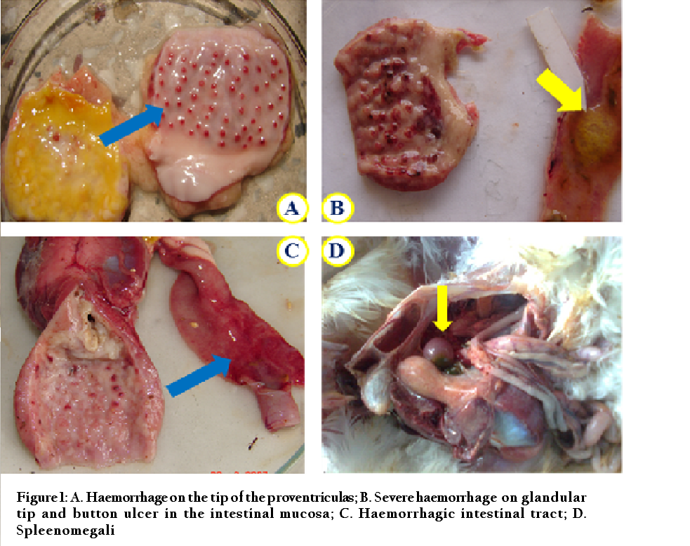
Figure 1: A. Haemorrhage on the tip of the proventriculas; B. Severe haemorrhage on glandular tip and button ulcer in the intestinal mucosa; C. Haemorrhagic intestinal tract; D. Spleenomegali
Clinical Signs
Signs of ND followed as sudden death without sign, ruffled feathers, depression, prostration, edematous head and wattles, nervous signs as paralysis, torticollis and respiratory signs as gasping and coughing.
Collection of Samples
The sample, 1ml of blood from 179 birds (8 samples per thousand birds) was collected from each bird through wing vein puncture; Sera were separated and stored at –20°C until analyzed. Avinew® ND, VG/GA antigen lentogenic strain (Advance Animal Science Co. Ltd. Dhaka, Bangladesh) was used in this study to conduct HI test to detect the immune status against NDV.
HI Test to Screen Level of Antibodies in Blood Sera
HI test was performed in V–bottomed microtitration plates. Test procedure was conducted according to the methodology of OIE Manual (2002). For preparation of 1% chicken Red Blood Cells (RBCs) used in the test, 5ml blood was taken from chicken that had not been primed with Newcastle Disease vaccine. RBCs were collected by centrifuging blood at 1500rpm for 15minutes and these collected RBCs were pooled in an equal volume of Alsever’s solution. After that, RBCs were washed thrice in phosphate buffered saline (PBS) before using as 1% (packed cell v/v) suspension. Serum was tested in two fold serial dilutions upto 10th well in microtiter plate. For virus suspension 4 Haemagglutinating Units (HAU) Avinew® ND, VG/GA antigen lentogenic strain was added upto 11th well. After keeping the plates at room temperature for 30minutes 1% chicken RBC suspension was added to each well. The 11th well contains antigen and RBCs as the positive control and the 12th well contains only RBCs as the negative control. After gentle mixing, the RBCs were allowed to settle at room temperature for 40minutes and agglutination was assessed by tilting the plates. Finally, at the end of 40minutes samples were showing central button shaped settling of RBCs which were recorded as positive and maximum dilution of each sample causing Haemagglutination Inhibition (HI). This was used to estimate the HI titer (Figure 2). The HI titer of each serum sample was expressed as reciprocal of the serum dilution.
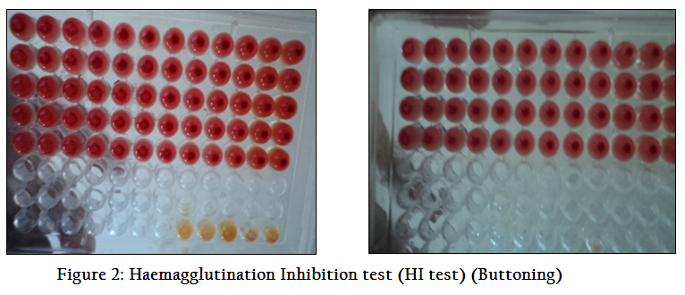
Figure 2: Haemagglutination Inhibition test (HI test) (Buttoning)
Data Analysis
Data that were primarily stored at MS excel (Microsoft office excel–2007, USA). Descriptive analysis was through by STATA version 11 (STATA Corporation, Texus, USA) and results were expressed as proportion ± SEM, 95% Confidence interval (CI) and also followed as logarithm table.