
The objective of the current study was to determine the prevalence of contagious ecthyma along with the association of various risk factors in goats and sheep through qualitative enzyme linked immunosorbent assay (ELISA). This technique is sensitive and cost-effective for detecting disease in large no of animals. For this purpose, serum samples from 350 goats and 91 sheep were collected on the 20th day post-infection for detection of antibodies, and essential information related to potential risk factors was collected through a questionnaire from fourteen districts of Punjab, Pakistan. Serologically positive samples were processed through PCR for confirmation using scab samples by identifying the GIF/IL-2 gene. This study found an overall 13.2% seroprevalence of contagious ecthyma infection, indicating a higher percentage in goats (14.6%) than in sheep (7.7%). In goats, the significant association (P<0.0001) of contagious ecthyma infection with seroprevalence was found in 4 districts; however, contagious ecthyma antibodies titers were found positive in sheep of Muzaffargarh only. The risk factors such as young age, female gender, grazing without stall feeding, presence of lesions on the lips, and injuries on the skin caused by prickly trees and cotton stubbles were found significantly (p<0.05) associated factors in goats; and age and injuries on the skin were important associated risk factors in sheep. The maximum occurrence of contagious ecthyma infection was found from September to December in both goats and sheep; having a significant association in goats (χ2=39.2) (P<0.0001). Likewise, the purchase of new animals and mixing up with the existing animals without quarantine measures cause contagious ecthyma to prevail in the vicinity. Moreover, contagious ecthyma antibodies were detected by ELISA kits up to 50th day post-infection in the current study. This is the first study that reported the seroprevalence of contagious ecthyma in Punjab, Pakistan. Comprehension of risk variables will not only be helpful to develop awareness amongst farmers but will also provide guidelines to government officials about the prevention and control of the disease.
Research Article · Volume 55, Issue 5, 31 July 2023 · Open Access
Seroprevalence of Contagious Ecthyma and Its Associated Risk Factors in Sheep and Goats of Punjab, Pakistan
· · · ·
Affiliations & Notes ⌄
- 1Department of Pathology, Faculty of Veterinary Sciences, University of Veterinary and Animal Sciences, Lahore, Pakistan, 54000, Pakistan 2Institute of Microbiology, Faculty of Veterinary Sciences, University of Veterinary and Animal Sciences, Lahore, Pakistan, 54000, Pakistan * Corresponding author: drniazi@uvas.edu.pk
Article Info ⌄
- Journal:
- Pakistan Journal of Zoology
- Published:
- 31 July 2023

From the article
- Our results showed that seroprevalence of contagious ecthyma were higher in goats (14.57%) than in sheep (7.71%).
- Besides, age was also observed to be a significant determinant of infection in goats and sheep and our study indicated that kids and lambs are more susceptible to ORF virus infection (Tables II, III).
- Our study also reported that brining new animals in a herd increased the susceptibility of animals to ORF virus infection.
- The occurrence of contagious ecthyma was found to be significantly associated with a number of risk factors, including species, gender, age, breed, presence or absence of lesions, seasons, and portable methods of transmission.
Selected from the article's own text.
Abstract
INTRODUCTION
Most of the world’s population relies on agriculture and livestock to make a living . In the tropical and subtropical areas, goats are becoming more attractive as a livestock species due to their low food costs and ease of rising with minimal maintenance needs . These animals also make up a large part of Pakistan’s livestock population and play a pivotal role in the provisions of hides, meat, and thus income for farmers in the rural areas of the country. Pakistan intends to boost livestock exports internationally . However, various infectious and non-infectious diseases pose a constant threat to the production of these animals and thereby economic losses. Contagious ecthyma, commonly identified as contagious pustular dermatitis or orf, is among one of these diseases.
Its causative agent, parapoxvirus belongs to the sub-family Chordopoxvirinae of the Poxviridae, is a persistent issue among goats and sheep in Punjab, Pakistan. The viral genome has a linear double-stranded DNA genome. It is a particular skin disease of a wide range of ruminants including sheep and goats and is endemic worldwide ; .
Animals that have been exposed to the ORF virus earlier might carry the virus in their hide or dried scabs and shed it into the environment. In the environment, the ORF virus has a higher survival, especially in tropical climates . Direct contact with infected animal and/or contaminated fomites containing the ORF virus causes transmission. The virus usually enters the skin through cuts or abrasions and establishes infection. From skin reddening (erythema) to macule, papule, vesicle, pustule, scab, and scar, the skin lesions develop and progress in a variety of stages ; . Although this disease is more common amongst young animals, few adult animals may also be distressed. Dehydration and malnutrition are the leading causes of death in young animals, owing to pain caused by oral cavity lesions that prohibit them from suckling their dam. Similarly, proliferative lesions, especially on the lips, badly affect the feed intake of adult animals.
Contagious ecthyma can be diagnosed in various ways like the detection of scabby lesions around the oral cavity, limbs, or teats, or through histopathology of epidermal lesions ; . The culture of the virus on cell lines ; ; is also good but it is considered as strenuous and much time-consuming procedure . Molecular tests for specific ORFV strains are also successfully used for its diagnosis but these tests are not effective for screening at the herd level . Contrarily, the qualitative enzyme linked immunosorbent assay (ELISA) approach is sensitive and affordable for detecting ORF viruses in a large population of animals in the field . The presence of antibodies (IgG) in a sero-converted animal indicates the existence of sustained infection of contagious ecthyma, which helps in distinguishing freshly infected animals from long-term contagious ecthyma infection . Therefore, ELISA was preferred for screening of ORFV in this study.
Normally, this disease cures on its own. Nevertheless, its economic burden is enormous in terms of mortality and wasting in severe cases due to secondary bacterial or fungal infections or delayed treatment . Moreover, its morbidity can also be as high as 100% ; . In Pakistan, owing to the lack of any study on this disease, nothing can be said for sure about its exact economic repercussions. However, the disease has a high economic cost as indicated by an estimate showing a national expense of approximately ten million pounds on ORFV in the British sheep sector . Because of the aforementioned context, this study was planned to estimate the rate of current ORF virus infection in goats and sheep based on detecting IgG antibodies in fourteen districts of Punjab, Pakistan. The study also attempted to estimate the association of potential risk factors with the occurrence of ORFV infection in goats and sheep. Seropositive samples were then submitted for molecular validation through PCR assay.
MATERIALS and METHODS
Ethical statement
All experimental procedures were carried out in accordance with guidelines approved by the University of Veterinary and Animal Sciences (UVAS), Lahore, Pakistan’s ethical review committee (Permit No. DR/452 dated 06/10/2020).
Study area and sample size
The study area comprised of 14 districts of Punjab having over 220,000 sheep and goats. Punjab is located at 31.1704° N, 72.7097° E.
There had been no previous reports on the prevalence of contagious ecthyma in Pakistan, consequently, the sample size was determined for a disease with a 50% predicted prevalence at a 95% confidence range and a 5% desirable absolute precision . According to elaboration, the sample size was calculated via sample size calculator available at (https://www.surveysystem.com/sscalc.htm), expecting the population of goats and sheep over 220,000 yielding a total sample size of 383.
Goats and sheep that appeared to have recovered from contagious ecthyma, as well as animals with nodular to diffuse proliferative skin lesions around the lips, muzzle, teats, and nostrils, were considered for serum sample collection. We studied n=350 goats and n=91 sheep. The study was carried out for a year from July 2020 to July 2021.
Sample collection and processing for ELISA
Blood samples (3 ml) were taken aseptically from the jugular vein using sterile syringes and transferred to anticoagulant-free test tubes for serum separation . Blood samples were collected at day 20 and day 50. After collection of the whole blood, allowed the blood to clot by leaving it undisturbed at room temperature for 10-20 min. The clot was removed by centrifuging at 2,000-3,000 rpm for 20 min. The serum was subsequently transferred to a fresh 1.5 ml micro-centrifuge tube and stored at -20°C until the indirect Sandwich-ELISA test was performed. The samples were again centrifuged, if any precipitations appeared during storage .
The serum samples were tested qualitatively for anti-contagious ecthyma IgG in animals presenting clinical indications and seemingly healthy animals in the herd using the enzyme-linked immunosorbent assay (ELISA) technique. The contagious ecthyma IgG was carried out according to the manufacturer’s instructions, using goat and sheep micro-ELISA strip plates (Catalogue Number: SL0049Gt, SL00097Sp; SunLong Biotech Co., LTD). In the microplate, micropores were labeled according to sample IDs; whereas, left two wells were designated as negative controls, two wells adjacent to negative controls as positive controls, and one empty well as a blank control. In the negative and positive control wells, 50 µl of respective controls were added. 40 µl sample dilution buffer and 10 µl sample were introduced to the sample wells. Without touching the well wall, samples were loaded onto the bottom. The samples were thoroughly mixed with gentle shaking before being sealed with the closure plate’s membrane and incubated at 37°C for 30 min. After incubation, the closure plate membrane was gently removed, and the reaction was rinsed 5 times with a wash solution at 30 second intervals. Except for the blank control well, each well received 50 µl of HRP-conjugate reagent after 5 washing cycles. The incubation and cleaning procedures were repeated once more. After that, 50 µl of each chromogen solution A and chromogen solution B were added to each well, stirred gently in a dark environment, and incubated at 37°C for another 15 min. By injecting 50 µl of stop solution into each well, the reaction was brought to a halt. In the well, there was a change in colour from blue to yellow.
A microtiter plate reader was used to read the absorbance Optical Density (O.D.) at 450 nm within 15 min of adding the stop solution (PR4100 Microplate Reader; Bio-Rad Laboratories). The test kit’s effectiveness was confirmed because the overall positive control result was larger than 1.00. The cut-off value was calculated using the following formula. Cut-off (CO) value = mean of two negative controls + 0.15 .
Study of risk factors
The well-structured questionnaire was distributed to farmers and personnel working in the field to study the prevalence of contagious ecthyma regarding the presence of IgG antibodies. The potential risk factors studied in goats and sheep included area (different districts), species, age, gender, breed, presence or absence of gross lesions, presence of orf lesions on different body parts, feeding, injuries on skin, faring system, purchase of new animals.
Molecular confirmation by PCR
The dermal tissue samples were collected from sheep and goats with possible diagnostic lesions on the oral commissure, muzzle, lips, and nose. Samples were taken from animals having nodular and widespread proliferative cutaneous lesions. Skin tissue samples exhibiting nodular lesions were gathered in a sterile polystyrene vial having antibiotics (penicillin 1,000 IU/mL, streptomycin 1,000 g/mL, kanamycin 500 g/mL) and antifungal (amphotericin B 25 mg/mL) reagents. The obtained samples were kept at -20°C until they were analyzed in the laboratory. The GeneJET Genomic DNA Purification Kit was used to extract genomic DNA (Thermo Fisher Scientific, Catalog Number: K0722). ORFV’s GIF/IL-2 gene was amplified using forward (5’-GCTCTAGGAAAGATGGCGTG-3’) and reverse (5’-GTACTCCTGGCTGAAGAGCG-3’) primers previously described .
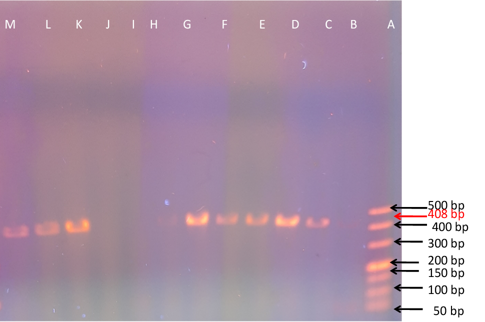
The PCR procerdure was carried out with a total volume of 40 µL reaction mixture, that included 20 µL master mix (PrimeSTAR Max DNA polymerase, Catalog Number: R045A), 2 µL of DNA template, 2 µL each forward and reverse primers and 14 µL PCR grade water. The PCR reaction setting were adjusted as initial denaturation (95°C, 5 min), afterwards 35 cycles of denaturation (94°C, 30 sec), annealing (58°C, 30 sec), and extension (72°C, 45 sec), subsequently a final extension (72°C, 7 min). Amplicons were seen on 1.5 percent agarose gel, stained with GelRed™ Nucleic Acid Gel Stain during gel electrophoresis (110V, 230mA, 30 min) and being recorded in a gel documentation system (Bio-Rad Laboratories, United States).
Statistical analysis
The Pearson Chi-square (χ2) test was applied to evaluate the association between potential risk factors. The odds ratio was calculated using Fisher’s exact test to evaluate the probability of risk factors within the research region. The level of significance was recognized at a 95 percent confidence interval in each procedure. For statistical analysis, SPSS version 22 was employed.
RESULTS
Overall sero-prevalence of contagious ecthyma
The sero-prevalence of contagious ecthyma was studied in a total of 441 sheep and goats samples from 14 different districts of Punjab province, Pakistan. The results revealed that the overall 58 samples were detected seropositive for orf virus, which show overall 13.2% seroprevalence in the Punjab province, Pakistan (Table I). Goats had a percentage positivity of 14.57, whereas sheep had a percentage positivity of 7.71. Moreover, the findings revealed that species were not significantly (χ2=2.9) (P=0.08) linked to contagious ecthyma. Goats had a 2.04 times higher chances of having contagious ecthyma than sheep. The commercially available ELISA kits detected IgG as early as 20th day post infection and level of antibodies was identified till 50th day post infection. Serologically positive samples were processed through PCR for confirmation. Positive amplified bands typical for GIF/IL-2 gene were revealed as shown in Figure 3 and Table I. Approximately 408 bp PCR amplified products of GIF/IL-2 gene of contagious ecthyma virus were produced in the procedure. Overall sero-prevalence of contagious ecthyma has been shown in Table I.
Sero-prevalence of contagious ecthyma virus among various districts
The results showed that the sero-positive samples for contagious ecthyma in goats were highest in districts Lahore, Lodhran and Dera Ghazi Khan (48.57, 42.42 and 41.38%, respectively). There was significant difference (χ2=112.5) (P<0.0001) in sero-positive samples of orf virus in goats among various districts, as shown in Figure 1A. The contagious ecthyma in sheep was sero-positive (53.8%) only in Muzaffargarh among various districts, as shown in Figure 2B.
Sero-prevalence of contagious ecthyma in goats based on risk factors
The higher sero-prevalence of contagious ecthyma in goats was found in 0-4 months kids (45.6%), as shown in Table II. The association amongst age groups for contagious ecthyma was significant (χ2=65.2) (P<0.0001) and young kids were 10.9 times more sero-positive than adults goats. The total of 46 (16.8%) female goats samples were sero-positive for contagious ecthyma, as shown in Table II. Gender based results showed significantly greater contagious ecthyma cases in female goats (χ2=4.9) (P=0.02). Female goats had 0.3 times larger probability of having contagious ecthyma than the males. The highest sero-prevalence of orf virus was in non-descript breed of goats (18.3%) and 5.4 times more orf virus sero-positive as compared to other breeds of goats, as shown in Table II. Whereas, the breed-wise prevalence was significantly associated with occurrence of contagious ecthyma in cases of goats (χ2=8.1) (P=0.04). The highest sero-prevalence of orf virus was in Non-descript breed of goats (18.3%) and non-descript breeds were 5.4 times more likely to have contagious ecthyma than Nachi, Deera Din Pananh, and Beetal, as shown in Table II. contagious ecthyma lesions, as well as their distribution on different body parts, were evaluated in relation to the occurrence of contagious ecthyma in goats. The higher percentage (21.8%) of contagious ecthyma was found in goats having gross lesions as shown in Table II. In goats, the presence of lesions were found to be significantly (χ2=19.9) (P<0.0001) associated with occurrence of contagious ecthyma. The percentage sero-positivity of contagious ecthyma in goats was significantly higher in animals that had lips lesion (19.5 %) as compared to infected animals having lesions on muzzle or nostril. The location of lesions on various body parts was significantly (χ2=11.3) (P=0.004) associated in goats. The occurrence of contagious ecthyma in goats was 3.7 times higher with lesions on their lips. Sero-prevalence of contagious ecthyma in goats based on risk factors has been shown in Table II.
Sero-prevalence of contagious ecthyma in sheep based on risk factors
The higher percentage (85.7 %) of sero-positivity was found in young lambs as shown in Table III. The association amongst age groups for contagious ecthyma was significant (χ2=65.2; P=0.0001) and the odd ratio (OR) showed that contagious ecthyma infect young lambs 498 times higher than adults, as shown in Table III.The association of sero-positivity of ORF virus among male
Table I. Overall sero-prevalence of contagious ecthyma.
| Species | Total sample (n) | Seropositive %(n) IgG | Confirmation through PCR (%) n | Chi-square value | P value | OR | CI |
| Goat | 350 | 14.6(51) | 14.6(51) | 2.9 | 0.08 | 2.04 | (0.8,4.6) |
| Sheep | 91 | 7.7(7) | 7.7(7) | ||||
| Overall | 441 | 13.2(58) | 13.2(58) |
OR show odd ratio, CI show confident intervals, which was 95%.
Table II. Association of sero-prevalence of contagious ecthyma with relative risk factors in goats.
| Variables | Categories | n | Sero-positive % (n) | Chi-square value | P value | OR | CI |
| Age | Young (0-4 months) | 68 | 45.6(31) | 65.2*** | 0.0001 | 10.9 | (5.6, 21.1) |
| Adults (5 months-3 years) | 282 | 7.1(20) | |||||
| Gender | Male | 76 | 6.6(5) | 4.9* | 0.02 | ||
| Female | 274 | 16.8(46) | 0.3 | (0.1, 0.9) | |||
| Breeds | Beetal | 51 | 3.9(2) | 8.1* | 0.04 | ||
| Dera Din Panah | 34 | 8.8(3) | 2.3 | (0.3, 15.0) | |||
| Nachi | 46 | 13(6) | 3.6 | (0.7, 19.1) | |||
| Non Descript | 219 | 18.3(40) | 5.4 | (1.2, 23.2) | |||
| Orf lesions | Present | 202 | 21.8(44) | 19.9*** | 0.0001 | 5.6 | (2.5, 13.2) |
| Absent | 141 | 4.7(7) | |||||
| Lesions on body parts | Lips | 214 | 19.5(42) | 11.3** | 0.004 | 3.7 | (1.1, 12.7) |
| Muzzle | 104 | 6.7(7) | 1.1 | (0.2, 5.5) | |||
| Nostril | 32 | 6.3(2) |
n, total sample size; OR, odd ratio; CI, 95% confident intervals; * indicated P=0.02-0.04; ** indicated P=0.004 and *** indicated P=0.0001.
Table III. Relative risk factors association with sero-prevalence of contagious ecthyma in sheep.
| Variables | Categories | n | Sero-positive % (n) | Chi-square value | P value | OR | CI |
| Age | Young (0-4 months) | 7 | 85.7 (6) | 65.2* | 0.0001 | 498 | (32.6, 851.1) |
| Adults (5 months-3 years) | 84 | 1.2 (1) | |||||
| Gender | Male | 12 | 8.3 (1) | 0.009 | 0.9 | ||
| Female | 79 | 7.6 (6) | 1.1 | (0.3, 10.9) | |||
| Breeds | Kajli | 9 | 22.2 (2) | 3.6 | 0.1 | ||
| Thalli | 7 | 14.3 (1) | 1.1 | (0.1, 25.1) | |||
| Non Descript | 75 | 5.3 (4) | 2.3 | (0.2, 16.8) | |||
| Orf lesions | Present | 68 | 8.8 (6) | 0.5 | 0.4 | 2.1 | (0.5, 7.2) |
| Absent | 23 | 4.3 (1) | |||||
| Lesions on body parts | Lips | 31 | 16.1 (5) | 4.7 | 0.09 | 5.5 | (0.6, 62.7) |
| Muzzle | 30 | 3.3 (1) | 1.1 | (0.2, 20.5) | |||
| Nostril | 30 | 3.3 (1) |
n, total sample size; OR, odd ratio; Cl, 95% confident intervals, * indicate P=0.0001
and female sheep was not significant (χ2=0.009) (P=0.9).Female sheep had 6 times larger probability of having contagious ecthyma than the males. Similarly, the association of sero-prevalence of contagious ecthyma was not significant among various breeds of sheep (χ2=3.6) (P=0.1). However, according to OR chances of contagious ecthyma was 2.3 times higher in Non-descript breed as compared to other breeds of sheep in Punjab Province. In sheep, the presence of lesions were found to be non-significantly (χ2=0.5) (P=0.4) associated with occurrence of contagious ecthyma. The location of lesions on various body parts was non-significantly (χ2=0.5) (P=0.4) associated in sheep. The percentage sero-positivity of contagious ecthyma in sheep was significantly higher in animals that had lips lesion (16.1 %) as compared to infected animals having lesions on muzzle or nostril. The occurrence of contagious ecthyma in sheep was 5.5 times higher with lesions on their lips. Sero-prevalence of contagious ecthyma in sheep based on risk factors has been shown in Table III.
Association of sero-prevalence of contagious ecthyma with months in sheep and goats
The higher percentage (78.4 and 71.4%) of sero-positivity was found in goats and sheep respectively from September to December months. The association for occurrence of contagious ecthyma was significant (χ2=78.4) (P=0.0001) in goats from September to December, as shown in Figure 2A. However, the association for prevalence of contagious ecthyma was non-significant (P=0.7) in sheep. Moreover, lower seroprevalence of contagious ecthyma from the months of May to August in goats, while in sheep sero-prevalence of contagious ecthyma was remain constant from January to August, as shown in Figure 2B. Association of sero-prevalence of contagious ecthyma with months has been shown in Figure 2A, B.
Association of sero-prevalence of contagious ecthyma with management risk factors in goats and sheep
The higher percentage (20 %) of sero-positivity was found in grazing goats. The association amongst various management risk factors for contagious ecthyma were significant (χ2=7.6) (P=0.002) as shown in Table IV and Figure 4. However, association for seroprevalence of contagious ecthyma in sheep was non-significant (χ2=0.42)
Table IV. Association of sero-prevalence of contagious ecthyma with management risk factors in goats.
| Risk factor | Categories | n | Seroprevalence % (n) | Chi square value | P value | OR | CI |
| Feeding | Grazing | 160 | 20(32) | 7.6* | 0.02 | 3.3 | (1.6, 9.1) |
| Grazing + Stall | 133 | 11.3(15) | 1.6 | (0.6, 5.4) | |||
| Stall feeding | 57 | 7(4) | |||||
| Injuries on skin | Present | 200 | 20(40) | 11.05** | 0.001 | 3.1 | (1.8, 12.6) |
| Absent | 150 | 7.3(11) | |||||
| Farming system (goat/sheep) | Mixed | 186 | 15.1(28) | 0.07 | 0.7 | 1.1 | (0.1, 3.4) |
| Separated | 164 | 14(23) | |||||
| Purchase of new animals | Yes | 250 | 19(40) | 8.4** | 0.004 | 2.7 | (1.1, 10.3) |
| No | 100 | 7.9(11) |
n, total sample size; OR, odd ratio; Cl, 95% confidant intervals; * indicate significance value P=0.02 and ** indicate significance value P=0.004-0.001
Table V. Association of sero-prevalence of contagious ecthyma with management risk factors in sheep.
| Risk factor | Categories | n | Seroprevalence % (n) | Chi square value | P value | OR | CI |
| Feeding | Grazing | 55 | 9.1(5) | 0.42 | 0.8 | 2.0 | (0.41, 5.22) |
| Grazing + Stall | 15 | 6.7(1) | 1.4 | (0.35, 4.61) | |||
| Stall feeding | 21 | 4.8(1) | |||||
| Injuries on skin | Present | 44 | 13.6(6) | 4.23* | 0.04 | 3.1 | (1.45, 8.11) |
| Absent | 47 | 2.1(1) | |||||
| Farming system (goat/sheep) | Mixed | 57 | 8.8(5) | 0.25 | 0.6 | 1.5 | (0.2, 3.8) |
| Separated | 34 | 5.9(2) | |||||
| Purchase of new animals | Yes | 44 | 13.6(6) | 4.23* | 0.04 | 3.1 | (1.45, 8.11) |
| No | 47 | 2.1(1) |
n, total sample; OR, odd ratio; CI, 95% confident intervals; * indicate significance value P= 0.04
(P=0.8) amongst various groups: grazing, stall feeding and grazing and stall feeding animals, as shown in Table V. The seroprevalence of contagious ecthyma highly depends on cuts on skin as shown in findings: The seroprevalence of contagious ecthyma was significantly high in injured goats (χ2=11.05) (P=0.001) and sheep (χ2=4.23) (P=0.04) as compared to non-injured animals, as shown in Tables IV, V. There was no significant (P>0.05) association between seroprevalence of contagious ecthyma with farming system in both goats and sheep. The purchase of new animals was highly associated (P<0.05) with sero-prevalence of contagious ecthyma a in both goats and sheep. Association of sero-prevalence of contagious ecthyma with management risk factors in goats and sheep has been shown in Tables IV, V. Animals grazing areas and lips lesion in infected animals as shown in Figure 4.
DISCUSSION
This is the first study in Pakistan to present a detailed description of the prevalence of contagious ecthyma in goats and sheep using ELISA to detect antibodies and potential risk factors. Lahore, Lodhran, Dera Ghazi Khan, Multan, and Muzaffargarh had the most seropositive goats and sheep. In order to gain a clearer picture, sampling from a wider population of different areas of Punjab, Pakistan was conducted.
The prevalence of contagious ecthyma is higher in other parts of the world as compared to our findings, i.e., 13.2 percent ORF infection in our study as compared to 19.51 percent of lambs in England , 34.89 percent in China , 98 percent in the Nilgiri Hills in Tamil Nadu, India , and 54 percent in Saudi Arabia .
Our results showed that seroprevalence of contagious ecthyma were higher in goats (14.57%) than in sheep (7.71%). The prior studies also showed this trend. For example, prevalence of contagious ecthyma in sheep was 1.9 percent in England , 34.9 percent in goats in China , 25.1-36.4 in Malaysia and 76.6 percent in Indian goats . The higher prevalence of contagious ecthyma in goats might be due to the fact that caprine are naturally more aggressive than ovine and consequently caprine are more likely to injure one another, making themselves more susceptible to ORF virus ; . In goats and sheep, age was found to be a key driver of infection, suggesting that young goats and sheep are sensitive to ORF virus infection (Tables II, III). This conclusion is consistent with that of , who discovered that contagious ecthyma is more common in lambs. Besides, age was also observed to be a significant determinant of infection in goats and sheep and our study indicated that kids and lambs are more susceptible to ORF virus infection (Tables II, III). This result is in agreement with the findings of , who found that contagious ecthyma is more prevalent in lambs. Because of their immature immune systems, young animals were more susceptible to infection and acquired more serious lesions, which sometimes resulted in death . However, another study reported higher prevalence of contagious ecthyma in goats older than 8 months . Therefore, further studies are required to establish the role of age in the occurrence of contagious ecthyma.
In our study, gender was also found a significant driver of infection with females having higher seroprevalence of contagious ecthyma than males. An older study conducted by ; also reported a similar observation. However, some researchers showed that ORF virus infection does not discriminate between males and females ; . So, future studies should focus on establishing the role of gender in the susceptibility of contagious ecthyma in small ruminants. Male animals are usually slaughtered earlier as compared to female which are kept for longer period for breeding purpose and for yielding milk, male animals’ number is usually low in the area as compared to female animals.
To some extent, breed was also established as a risk factor for ORF virus infection in our study. Non-Descript goats and sheep had a greater prevalence of contagious ecthyma in this investigation. Worldwide, the findings of different researchers showed that contagious ecthyma is more common in some distinct breeds. For example, the seroprevalence of contagious ecthyma in Damara sheep and Indian, Chinese, and Boer goat breeds showed their higher susceptibility to ORF virus infection ; ; . Therefore, future studies based on sampling from a wider population of various breeds would provide necessary data to establish the role of breed as a risk factor of contagious ecthyma.
As far as association of lesions with occurrence of disease is concerned, very interesting results were found. In few animals, gross lesions were not found but the animals found positive for contagious ecthyma via ELISA test. This might be due to the presence of IgG antibodies in animals as these antibodies are usually produced after three weeks of initiation of infection and wounds are mostly cured till that time. Conversely, a large no of animals was found negative although gross lesions were present. The reason in those cases would also be the same i.e., IgG did not start developing as samples of sera were taken in the first week of the disease outbreak.
The results of different months showed the highest occurrence of contagious ecthyma in the months from September to December (Fig. 2). Bora et al. (2016) and Chi et al. (2013) reported that production of antibodies against orf infection in infected animals does not give long-term protection. Higher incidence in these months might be due to the fact that cotton crop is ready in the month of September and cotton stubbles are present along with the leaves to be consumed by small ruminants in our study area. These cotton stubbles cause wounds, cuts, and abrasions, all of which serve as a predisposing factor for the virus penetration through the skin ; (Fig. 4). The high prevalence of orf infection might be due to shedding of viruses from injure animals in the environment, which is viable for long time and they served as a source of seasonal outbreaks among same herd and also transmitted to neighbouring herds . The high prevalence of this disease highlights the infectious nature of ORF virus and its impact on the small ruminant industry ; . The high prevalence of this disease highlights the infectious nature of ORF virus and its impact on the small ruminant industry (Abd Elgowad et al., 2021; . Our study also reported that brining new animals in a herd increased the susceptibility of animals to ORF virus infection. We propose that the stress of traveling caused sickness and these stressed animals become more prone to ORF virus infection. However, Bala et al. (2019) reported that the infection could spread to other herds by moving infected animals. The both possibilities might be true in different cases.
PCR assay was used as confirmatory diagnostic tool for diseases. This assay has helped to distinguish ORF virus infection from other similar diseases affecting sheep and goats. All the serologically positive animals were confirmed through PCR assay on the scab or skin tissues samples . Skin/scab tissue from the nostrils, lips, oral commissure, and muzzle showing nodular and diffuse proliferative cutaneous lesions (various stages of lesions formation like papules followed by vesicles, pustules, and finally scab formation) were collected for sampling.
It is concluded that the prevalence of contagious ecthyma infection by detecting IgG antibodies in sheep and goats in Punjab, Pakistan, was (13.2%). It was also noted that goats (14.57%) had higher prevalence of contagious ecthyma than sheep (7.71%). The occurrence of contagious ecthyma was found to be significantly associated with a number of risk factors, including species, gender, age, breed, presence or absence of lesions, seasons, and portable methods of transmission. The findings of study will serve as a baseline for future studies and will provide essential information for policy makers and epidemiologist for controlling contagious ecthyma in study area and beyond.
ACKNOWLEDGEMENTS
Authors are highly thankful to Prof. Dr. Aneela Zameer Durrani, Department of Clinical Medicine, Dr. Arfan Ahmed, University Diagnostic Laboratory, Institute of Microbiology, University of Veterinary and Animal Sciences Lahore for technical assistance.
Financial support
This study was supported by the Higher Education Commission of Pakistan under the project Faculty Development Program for Pakistani Universities Batch II (17-5/FVS2-001/HEC/Sch-FDP/2019).
Statement of conflict of ınterest
The authors have declared no conflict of interest.
Figures
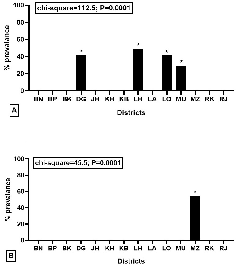
Sero-prevalence of contagious ecthyma virus among various districts in (A) goats and (B) sheep has been shown. BN, Bahawal Nagar; BP, Bahawalpur; BK, Bakkar; DG, Dera Ghazi Khan; JH, Jhang; KH, Kasur; KB, Khushab; LH, Lahore; LA, Layyah; LO, Lodhran; MU, Multan; MZ, Muzaffargarh; RK, Rahim Yar Khan; and RJ, Rajan Pur; *, 0.0001.
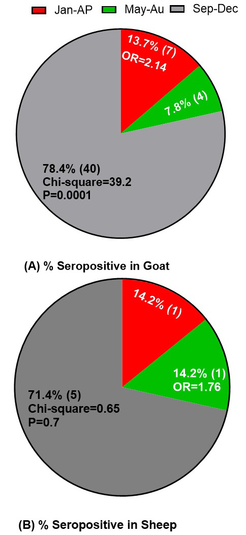
Association of sero-prevalence of contagious ecthyma with months in (A) goats and (B) sheep has been shown: “Jan-Ap” indicated from January to April, “May-Au” indicated from May to August and “Sep-Dec” indicated from September to December.
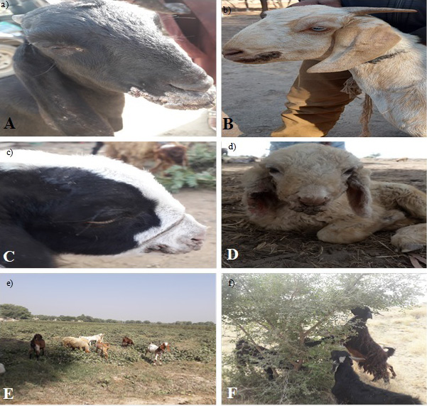
Lesions of contagious ecthyma and predisposing factors for disease. A and B, scab formation on lips in adult goats; C, scab formation in goat kid; D, scab formation in sheep kid affected with contagious ecthyma; E, goats were grazing in cotton field having cotton leaves and stubbles; F, goats were eating leaves of prickly tree.
References
- Abd-Elgowad, K.E.A., Aboezz, Z.R.A., El-mayet, F.S., El-Habbaa, A.S. and Ali, S.S., 2021. Molecular and serological diagnosis of Orf virus from sheep and goats in Minufiya governorate, Egypt. Benha Vet. Med. J., 41: 55-59. https://doi.org/10.21608/bvmj.2021.86728.1451[CrossRef]
- Amann, R., Rohde, J., Wulle, U., Conlee, D., Raue, R., Martinon, O. and Rziha, H.J., 2013. A new rabies vaccine based on a recombinant ORF virus (parapoxvirus) expressing the rabies virus glycoprotein. J. Virol., 87: 1618-1630. https://doi.org/10.1128/JVI.02470-12[CrossRef]
- Ashraf, M., Farid, I., Shehzad, W., Yousaf, M.R., Ahmad, N., Ihsan, A., Riaz, A., 2021. Effects of different nutritional regimens during pre-weaning period on body and testis growth, sexual behavior, hormonal profile and genes expression in male beetal goat kids. Pak. Vet. J., 41: 160-162.
- Bala, J., Balakrishnan, K., Abdullah, A., Yi, L., Bitrus, A., Abba, Y., and Mohamed, R.B., 2018. Sero-epidemiology of contagious ecthyma based on detection of IgG antibody in selected sheep and goats farms in Malaysia. Adv. Anim. Vet. Sci., 6: 219-226. https://doi.org/10.17582/journal.aavs/2018/6.5.219.226[Full text][CrossRef]
- Bala, J.A., Balakrishnan, K.N., Abdullah, A.A., Adamu, L., Noorzahari, M.S., May, L.K., Noordin, M.M., 2019. An association of Orf virus infection among sheep and goats with herd health programme in Terengganu state, eastern region of the peninsular Malaysia. BMC Vet. Res., 15: 1-15. https://doi.org/10.1186/s12917-019-1999-1[CrossRef]
- Balakrishnan, K.N., Abdullah, A.A., Bala, J., Abba, Y., Sarah, S.A., Jesse, F.F.A., Mohd-Azmi, M.L., 2017. Identification and comparison of RCMV ALL 03 open reading frame (ORF) among several different strains of cytomegalovirus worldwide. Infect. Genet. Evol., 54: 81-90. https://doi.org/10.1016/j.meegid.2017.06.020[CrossRef]
- Bora, M., Bora, D.P., Barman, N.N., Borah, B. and Das, S., 2016. Seroprevalence of contagious ecthyma in goats of Assam: An analysis by indirect enzyme-linked immunosorbent assay. Vet. World, 9: 1028. https://doi.org/10.14202/vetworld.2016.1028-1033[CrossRef]
- Chan, K.W., Lin, J.W., Lee, S.H., Liao, C.J., Tsai, M.C., Hsu, W.L., and Shih, H.C., 2007. Identification and phylogenetic analysis of orf virus from goats in Taiwan. Virus Genes, 35: 705-712. https://doi.org/10.1007/s11262-007-0144-6[CrossRef]
- Chan, K.W., Yang, C.H., Lin, J.W., Wang, H.C., Lin, F.Y., Kuo, S.T., and Hsu, W.L., 2009. Phylogenetic analysis of parapoxviruses and the C-terminal heterogeneity of viral ATPase proteins. Gene, 432: 44-53. https://doi.org/10.1016/j.gene.2008.10.029[CrossRef]
- Chi, X., Zeng, X., Hao, W., Li, M., Li, W., Huang, X., and Luo, S., 2013. Heterogeneity among orf virus isolates from goats in Fujian Province, Southern China. PLoS One, 8: e66958. https://doi.org/10.1371/journal.pone.0066958[CrossRef]
- Delhon, G., Tulman, E., Afonso, C., Lu, Z., De la Concha-Bermejillo, A., Lehmkuhl, H., Rock, D., 2004. Genomes of the parapoxviruses ORF virus and bovine papular stomatitis virus. J. Virol., 78: 168-177. https://doi.org/10.1128/JVI.78.1.168-177.2004[CrossRef]
- Fleming, S.B., Wise, L.M. and Mercer, A.A., 2015. Molecular genetic analysis of orf virus: A poxvirus that has adapted to skin. Viruses, 7: 1505-1539. https://doi.org/10.3390/v7031505[CrossRef]
- Gao, Y., Zhao, Y., Liu, J., Zhou, M., Liu, H., Liu, F., and Chen, D., 2016. Orf in goats in China: Prevalence and risk factors. J. Agric. Sci. Technol. A, 6: 116-123. https://doi.org/10.17265/2161-6256/2016.02.005[CrossRef]
- Hota, A., Biswal, S., Sahoo, N., Venkatesan, G., Arya, S., Kumar, A., and Rout, M., 2018. Seroprevalence of Capripoxvirus infection in sheep and goats among different agro-climatic zones of Odisha, India. Vet. World, 11: 66. https://doi.org/10.14202/vetworld.2018.66-70[CrossRef]
- Housawi, F., Elzein, E.A., Al Afaleq, A. and Amin, M., 1992. Sero-surveillance for Orf antibodies in sheep and goats in Saudi Arabia employing the ELISA technique. J. comp. Pathol., 106: 153-158. https://doi.org/10.1016/0021-9975(92)90044-U[CrossRef]
- Hussain, M.H., Asi, M.N., Al-Uahmadi, S.S.R., Al-Subhi, A.H.A., Al-Senaidi, N.Y.A., Al-Subhi, R.S.N., Mansoor, M.K., 2021. Seroprevalence and associated risk factors of contagious caprine pleuropneumonia in the small ruminants of Oman. Pak. Vet. J., 41: 45-50. https://doi.org/10.29261/pakvetj/2020.086[CrossRef]
- Jesse, F.F.A., Latif, S.N.A.A., Abba, Y., Hambali, I.U., Bitrus, A.A., Peter, I.D., Abdullah, A.A., 2018. Seroprevalence of orf infection based on IgM antibody detection in sheep and goats from selected small ruminant farms in Malaysia. Comp. clin. Pathol., 27: 499-503. https://doi.org/10.1007/s00580-017-2619-8[CrossRef]
- Karki, M., Venkatesan, G., Kumar, A., Kumar, S. and Bora, D.P., 2019. Contagious ecthyma of sheep and goats: A comprehensive review on epidemiology, immunity, diagnostics and control measures. Vet. Arh., 89: 393-423.
- Kinley, G.E., Schmitt, C.W. and Stephens-Devalle, J., 2013. A case of contagious ecthyma (Orf virus) in a nonmanipulated laboratory dorset sheep (Ovis aries). Case Rep. Vet. Med., 2013: 210854. https://doi.org/10.1155/2013/210854[CrossRef]
- Kumar, R., Trivedi, R., Bhatt, P., Khan, S., Khurana, S., Tiwari, R., and Chandra, R., 2015. Contagious pustular dermatitis (Orf disease) epidemiology, diagnosis, control and public health concerns. Adv. Anim. Vet. Sci., 3: 649-676. https://doi.org/10.14737/journal.aavs/2015/3.12.649.676[Full text][CrossRef]
- Li, W., Ning, Z., Hao, W., Song, D., Gao, F., Zhao, K., Luo, S., 2012. Isolation and phylogenetic analysis of orf virus from the sheep herd outbreak in northeast China. BMC Vet. Res., 8: 1-13. https://doi.org/10.1186/1746-6148-8-229[CrossRef]
- Onyango, J., Mata, F., McCormick, W. and Chapman, S., 2014. Prevalence, risk factors and vaccination efficacy of contagious ovine ecthyma (orf) in England. Vet Rec., 175: 326. https://doi.org/10.1136/vr.102353[CrossRef]
- Orgeur, P., Mimouni, P. and Signoret, J., 1990. The influence of rearing conditions on the social relationships of young male goats (Capra hircus). Appl. Anim. Behav. Sci., 27: 105-113. https://doi.org/10.1016/0168-1591(90)90010-B[CrossRef]
- Rafique, A., Nasir, S., Ashraf, A., Nawaz, Z., Zahid, F.M., Abbas, A. and Masood, S., 2021. Sero-surveillance and risk factors analysis of caprine toxoplasmosis in Faisalabad Punjab, Pakistan. Pak. Vet. J., 41.
- Ramesh, A., Vadivoo, V., Babu, S.S. and Saravanabava, K., 2008. Confirmatory diagnosis of contagious ecthyma by amplification of the GIF/IL-2 gene by PCR. J. Vet. Anim. Sci., 4: 208-210.
- Rehman, H.U., Rabbani, M., Ghafoor, A., Riaz, A., Awan, F.N. and Raza, S., 2020. First isolation and genetic characterization of bovine herpesvirus 1 from cattle in Pakistan. Pak. Vet. J., 40: 163-165. https://doi.org/10.29261/pakvetj/2020.092[CrossRef]
- Riaz, A., Javid, B., Shah, M.A.A., Raza, S., Yousaf, A., Sial, A. and Abbas, S., 2021. First report on the detection and molecular characterization of bovine herpesvirus 1 from a clinical case of infectious bovine rhinotracheitis in Pakistan. Pak. Vet. J., 41: 160-162. https://doi.org/10.29261/pakvetj/2020.084[CrossRef]
- Sadiq, M.A.A., Abdullah, F.F.J.C., Bitrus, A.A.A., Balakrishnan, K.N.B., and Lila, M., 2017. Severe persistent case of con-tagious ecthyma (Orf) in goats. J. Anim. Hlth. Prod., 5: 24-28. https://doi.org/10.14737/journal.jahp/2017/5.1.24.28[Full text][CrossRef]
- Spyrou, V. and Valiakos, G., 2015. Orf virus infection in sheep or goats. Vet. Microbiol., 181: 178-182. https://doi.org/10.1016/j.vetmic.2015.08.010[CrossRef]
- Thurman, R.J. and Fitch, R.W., 2015. Contagious ecthyma. N. Engl. J. Med., 372: E12. https://doi.org/10.1056/NEJMicm1304779[CrossRef]
- Zhang, K., Xiao, Y., Yu, M., Liu, J., Wang, Q., Tao, P., and Ning, Z., 2015. Phylogenetic analysis of three orf virus strains isolated from different districts in Shandong Province, East China. J. Vet. Med. Sci., 15: 0368. https://doi.org/10.1292/jvms.15-0368[CrossRef]
Cited by 1 article
In journals published by ResearchersLinks.
Services
Article usage statistics
Loading…