
The current study was conducted in district DG Khan of Punjab that is situated in the mid junction of all provinces of Pakistan. Owing to location at boundaries of provinces a large number of nomads moves from Baluchistan and Sindh to Punjab via DG Khan. Harsh climate and relatively less rain fall also contributing factors towards the establishment of infection. Contagious Ecthyma is an extremely contagious, highly communicable, economically important viral disease of skin that affects most of animals. The purpose of current study was to establish and familiarize a precise and rapid detection approaches with molecular diagnostic tool. The method relies on periodically thermo cycling heating and cooling of DNA in presence of certain chemical reagents. In order to detect and differentiate parapox infections from those induced by pox virus. A new parapox specific and sensitive PCR assay based on amplification of B2L gene was applied against conserved gene of Orf virus. Crusted scraps and biopsies from different portion of skin and blood were taken as sample for PCR. Gross, histopathological, hematological examination was carried out to rule out possible cause of infection and adopt preventive measure so that further losses may be avoided. 96 out of total were clinically positive with strong clue of contagious ecthyma. In current study 12 (25%) goats out of 48 were PCR positive. In case of sheep, 7 (14.58%) out of 48 were PCR positive. Tissues were taken for histopathological studies. Gross pathological finding includes scraps on commissure, ulcerations, vesicles on in oral cavity, erosion on dental pads, warts on external ears, ulceration on udder and scrotum. Increase in WBC, lymphocytes, monocytes count while decrease in RBC, platelets count and Hb concentration in diseased animals representing hematological investigation.
Research Article · Volume 6, Issue 1, 14 May 2019 · Open Access
Pathological Studies on Contagious Ecthyma in Naturally Infected Small Ruminants
· · · · ·
Affiliations & Notes ⌄
- 1Department of Pathology, Faculty of Veterinary Science, University of Veterinary and Animal Sciences, Lahore, Pakistan
- 2Department of Microbiology, Faculty of Veterinary Science, University of Veterinary and Animal Sciences, Lahore, Pakistan
- 3College of Wildlife Recourse, Northeast forestry University 26 Hexing Rd, Xiangfang Qu, Haerbin Shi, Heilongjiang
- 4Shanghai Veterinary Research Institute Graduate School Chines Academy of Agriculture Science. drbaloch466@hotmail.com
Article Info ⌄
- Journal:
- Hosts and Viruses
- Published:
- 14 May 2019
From the article
- Initially PCR was only tool to differentiate Orf Positive or negative animals.
- Eleven (91.6%) goats and six (85.71%) sheep developed ulceration and erosion on dental pad.
- Due to foot lesion, two (28.5%) sheep were found lamed throughout the study and showing FMD like disease.
- Different parameters were estimated for hematological investigation by complete blood count such as white blood cells (WBC) count, red blood cell (RBC) count, hemoglobin estimation platelets (PLT) count, packed cell volume (PCV) analysis from PCR positive animals.
Selected from the article's own text.
Abstract
Introduction
Small ruminants have a significant role in terms of generating income in the form of meat, hide, wool and milk to landless and small or marginal farmers particularly in arid and semi-arid regions of the country. However, health challenges deterring by infectious diseases are still hindering the productivity and causes huge losses to economy ; Koufakiset al., 2014). It is an acute, contagious, transferable and economically significant skin disease that puts detrimental effects on animals, principally small ruminants and other native and wild ruminants. Contagious ecthyma also known as infectious labial dermatitis, pustular dermatitis, sore mouth or scabby mouth disease with a worldwide distribution in sheep and goats . Infection spreads to alimentary tract and trachea and sever immunodiffusion occurs during systemic invasion of virus. Dried spiny feed erode the tissue of lips, mouth and nostrils during grazing. Virus penetrates from such abrasion of skin and leads to degenerative changes. Gingiva and adjoining incisors, tongue, palates develop lesions and categorized by single or numerous amalgamating papules concealed with yellowish exudate []. Problematic young one suffer severely due to insufficient suckling and feeding . Although, the disease has been considered as a benign but it has taken a serious turn in last decade in certain countries due to its severe malignancy ; ; Ishii et al., 1953; ; ; . Numerous escalative lesions particularly on muzzle, mouth and face region of animals are obvious. Animals of various ages have very painful erythematous macule, pustule, vesicle, papule, and then scab development. Although, ailment has been reflected as a benign but it has taken a serious turn as severe malignant form reported in some countries in last decade. Infection is notifiable viral disease caused by Orf virus from poxviridae family and have a zoonotic importance based on the classification of ICTV (international committee on taxonomy of viruses) ; ; Pal et al.,2003). Morbidity and mortality is variable. Host immune response has a vital role in development and multiplication of Orf virus. Dynamics of immune cells, cytokines activity and antibodies of host plays a significant role in type and severity of infection. Environmental contamination and direct contact with infected animals are predisposing factor for spread of infection ; . The virus is linear double-stranded DNA ranging from 134 up to 139 kb . B2L is an envelope gene of the virus that encodes a highly immunogenic envelope protein and amplification of B2L gene has been used for the detection of ORF Virus with help of PCR ; , 2004).
Live vaccine is used to control the disease but it also has potential to spread the infection to healthy animals . Several outbreaks of contagious ecthyma has been occurred here in subcontinent. However, there is detailed report on molecular identification, gross, histopathological and hematological investigation is missing. The study was aimed to identify the status of the disease in the study area.
Materials and Methods
Sample collection
A total of 120 animals were selected. Ninety-six out of total animals were selected with strong indication of contagious ecthyma from slaughter houses of district DG Khan (Kot Chutta. Taunsa Sharif and D.G. Khan Tehsils) in Punjab. On the basis of published Orf virus (ORFVNZ-2 strain), a set of primers were designed with genomic sequence available in NCBI GenBank. (www.ncbi.nlm.nih.gov) with Accession code KF913722.1. For all clinical samples, a Full-length B2L gene was amplified for PCR.
Forward Primer ATGTGGCCGTTCTCCTCCAT
Reverse Primer CGGACCTTCCGCGCTTTAAT
Twenty-four healthy animals were selected as negative control. Four groups, based on age, were devised namely group A, B, C with sixteen animals in each group in case of goats (n=16). Groups a, b and c contained diseased animals with age 1-12, 12-24 and above 24 months respectively in case of sheep. While group D in goats and d in sheep serving as control, were comprised of 24 healthy animals. Skin scrapings from animals after slaughtering from effected areas were collected for molecular identification of virus, without reflection of sex and breed. For hematological analysis, 5.0 ml blood was collected from each of the animal in lithium Heparin vacationers (BIO-VAC). Different tissues from grossly infected animals from slaughter houses were collected in clean tissue containers and were fixed in 10% buffer formalin solution.
Results and Discussion
Polymerase chain reaction
DNA extraction was done with tissue DNA extraction Kits with Catalog # 732-6343, at Postgraduate Laboratory of the Department of Pathology, UVAS, Lahore. Skin scabs of 5 to 10 mg were collected for DNA extraction from infected sheep and goats showing clinical clue of contagious ecthyma without reflection of sex and breed.
In group A only five animals (31.25%) out of 16 were found positive for Orf infection While in group B only five animals (31.25%) out of 16 were found to be positive for Orf infection. Two animals (12.5%) belonging to group C were pcr positive out of 16. Tissues from Group D were also processed in parallel with above mentioned groups and they were negative for Orf and used as negative control.
Table 1: Prevalence of Orf virus with help of polymerase chain reaction in Goats.
| Age group | No of Animals | PCR positive Animals | %positive |
| 1-12 month | 16 | 5 | 31.25 |
| 12-24 month | 16 | 5 | 31.25 |
| Above 24 month | 16 | 2 | 12.5 |
In sheep, groups were devised as a, b, c and d. Numbers of animals and ages were same as in groups of goats. In group a there were three sheep (18.75%), found to be positive for contagious ecthyma infection. Two sheep (12.5%) were positive for contagious ecthyma in group b. In group c there were two sheep (12.5%) were positive for infection.
Table 2: Prevalence of orf virus with help of polymerase chain reaction in Sheep.
| Age group | No of Animals | PCR positive Cases | %positive |
| 1-12 month | 16 | 3 | 18.75 |
| 12-24 month | 16 | 2 | 12.5 |
| Above 24 month | 16 | 2 | 12.5 |
Gross pathological examination in PCR positive goats
Gross examination of animal was done during entering in slaughter house by observing the gait of animal then examined them by approaching the animals. All small ruminants in experiment were palpated from front to back then limbs and udder. Skin of various parts of the body was carefully inspected to sort out any lesion. Following points were considered and documented for record during inspection. The most demarcating gross pathological lesions in PCR positive animals were seen on commissure in ten (83.33%) goats. Warts were found on commissure seven (58.33%) goats. In ten (83.33%) goats had other lesion such as vesicles and papules development on dental pad and around gum. Severe proliferative lesions were observed in nine (75%) goats. Lesion like hyperkeratosis was present on face and around the nose in seven (58.33%) goats. Eleven (91.6%) goats developed ulceration and erosion on dental pad. Crusted lesions on ear pinnae were present eight (66.6%) goats. Development of scrotal ulceration was present in four (33.3%) goats. One (8.1%) goat showed lesion on thigh region. Similarly, ulcer formation on udder in four (33.3%) goats was also present.
Gross pathological examination in PCR positive sheep
Gross pathological examination of PCR positive sheep revealed the development of lesions on skin surface of face, ears, feet, udder, flanks, and scrotum, ranging from few to several centimeters. The most demarcating gross pathological lesions in PCR positive animals were seen on commissure in six (85.71%) sheep. Warts were found on commissure in four (57.14%) sheep. In six (85.71%) sheep had other lesion such as vesicles and papules development on dental pad and around gum. Severe proliferative lesions were observed on tongue in five (71.14%) sheep. Six (85.71%) sheep developed ulceration and erosion on dental pad. Crusted lesions on ear pinnae were present in five (71.4%) sheep presenting in figure 4.1(B). Due to foot lesion, two (28.5%) sheep were found lamed throughout the study. One (14.1%) sheep showed lesion on thigh region. Similarly, ulcer formation on udder in two (42.85%) sheep was also present. Lesion like hyperkeratosis was present on face and around the nose in four (57.14%) sheep. Development of scrotal ulceration was present in three (42.85%) sheep.
Histopathological finding in PCR positive goats
Histopathology investigations were done on twelve PCR positive goats. Eleven animals (91.66%) showed infiltration by mononuclear cells. Seven (58.33%) animals were evident of ballooning degeneration in stratum granulosum and ten (83.3%) animals developed acanthosis in stratum spinosum. Five animals (41.6%) revealed presence of neutrophils and lymphocytes in their lesions. Hyperkeratosis was present in nine (75%) animals. Fibrosis was present in six (50%) animals. Other Changes like granulomatous inflammation and keratin sloughing were also obvious.
Histopathological finding in PCR positive goats
Histopathology investigations were done on twelve PCR positive goats. Eleven animals (91.66%) showed infiltration by mononuclear cells. Seven (58.33%) animals were evident of ballooning degeneration in stratum granulosum and ten (83.3%) animals developed acanthosis in stratum spinosum. Hyperkeratosis was present in nine (75%) animals. Fibrosis was present in six (50%) animals. Five animals (41.6%) revealed presence of neutrophils and lymphocytes in their lesions. Other Changes like granulomatous inflammation and keratin sloughing were also obvious.
Table 3: P-Values of histopathological changes in tissues of sheep and goats.
| S/N | Histopathological lesion | PCR positive Goats | PCR positive Sheep | P value |
| 1. | Ballooning degeneration | 2/12 | 2/7 | 0.22 |
| 2. | Acanthosis | 10/12 | 5/7 | 0.47 |
| 3. | Hyperkeratosis | 9/12 | 4/7 | 0.38 |
| 4. | Fibrosis | 6/12 | 3/7 | 0.57 |
| 5 | Infiltration of Neutrophils and lymphocytes | 5/12 | 5/7 | 0.22 |
The value (P<0.05) represent statistically significant data.
Statistical investigation was done with Chi Square with SPSS version 17.0 to associate the histopathological variations between sheep and goats. The P values were fall between 0.22 to 0.57 shown statistically insignificant results representing that there was no significant variation in intensities of histopathological lesion between sheep and goats.
Hematological examination
5 ml of blood from each animal was collected in lithium Heparin vaccutainer (BIO-VAC) under aseptic condition. The blood samples were stored in accordance with recommended methods and immediately brought to department for analysis. Complete blood count such as white blood cells (WBC) count, red blood cell (RBC) count, hemoglobin estimation platelets (PLT) count, packed cell volume (PCV) analysis were blood-oriented parameters. Hematology of PCR positive animals was investigated.
Table 4: Hematological comparison of healthy and infected Goats.
| Parameter | Healthy | Infected | Unit | P- Value |
| WBC count* | 9.92 ±0.35 | 21.17±1.09 | x 103/uL | 0.01* |
| RBC count* | 12.62±0.42 | 8.75±0.74 | x 106/uL | 0.01* |
| Hb concentration* | 9.23±0.36 | 7.33±0.36 | g/dl | 0.01* |
| MCV | 23.46±0.98 | 22.92±1.02 | fL | .70 |
| PCV | 30.17±0.77 | 29.28±0.94 | % | 0.790 |
| MCH | 7.08±0.26 | 6.63±0.00 | Pg | 0.53 |
| MCHC | 31.15±0.53 | 31.17±0.63 | % | 0.99 |
| Platelets* | 575.08±11.46 | 436.50±24.56 | x 103/uL | 0.02* |
| Monocytes count* | 3.13±0.22 | 6.50±0.26 | x 103/uL | 0.02* |
| Lymphocyte count* | 5.92±0.46 | 11.25±0.46 | x 103/uL | 0.01* |
| Eosinophil count* | 6.46±0.29 | 6.42±0.31 | x 103/uL | 0.92 |
The value (P<0.05) symbolize statistically significant data.
Superscripts in rows represent statistically significant results between variables.
Results were statistically investigated with unpaired T test with help of SPSS version 17.0. The comparisons were made between healthy and infected goats. Significant results represent that there were increase in WBC, lymphocytes, monocytes count while decrease in RBC, platelets count and Hb concentration in diseased goats.
Table 5: Hematological comparison of healthy and infected Sheep.
| Hematological parameter | Healthy | Infected | Unit | P- Value |
| WBC count* | 10.57 ±0.78 | 17.19±0.57 | x 103/uL | 0.00* |
| RBC count | 12.17±0.83 | 12.00±0.72 | x 106/uL | 0.82 |
| Hb concentration* | 12.00±0.31 | 7.00±0.31 | g/dl | 0.00* |
| PCV | 36.43±1.43 | 36.43±1.43 | % | 1.00 |
| MCV | 31.43±1.54 | 31.29±1.58 | fL | 0.95 |
| MCH | 9.00±0.37 | 8.86±0.34 | Pg | 0.78 |
| MCHC | 30.86±0.95 | 30.86±0.95 | % | 1.00 |
| Platelets | 578.29±13.42 | 446.51±27.56 | x 103/uL | 0.22 |
| Lymphocyte count* | 6.00±0.31 | 13.00±0.44 | x 103/uL | 0.00* |
| Monocytes count* | 3.00±0.54 | 8.14±0.26 | x 103/uL | 0.00* |
| Eosinophil count | 8.43±0.36 | 8.43±0.36 | x 103/uL | 1.00 |
The value (P<0.05) denote statistically significant data.
Superscripts in above mentioned table showed statistically significance values between variables.
Data was statistically analyzed with unpaired T test with help of SPSS version 17.0.
The comparisons were made between healthy and infected sheep. Statistically significant results represent that there were increase in WBC, lymphocytes, monocytes count while decrease in Hb concentration infected sheep.
Discussion
The current study was conducted at three Tehsils of district DG Khan in Punjab. Due to shortage of fodder mostly inhabitants of this district are involved in production of small ruminants. Orf virus is common in Pakistan, but seldom reported in the literature, because of its low mortality and correlation with other infections like pox and FMD as discussed by . Study using PCR as molecular diagnostic tool is a fast, elusive and definite tool for most of diseases in veterinary importance . B2L gene was selected in this study for primers designing from NCBI GenBank as done by and . Primer designing was based on the presence of immunogenicity in envelop protein . In current study an agreement is found with , amplification of the B2L gene is best method for identifications and discriminating the true Orf infection from infection like Capri pox, goat pox and sheep pox.
Initially PCR was only tool to differentiate Orf Positive or negative animals. Grouping was made on bases of age without discrimination of sex and breed. Lesions on tongue ulceration and erosion on dental pads. As described earlier by that kids and lambs are more prone to infection due to less immunity and study was in line, five kids (31.25%) out of 16 goats with age of 1-12 month were found positive for orf infection. Crusted scabs on ear pinnae animals were obvious as describe by . Eleven (91.6%) goats and six (85.71%) sheep developed ulceration and erosion on dental pad. Infected animals were reluctant to feeding because of oral lesion in ten (83.33%) goats had other lesion such as vesicles and papules development on dental pad and around gum and six (85.71%) sheep have same sort of lesion as reported by . Skin from different parts of the body was closely inspected to sort out any lesion. Due to foot lesion, two (28.5%) sheep were found lamed throughout the study and showing FMD like disease.
Fresh blood samples were stored BIO-VAC container. Different parameters were estimated for hematological investigation by complete blood count such as white blood cells (WBC) count, red blood cell (RBC) count, hemoglobin estimation platelets (PLT) count, packed cell volume (PCV) analysis from PCR positive animals.
Tissues from grossly infected animals from slaughter houses were collected in clean tissue containers and were fixed in 10% buffer formalin solution. Histopathological investigation was made with standard technique for fixation, dehydration, cleaning, embedding, sectioning and staining. An agreement is found with that there was ballooning degeneration in stratum granulose.
Authors Contribution
Different authors have played various role from beginning to current version of manuscript: Muhammad Usman Ghani designed and carried out the experiment and drafted the manuscript; Dr. Muti Ur Rehman Khan supervised whole study, provide recommendation and revised the whole MS. From the beginning to the current version of this Manuscript; Dr. Li Bo participated in conceptualization of MS; Dr. Naveed Anwar provide suggestion and writing MS.
Ethical Declaration: Animals used in experiment were approval from appropriate Ethical committee in accordance with “Principles of Laboratory Animal Care”.
Figures
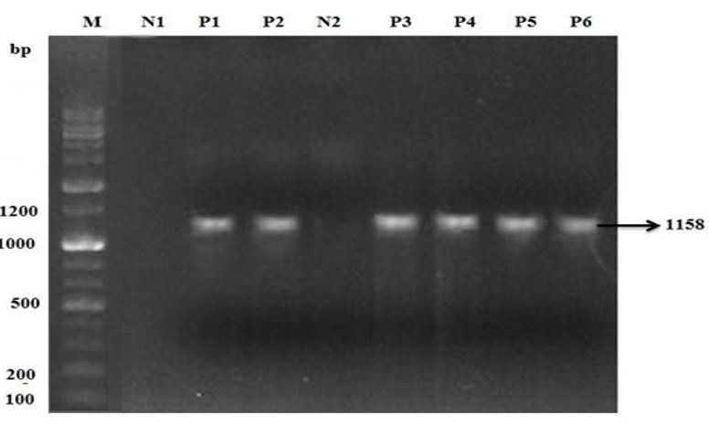
Agrose gell electrophoresis of PCR product of Orf. M is 2 kb (+) DNA ladder marker. N1 is negative control. P1 and P2 are positive sample of sheep. N2 is negative control used for goat samples. P3, P4, P5 and P6 are positive sample of goats for Orf infection.
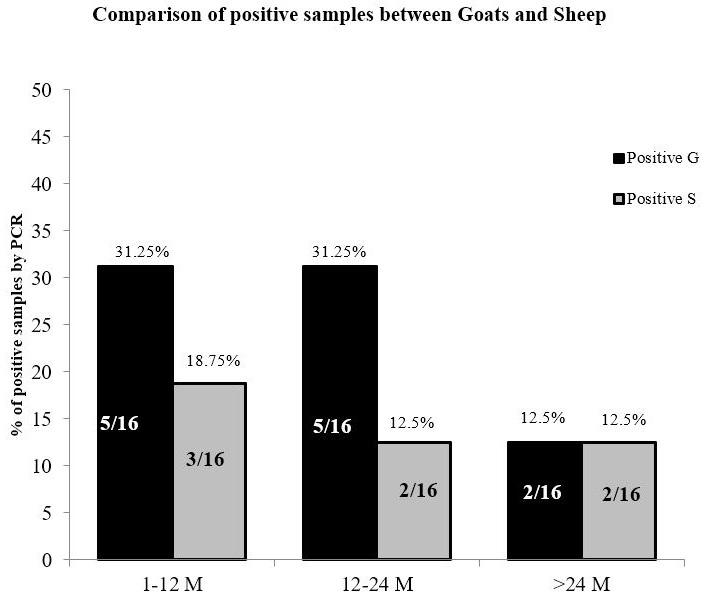
Comparison of PCR positive sheep and goats with Bar chart.
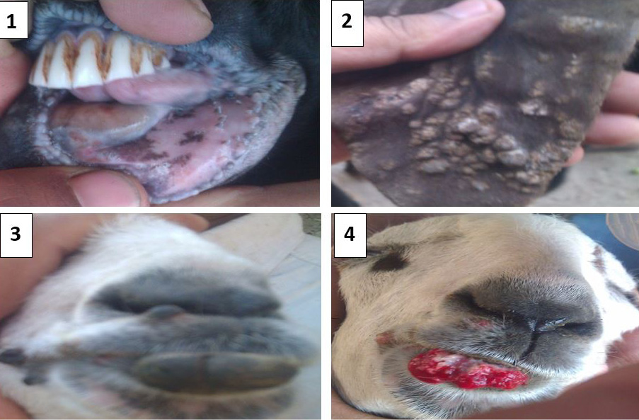
Gross pathological lesions in sheep and goats. (1) Ulceration and hemorrhages on soft tissues of oral cavity. (2) Nodules formation on ear pinnae of external ear of sheep. (3) Warts on lower lip and commissure of goat. (4) Oozing of blood and ulceration upon removal of warts.
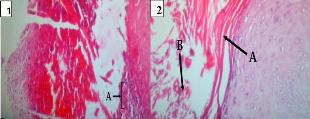
Lesion development in soft tissue of oral cavity. In (1): Necrotic dermatitis with zone of dead and dying neutrophils in soft tissue of oral cavity (A) Abscess formation; (2): (A) Acanthosis of stratum spinosum. (B) Hyperkeratosis of epidermis.
References
- Almagro, M., J.R. Maestre, P. Martinez, I. Malagon, E. Perez and I. Herrera. 1991. Milker’s nodes: transmission by fomites and virological identification. Enferm. Infecc. Microbiol. Clin. 9: 286–8.
- Chan, K.W., J.W. Lin, S.H. Lee, C.J. Liao, M.C. Tsai, W.L. Hsu, M.L. Wong and H.C. Shih. 2007. Identification and phylogenetic analysis of orf virus from goats in Taiwan. Virus Genes. 35: 705-712. https://doi.org/10.1007/s11262-007-0144-6[CrossRef]
- Damon, I. 2007. Poxviridae and their replication. In: Fields virology. New York: Raven press Ltd. pp. 2079–81.
- Elzein, J.W. Lin, S.H. Lee, C.J. Liao, M.C. Tsai, W.L. Hsu, M.L. Wong and H.C. Shih. Identification and phylogenetic analysis of orf virus from goats in Taiwan. Virus Genes. 35: 705-712. https://doi.org/10.1007/s11262-007-0144-6[CrossRef]
- Elzein, A.E.M.E. and F.M.T. Housawi. 1997. Severe long lasting contagious ecthyma infection in a kid. J. Vet. Med. 44: 561-564. https://doi.org/10.1111/j.1439-0450.1997.tb01008.x[CrossRef]
- Guo, J., Z. Zhang, J.F. Edwards, R.W. Ermel, C. Taylor and A. Concha- Bermejillo. 2003. Characterization of a north American orf virus isolated from a goat with persistent, proliferative dermatitis. Virus Res. 93:169-179. https://doi.org/10.1016/S0168-1702(03)00095-9[CrossRef]
- Guo, J., J. Rasmussen, A. Wunschmann, A. de La Concha-Bermejillo. 2004. Genetic characterization of orf viruses isolated from various ruminant species of a zoo. Vet. Microbiol. 99: 81-92. https://doi.org/10.1016/j.vetmic.2003.11.010[CrossRef]
- Haig, D.M., A.A. Mercer. 1998. Ovine diseases. Orf. Veterinary research. 29: 311-326.
- Housawi, F.M.T., E.M.E. Abu-Elzein. 1992. (Orf infection following ear tagging in goats) Revue d’Elevage et de Med. Vet. Trop. 44: 277-278.
- Hosamani, M., V. Bhanuprakash, A. Scagliarini and R.K. Singh. comparative sequence analysis of major envelope protein gene (B2L) of Indian orf viruses isolated from sheep and goats. Vet. Microbiol. 116: 317-324. https://doi.org/10.1016/j.vetmic.2006.04.028[CrossRef]
- Inoshima, Y., A. Morooka and H. Sentsui. 2000. Detection and diagnosis of parapoxvirus by the polymerase chain reaction. J. Virol. Methods. 84: 201-8. https://doi.org/10.1016/S0166-0934(99)00144-5[CrossRef]
- Ishii, S., Y. Kawakami and S. Fukuhara. 1953. Pustular dermatitis of sheep and goats.1. The property of the comparative agent. Exp. Rep. Govt. Exp. Stat. Anim. Hyg. 27: 141-150.
- Koufakis, T., P. Katsaitis, I. Gabranis. 2014. Orf disease: a report of a case. Braz. J. Infect. Dis. 18(5):568–9. https://doi.org/10.1016/j.bjid.2014.04.001[CrossRef]
- Kumar, R. 2015. Contagious pustular dermatitis (Orf disease), epidemiology, diagnosis, control and public health concerns. Adv. Anim. Vet. Sci. 3(4): 560–9. https://doi.org/10.14737/journal.aavs/2015/3.12.649.676[Full text][CrossRef]
- Li, H., X. Zhu, Y. Zheng, S. Wang, Z. Liu, Y. Dou, X. Cai and X. Luo. 2013. Phylogenetic analysis of two Chinese orf virus isolates based on sequences of B2L and VIR genes. Arch. Virol. 158: 1477-1485. https://doi.org/10.1007/s00705-013-1641-7[CrossRef]
- McInnes, C.J., A.R. Wood and A.A. Mercer. 1998. Orf virus encodes a homolog of the vaccinia virus interferon-resistance gene E3L. Virus Genes. 17: 107-115. https://doi.org/10.1023/A:1026431704679[CrossRef]
- McElroy, M.C. and H.F. Bassett. 2007. The development of oral lesions in lambs naturally infected with orf virus. Vet. J. (London, England: 1997) 174: 663-664. https://doi.org/10.1016/j.tvjl.2006.10.024[CrossRef]
- Mangana-Vougiouka, O., P. Markoulatos, G. Koptopoulos, K. Nomikou, N. Bakandritsos and O. Papadopoulos. 1999. Sheep poxvirus identification by PCR in cell cultures. J. Virol. Methods. 77: 75-79. https://doi.org/10.1016/S0166-0934(98)00138-4[CrossRef]
- Nandi, S., U.K. De and S. Choudhary. 2011. Current status of contagious ecthyma or orf disease in goat and sheep- A global perspective. Small Rum. Res. 96: 73–82. https://doi.org/10.1016/j.smallrumres.2010.11.018[CrossRef]
- Nadeem, M., P. Curran, R. Cooke, C.A. Ryan and K. Connolly. 2010. Orf: contagious pustular dermatitis. Ir. Med. J. 03(5): 152–3.
- Pal, M., S. Tesfaye and P. Dave. 2013. Zoonoses occupationally acquired by abattoir workers. J. Environ. Occu. Sci. 2(3): 155–62. https://doi.org/10.5455/jeos.20131013121517[CrossRef]
- Ropp, S.L., Q. Jin, J.C. Knight, R.F. Massung and J.J. Esposito. 1995. PCR strategy for identification and differentiation of small pox and other orthopoxviruses. J. Clin. Microbiol. 33: 2069-2076.
- Sullivan, J.T., A.A. Mercer, S.B. Fleming and A.J. Robinson. 1994. Identification and characterization of an orf virus homologue of the vaccinia virus gene encoding the major envelope antigen p37K. Virol. 202: 968-973. https://doi.org/10.1006/viro.1994.1420[CrossRef]
- Venkatesan, G., V. Balamurugan and V. Bhanuprakash. 2014. Multiplex PCR for simultaneous detection and differentiation of sheeppox, goatpox and orf viruses from clinical samples of sheep and goats. J. Virol. Methods. 195: 1-8. https://doi.org/10.1016/j.jviromet.2013.10.009[CrossRef]
- Zhao, K., D. Song, W. He, H. Lu, B. Zhang, C. Li, K. Chen and F. Gao. 2010. Identification and phylogenetic analysis of an Orf virus isolated from an outbreak in sheep in the Jilin province of China. Vet. Microbiol. 142: 408-415. https://doi.org/10.1016/j.vetmic.2009.10.006[CrossRef]
Services
Article usage statistics
Loading…