Research Article · Volume 12, Issue 3, 29 January 2024 · Open Access
Sequence and Morphology of Cysticercus pisiformis in Local Breed Rabbits
·
Affiliations & Notes ⌄
- 1Department of Veterinary, Karbala Veterinary Hospital, Ministry of Agriculture, Iraq
- 2Department of Parasitology, College of Veterinary Medicine, University of Baghdad, Iraq. *Correspondence | Kadhim Kh. K. Al-Khayat, Department of Veterinary, Karbala Veterinary Hospital, Ministry of Agriculture, Iraq
- 3Email: kadhimalkhayat@gmail.com
Article Info ⌄
- Journal:
- Advances in Animal and Veterinary Sciences
- Published:
- 29 January 2024

From the article
- Fourteen of 60 (23.33%) necropsied local bred rabbits was found infected naturally with C. pisiformis (Table 3).
- According to the sex a total rate of infection in 27.90% (12/43) females was which was more than males’ infection 11.76% (2/17) without significant differences (P≥0.05) (Table 3).
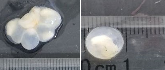
- Fifty-five cysts were isolated from peritoneal cavity of 14 positive samples (single or group) as balloon filled with clear fluid with white floating scolex (evagenated or invagenated) or pus-filled in others.
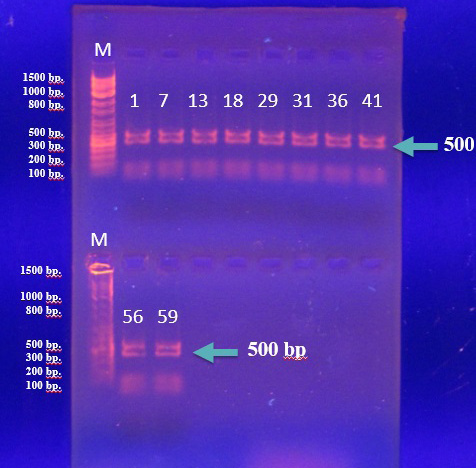
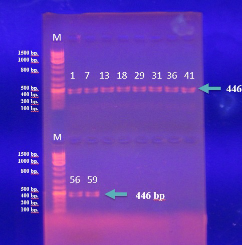
- The result of convential PCR technique to which used for confirm C. pisiformis based on using (NAD1) genes and primer 500bp the result was 100(14/14) (Figure 3) , also 14 DNA sample were subgected to PCR to amplification (COX1) genes with primer 446 bp the result was was 100(14/14) (Figure 4).
Selected from the article's own text.
Abstract
Introduction
Since antiquity, tapeworm has caused considerable damage to wild and domestic animals, as well as human health, resulting in economic losses due to food safety, livestock output, and serious public health implications . Rabbits are affected by many different species of parasites . Taenia pisiformis which is one of these cestodes belonged to family Taeniidae and to the genus Taenia . The final hosts of the parasite are canines and rarely felines, while their intermediate hosts are some rodents .This parasite distributed in different areas of the world, Lahmar et al. (2008) found the prevalence with T. pisiformis of stray dogs in Tunisia was 6.36%, 3.33% in East-Azerbaijan Province, north-west Iran , 26% in New Zealand and 9.4% in Ukraine and in Iraq, 8. 6% in Mosul , 38. 75% in Baghdad Province and 54.55% in Al-Diwaniyah Province .
The adult worm colonizes the small intestine of dogs , and the larval stage causes cysticercosis in rabbits . It mostly produces blood poisoning, depression, lethargic, loss of appetite and emaciation, slate themselves from normal activities of the flock, a syndrome that has been defined as sickness behaviors, but it can also impair resistance to other illnesses and may even result in mortality ; ; .
Because there is no phylogenetic analysis of C. pisiformis in local bred rabbits in Iraq, this study was achieved for confirmation and identification of this parasite from rabbits.
Materials and Methods
Animals of the study
Sixty local breed rabbits were purchased from Al-Ghazel market in the center of Baghdad city, visited weekly during a period 1/March/ until 31/May/2022, the animals were placed under the optimal conditions (room temperature regulated at 24˚C, dry food and clean tap water) of rearing the laboratory animals in the Animal House, College of Veterinary Medicine/ University of Baghdad.
Necropsy finding
Five rabbits humanely euthanized weekly. The skin and muscles of abdomen of necropsied rabbits were opened longitudinally and the looking for any cyst may be founded grossly on the abdominal cavity or on the visceral organs, recorded the location of cysts and numbers . The isolated cysts were stored in clean containers before being sent to the Parasitology Department, College of Veterinary Medicine, University of Baghdad Laboratory for further analysis.
Identification of cysts
All cysts were examined for their structures scolex, membrane and the size measurements. The fluid was collected from each cyst for measurement the volume ; . The cysts were identified as C. pisiformis according to the Loos-Frank (2000) description, and some cysts were stained with a cetoacetic acid carmine stain to seen the structure of cysts . To confirm the diagnosis of C. pisiformis (scolex and part of membrane) from these cysts were put in a 70% ethanol for further used in conventional molecular diagnosis.
Molecular identification
Molecular confirmation was done on fourteen C. pisiformis isolated from naturally infected rabbits, they were achieved by DNA extraction from cyst tissue (scolex and part of membrane) by using commercially available kit (Add Prep Genomic DNA Extraction Kit/ Add Bio/ Korea) for tissue DNA extraction and according to instruction of manufacture’s ; .
Conventional polymerase chain reaction (PCR) technique were performed for (14 DNA samples) by using specific primers for T. pisiformis, first amplification of gene NADH dehydrogenase subunit1 (NAD1) using the primers JB11F (5’-AGATTCGTAAGGGGCCTAATA-3’) forward and JB12R (5’-ACCACTAACTAATTCACTTTC-3’) reveres, that amplify a fragment of about 500bp., the second conventional PCR was carried out for amplification of cytochrome oxidase subunit1 (COX1) using the primers CO1F (5’-TTTTTTGGCCATCCTGAGGTTTAT-3’) forward and CO1R (5’-TAACGACATAACATAATGAAAATG-3’) reveres, which amplifies a fragment of 446bp. according to (Samorek-Pier et al., 2021). Reaction of PCR technique Master Mix were performed by added 10μl of Master Mix to 3μl DNA template, 1.5μl primer forward, 1.5μl reverse, and completed by 4μl distilled water to make total volume of 20μl (Table 1).
Table 1: Contents for reaction of PCR technique.
| Mixture PCR | Volume |
| Master-Mix | 10 µL |
| DNA Template | 3 µL |
| Primer Forward | 1.5 µL |
| Primer Reverse | 1.5 µL |
| Free nuclease water | 4 µL |
| Total volume | 20 µL |
Thermocycler conditions were carried out as the following steps for both genes and for each sample (Table 2).
DNA sequencing and phylogenetic tree analysis
DNA sequencing was used to validate the presence of local C. pisiformis and to create a phylogenetic connection tree between local isolates and NCBI-Blast submissions, as well as to submit local isolates to NCBI-Gen Bank. Three PCR positive isolate results for each gene were delivered in an ice bag to Bioneer Company in Korea for DNA sequencing using Applied Biosystem (AB) of DNA sequencing system. The NCBI-Gen Bank submission was completed using the Ban kit submission tool. Six PCR products’ sequences were submitted to the National Center for Biotechnology Information (NCBI) under specific accession numbers for each strain. The sequences data were arranged into a FASTA format by saving the consensus as a single file . The DNA sequencing analysis was conducted by using molecular evolutionary genetics analysis version 6.0. (Mega 6.0) and multiple sequence alignment analysis of the partial nicotinamide adenine dinucleotide dehydrogenase subunit1 (NAD1) and cytochrome oxidase subunit1 (COX1) genes, internal transcribed spacer each gene based Clustal W alignment analysis, the evolutionary distances were computed using the maximum composite likelihood method by phylogenetic tree UPGMA (unweighted pair group method with arithmetic mean) method . Using molecular evolutionally genetics analysis version 6.0 (Mega 6.0) multiple sequencing alignment analysis of the partial NAD1 gene and COX1 gene. Sequence analysis of the NAD1 and COX1 genes has been used to show the similarity and differences between local and international genes sequence.
Table 2: Thermocycler conditions of PCR.
| PCR steps | Temperature | Time | Repeated |
| Initial denaturation | 94°C | 5 minutes | 1 |
| Denaturation | 94°C | 30 Seco. | 35 cyles |
| Annealing | 52°C | 30 Seco. | |
| Extension | 72°C | 60 Seco. | |
| Final extension | 72°C | 5minutes | 1 |
| Hold | 12°C | ∞ |
Statistical analysis
To determine the significance of different components in research parameters, the Statistical Analysis System- SAS 9.3 (2012) application was utilized. In this study, the Chi-square test was utilized to compare percentages (0.05 and 0.01 likelihood).
Results and Discussion
Fourteen of 60 (23.33%) necropsied local bred rabbits was found infected naturally with C. pisiformis (Table 3). The result of current study was less than result of Athraa (2014) who recorded 38.75% (31/80) infection rate of in local rabbits in Baghdad city-Iraq, also Jori (2016) who founded 40% (4/10) infection rate from domestic rabbits in Basrah city-Iraq, and less than Marhoon et al. (2018) recorded 54.55% (30/55) of C. pisiformis in a wild rabbits collected from the local animals market in Al-Diwaniyah Province-Iraq and less than Roldan (2019) in Spain 67.7% (21/31) infection rate with metacestodes C. pisiformis from the capturing wild rabbits, while the results enclose with study of Hajipour and Zavarshani (2020) in Iran which found the prevalence rate 26% (13/50) in new Zealand white rabbits. The results are higher than Al-Moula (2005) which recorded 8.6% (2/30) infection rate when examined domestic rabbits in Mosul city-Iraq. Also, more than Foronda et al. (2005) in Canary Islands which found the prevalence rate 16% in total 244 wild rabbits. Also higher than Yagoob and Hossein (2011) which isolated 3.33% (2/60) from abdominal cavity of wild rabbits in East Azerbaijan province of Iran.
According to the sex a total rate of infection in 27.90% (12/43) females was which was more than males’ infection 11.76% (2/17) without significant differences (P≥0.05) (Table 3). This result agree with Roldan (2019) in Spain which recorded high infection rate in females 80% (17/21) than males 40% (4/10) with significant difference (P≤0.05).In contrast to Hajipour and Zavarshani (2020) in Iran who found males affected more than females and recorded infection rate 23.80% (5/21) in females and 27.58% (8/29) males without significant difference (P≥0.05). The disparities between the current study and prior studies might be attributed to variances in the quantity of samples obtained between investigations, difference in type of animals (domestic or wild), as well as differences in the distribution and proliferation of stray dogs.
Table 3: Total Infection rate with C. pisiformis in naturally infected rabbits according to the sex.
| Sex | Number of rabbits | Number of infected | Ratio |
| Females | 43 | 12 | 27.90% |
| Males | 17 | 2 | 11.76% |
| Total | 60 | 14 | 23.33% |
| Statistical analysis | χ2= 1.17 DF=1 P value =0.27 Non-Significant (P≥0.05) | ||
Fifty-five cysts were isolated from peritoneal cavity of 14 positive samples (single or group) as balloon filled with clear fluid with white floating scolex (evagenated or invagenated) or pus-filled in others. The range of diameters of cyst between 7 to 21 in length and 4 to 10 mm in width. (Table 4) and (Figure 1), this cysts were confirmed to the Loos-Frank (2000) description as C. pisiformis.
According to the location of cyst into internal organs of 55 cysts, large number of larval cysts attached to the mesentery 47.27% (26/55), liver 21.82% (12/55), other cysts found free within the abdominal cavity 21.82% (12/55) and on omentum only 9.09% (5/55) with Significant deffrences (P<0.05) (Table 5).
Table 4: Measurements of cysts size and volume.
| Number of cysts | Range of size | Volume of fluid (µl) | Percentage (%) | |
| Range length (mm) | Range width (mm) | |||
| 6 | 18-21 | 9-10 | 12-17 | 10.90 |
| 17 | 12-15 | 4-10 | 8-11 | 30.90 |
| 27 | 7-10 | 4-9 | 4-7 | 49.09 |
| 5 | 5-6 | 3-4 | 2-3 | 9.09 |
Table 5: The site and number of cysts in fourteen naturally infected rabbits.
| Site No. | Mesentery | Liver | Free in abdominal cavity | Omentum |
| 1 | 7 | |||
| 7 | 4 | |||
| 13 | 2 | |||
| 14 | 1 | |||
| 18 | 4 | |||
| 29 | 3 | |||
| 30 | 2 | |||
| 31 | 3 | 6 | ||
| 36 | 1 | 2 | 2 | |
| 38 | 2 | |||
| 39 | 2 | |||
| 41 | 2 | 3 | ||
| 56 | 2 | 2 | ||
| 59 | 3 | 2 | ||
| Total cysts | 26 | 12 | 12 | 5 |
| Ratio | 47.27% | 21.82% | 21.82% | 9.09% |
| Statistical analysis | X2= 41.13 DF=4 P value =0.001 Significant (P<0.05) | |||
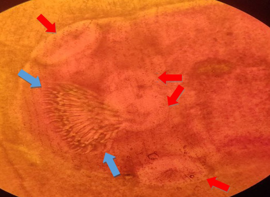
Cysts staining with acetoacetic carmine stain appeared scolex with armed rostellum with tow raw of hooks and four suckers (Figure 2) like the method of .
Result of moleculer study
Results of DNA sequencing and phylogenetic tree construction
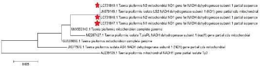
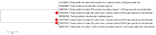
The nucleotide sequences of 3 local C. pisiformis isolates for each genes were submitted to GenBank under the accession numbers (LC731847.1, LC731848.1 and LC731849.1) for NAD1 gene (Table 6) and (OP274120.1, OP277616.1 and OP277618.1) for COX1 gene (Table 7).
Three sequences of NAD1 gene (LC731847.1, LC731848.1 and LC731849.1) was 100% identical to the China sequence JN870149.1 while less identically (97.15-99.47%) in Australia and Poland (Table 6 and Figure 5), and sequences of COX1 gene (OP274120.1, OP277616.1 and OP277618.1) were matching variance between 100% to 99.56% in the China, Portugal and Poland (Table 7 and Figure 6).
Table 6: Isolates of T. pisiformis from natural infected by NAD1 gene sequencing ID in gene bank and nucleotide sequence identity from NCBI.
| Local isolates | Accession numbers for local isolates | NCBI-BLAST homology sequence Identity(%) | ||
| Accession numbers | Country | Identity (%) | ||
| T. pisiformis No.1 | LC731847.1 | JN870149.1 | China | 100 |
| MW350140.1 | China | 99.44 | ||
| GU569096.1 | China | 99.44 | ||
| MZ287427.1 | Poland | 99.16 | ||
| JX677976.1 | China | 98.88 | ||
| AJ239109.1 | Australia | 97.21 | ||
| T. pisiformis No.2 | LC731848.1 | JN870149.1 | China | 100 |
| MW350140.1 | China | 99.47 | ||
| GU569096.1 | China | 99.47 | ||
| MZ287427.1 | Poland | 99.20 | ||
| JX677976.1 | China | 98.93 | ||
| AJ239109.1 | Australia | 97.34 | ||
| T. pisiformis No.3 | LC731849.1 | JN870149.1 | China | 100 |
| MW350140.1 | China | 99.43 | ||
| GU569096.1 | China | 99.43 | ||
| MZ287427.1 | Poland | 99.14 | ||
| JX677976.1 | China | 98.86 | ||
| AJ239109.1 | Australia | 97.15 | ||
Table 7: Isolates of T. pisiformis from natural infected by COX1 gene sequencing ID in gene bank and nucleotide sequence identity from NCBI.
| Local isolates | Accession numbers for local isolates | NCBI-BLAST Homology sequence | ||
| Accession numbers | Country | Identity (%) | ||
| T. pisiformis No.1 | OP274120.1 | MW350140.1 | China | 99.56 |
| JN870104.1 | China | 99.56 | ||
| GU569096.1 | China | 99.56 | ||
| KC020696.2 | Portugal | 99.56 | ||
| MZ287426.1 | Poland | 99.56 | ||
| T. pisiformis No.2 | OP277616.1 | MW350140.1 | China | 100 |
| JN870104.1 | China | 100 | ||
| GU569096.1 | China | 100 | ||
| KC020696.2 | Portugal | 100 | ||
| T. pisiformis No.3 | OP277618.1 | MZ287426.1 | Poland | 100 |
| MW350140.1 | China | 99.67 | ||
| JN870104.1 | China | 99.67 | ||
| GU569096.1 | China | 99.67 | ||
| KC020696.2 | Portugal | 99.64 | ||
This study is the first one by using conventional PCR and sequence identification of Cysticercus. pisiformis in Baghdad, Iraq.
Acknowledgement
Authors would like to express their special thanks and gratitude to the Department of Parasitology/College of Veterinary Medicine/University of Baghdad for all their kind and helpful support during the study period.
Novelty Statement
This study is the first one by using conventional PCR and sequence identification of Cysticercus pisiformis in Baghdad, Iraq and study the highly distribution of the infection of local bred rabbits in Souq al-Ghazal in the Baghdad city.
Author’s Contribution
Both authors conceived and designed the study, reviewed and approved the final manuscript. Kadhim Kh. K. Al-Khayat performed the experiments. Athmar K. A. Al-Azawi analyzed the data. Kadhim Kh. K. Al-Khayat contributed materials and wrote the manuscript.
Ethics statement
All animals in this study were handled and cared for according to the appropriate biosecurity procedures. The study was performed in accordance with the rules of the ‘Guide for the Care and Use of Laboratory Animals that were approved by the Ethics Committee of the College of Veterinary Medicine, University of Baghdad, Iraq (Number 472 P.G at 28/2/2023) before starting this study.
Conflict of interest
The authors have declared no conflict of interest.
Figures

Shape and measurements of Cysticercus pisiformis cysts, grossly.

Structure of Cysticercus pisiformis Scolex staining by acetoacetic carmine stain, Suckers (red arrow) and Hooks (blue arrow), X40.

Agarose (1%) gel electrophoresis of PCR products for (NAD1) gene of C. pisiformis of rabbits and DNA Marker Ladder (M) (100- 1500bp), PCR products amplicons band lances (1, 7, 13, 18, 29, 31, 36, 41, 56 and 59) sized 500bp. (1h, 7 volt/cm2 TBE).

Agarose(1%) gel electrophoresis of PCR products for (COX1) gene of C. pisiformis of rabbits and DNA Marker ladder (M) (100- 1500bp), PCR products amplicons band lances (1, 7, 13, 18, 29, 31, 36, 41, 56 and 59) sized 446bp. (1h, 7 volt/cm2 TBE).

Phylogenetic tree study based on Taenia pisiformis NAD1 gene partial sequence.

Phylogenetic tree study based on Taenia pisiformis COX1 gene partial sequence.
References
- Abbas AK (1998). Study in the epidemiology of cysticercus tenuicollis of sheep and goats. College of Veterinary Medicine, University of Baghdad.
- Abbas AK (2017). Detection some chemical and biochemical constituents of Cysticercus tenuicollis cyst fluid of Iraqi goat. J. Entomol. Zool. Stud., 5(2): 814–818.
- Abbas HB, and Abbas AK (2017). Analysis of chemical and biochemical composition of fertile and sterile hydatid cyst fluids of human origin. Iraqi J. Vet. Med., 41(1): 66–70. https://doi.org/10.30539/iraqijvm.v41i1.82[CrossRef]
- Abdulkareem MH, Razook BRF, Ameer AHA, Alrubaye B (2022). Histopathological changes of rabbits’ vital organs associated with corynebacterium pseudotuberculosis infection. Int. J. Special Educ., 37(3). https://doi.org/10.51470/bca.2022.22.2.3743[CrossRef]
- Al-Azawi AKA, Al-Biatee ST (2019). Prevalence and phylogeny of Cysticercus tenuicollis sp. determined by PCR from 6909 slaughtered sheep over 12 months. Online J. Vet. Res., 23(6): 594–605.
- Al-Moula IDH (2005). Study of some endo and ecto parasites in domestic rabbits in Mosul/Iraq. Iraqi J. Vet. Sci., 19(2): 143–153. https://doi.org/10.33899/ijvs.2005.46707[CrossRef]
- Athraa TW (2014). Study of Prevalence of gastrointestinal and blood parasites with some histopathological changes of local rabbits in Baghdad province. M. Sc. thesis, Coll. Vet. Med. Univ. Baghdad.
- Bagrade G, Kirjušina M, Vismanis K, Ozoliņš J (2009). Helminth parasites of the wolf Canis lupus from Latvia. J. Helminthol., 83(1): 63–68. https://doi.org/10.1017/S0022149X08123860[CrossRef]
- Bajalan MMM (2010). Prevalence of intestinal helminths in stray dogs of Kalar city, Sulaimani province. Iraqi J. Vet. Med., 34(1): 151–157. https://doi.org/10.30539/iraqijvm.v34i1.673[CrossRef]
- Bogach MV, Paliy AP, Horobei OO, Perotska LV, Kushnir VY, Bohach DM (2022). Endoparasites of rabbits (Oryctolagus cuniculus domesticus) in Southern Ukraine. Biosyst. Diver., 30(2): 173–178. https://doi.org/10.15421/012218[CrossRef]
- Borji H, Azizzadeh M, Kamelli M (2012). A retrospective study of abattoir condemnation due to parasitic infections: economic importance in Ahwaz, southwestern Iran. J. Parasitol., 98(5): 954–957. https://doi.org/10.1645/GE-2988.1[CrossRef]
- Chilton NB, Gasser RB, Beveridge I (1997). Phylogenetic relationships of Australian strongyloid nematodes inferred from ribosomal DNA sequence data. Int. J. Parasitol., 27(12): 1481–1494. https://doi.org/10.1016/S0020-7519(97)00134-3[CrossRef]
- El-Ashram S, Aboelhadid SM, Abdel-Kafy ESM, Hashem SA, Mahrous LN, Farghly EM, Kamel AA (2020). Investigation of pre-and post-weaning mortalities in rabbits bred in Egypt, with reference to parasitic and bacterial causes. Animals, 10(3): 537. https://doi.org/10.3390/ani10030537[CrossRef]
- Ezenwa VO (2016). Helminth–microparasite co‐infection in wildlife: lessons from ruminants, rodents and rabbits. Parasit. Immunol., 38(9): 527–534. https://doi.org/10.1111/pim.12348[CrossRef]
- Foronda PR, Figueruelo EO, Ortega AR, Abreu NA, Casanova JC (2005). Parasites (viruses, coccidia and helminths) of the wild rabbit (Oryctolagus cuniculus) introduced to Canary Islands from Iberian Peninsula. Acta Parasitol., 50(1): 80–84.
- Garcia LS, Ash LR (1979). Diagnostic parasitology: Clinical laboratory manual CV Mosby Co. St. Louis, Missouri, USA, pp. 145–146.
- Hadi AM, Faraj AA (2016). Prevalence of gastrointestinal Helminthes and protozoa among stray dogs in Baghdad. Iraqi J. Vet. Med., 40(1): 1–4. https://doi.org/10.30539/iraqijvm.v40i1.129[CrossRef]
- Hajipour N, Zavarshani M (2020). Ectoparasites and endoparasites of New Zealand white rabbits from North West of Iran. Iran. J. Parasitol., 15(2): 266. https://doi.org/10.18502/ijpa.v15i2.3310[CrossRef]
- Hart BL (1991). The behavior of sick animals. The Veterinary Clinics of North America. Small Anim. Pract., 21(2): 225–237. https://doi.org/10.1016/S0195-5616(91)50028-0[CrossRef]
- Hoberg EP (2002). Taenia tapeworms: Their biology, evolution and socioeconomic significance. Microb. Infect., 4(8): 859–866. https://doi.org/10.1016/S1286-4579(02)01606-4[CrossRef]
- Jori MM (2016). The effect of Cysticercus pisiformis on the haematological and biochemical parameters of rabbits in Basrah Province. Life Sci. Arch., 2(January): 458–463.
- Kakai R (1994). A study of factors associated with intestinal immunoglobulin A in children with diarrhoea only and those with diarrhoea and malnutrition. University of Nairobi.
- Karamon J, Sroka J, Dąbrowska J, Bilska-Zając E, Zdybel J, Kochanowski M, Różycki M, Cencek T (2019). First report of Echinococcus multilocularis in cats in Poland: A monitoring study in cats and dogs from a rural area and animal shelter in a highly endemic region. Parasit. Vectors, 12: 1–8. https://doi.org/10.1186/s13071-019-3573-x[CrossRef]
- Khademvatan S, Majidiani H, Foroutan M, Tappeh KH, Aryamand S, Khalkhali HR (2019). Echinococcus granulosus genotypes in Iran: A systematic review. J. Helminthol., 93(2): 131–138. https://doi.org/10.1017/S0022149X18000275[CrossRef]
- Kornyushin VV, Malyshko EI, Malega AM (2011). The helminths of wild predatory mammals of Ukraine. Cestodes. Vestnik Zool., 45(6): 483–490. https://doi.org/10.2478/v10058-011-0031-4[CrossRef]
- Lahmar S, Sarciron M, Rouiss M, Mensi M (2008). Echinococcus granulosus and other intestinal helminths in semi-stray dogs in Tunisia: infection and re-infection rates. La Tunisie Méd., 86(3): 279–286.
- Loos-Frank B (2000). An up-date of Verster’s (1969) Taxonomic revision of the genus Taenia Linnaeus’(Cestoda) in table format. Syst. Parasitol., 45(3): 155–184. https://doi.org/10.1023/A:1006219625792[CrossRef]
- Marhoon IA, Mattar KT, Mohammad FI (2018). Parasitic infection in wild rabbits Oryctolagus cuniculus. Eur. J. Anal. Chem., 13(5): 55. https://doi.org/10.29333/ejac/95252[CrossRef]
- Mraidi DA, Lafta IJ (2021). RAPD-PCR and phylogenetic analysis of E. coli isolated from human and cattle urinary tract infections. Iraqi J. Vet. Sci., 35: 59–66. https://doi.org/10.33899/ijvs.2021.131100.1918[CrossRef]
- Nakao M, Yanagida T, Okamoto M, Knapp J, Nkouawa A, Sako Y, Ito A (2010). State-of-the-art Echinococcus and Taenia: Phylogenetic taxonomy of human-pathogenic tapeworms and its application to molecular diagnosis. Infect. Genet. Evol., 10(4): 444–452. https://doi.org/10.1016/j.meegid.2010.01.011[CrossRef]
- Overgaauw PAM (2020). Parasite risks from raw meat-based diets for companion animals. Compan. Anim., 25(11): 261–267. https://doi.org/10.12968/coan.2020.0065[CrossRef]
- Roldan RD (2019). Aspectos de dimorfismo sexual, reproductivos e histológicos asociados a la teniosis (taenia pisiformis) cisticercosis en dos especies de lagomorfos y en el hámster dorado.
- Samorek-Pieróg M, Karamon J, Brzana A, Bilska-Zajacą E, Zdybel J, Cencek T (2021). Molecular confirmation of massive taenia pisiformis cysticercosis in one rabbit in Poland. Pathogens, 10(8).
- Steele JH (1973). A bookshelf on veterinary public health. Am. J. Publ. Health, 63(4): 291–311. https://doi.org/10.2105/AJPH.63.4.366[CrossRef]
- Tamura K, Peterson D, Peterson N, Stecher G, Nei M, Kumar S (2011). MEGA5: Molecular evolutionary genetics analysis using maximum likelihood, evolutionary distance, and maximum parsimony methods. Mol. Biol. Evol., 28(10): 2731–2739. https://doi.org/10.1093/molbev/msr121[CrossRef]
- Yagoob G, Hossein H (2011). Prevalence rate of endoparasites in wild rabbits of East-Azerbaijan Province, Iran. Ann. Biol. Res., 2: 31–35.
- Yang D, Ren Y, Fu Y, Xie Y, Nie H, Nong X, Gu X, Wang S, Peng X, Yang G (2013). Genetic variation of Taenia pisiformis collected from Sichuan, China, based on the mitochondrial cytochrome B gene. Korean J. Parasitol., 51(4): 449. https://doi.org/10.3347/kjp.2013.51.4.449[CrossRef]
- Zhou YX, Du AF, Zhang XJ, Wu YM, Tong FY, Wu GY (2008). Research of harmfulness of Cysticercus pisiformis in rabbit. J. Zhejiang Agric. Sci., 3: 372–373.
- Zhu GQ, Li L, Ohiolei JA, Wu YT, Li WH, Zhang NZ, Fu BQ, Yan HB, Jia WZ (2019). A multiplex PCR assay for the simultaneous detection of Taenia hydatigena, T. multiceps, T. pisiformis, and Dipylidium caninum infections. BMC Infect. Dis., 19: 1–7. https://doi.org/10.1186/s12879-019-4512-3[CrossRef]
Services
Article usage statistics
Loading…