Case Report · Volume 11, Issue 11, 6 November 2023 · Open Access
Rare Cases of Rumen Impaction in Two Sokoto Gudali Calves
· · · · · ·
Affiliations & Notes ⌄
- 1Department of Veterinary Surgery and Radiology, Usmanu Danfodiyo University, Sokoto
- 2Department of Veterinary Medicine, Collage of Applied Health and Sciences, A’Sharqiyah University, Sultanate of Oman
- 3Department of Veterinary Medicine, Usmanu Danfodiyo University, Sokoto. *Correspondence | Abubakar Adamu Abdul, Department of Veterinary Surgery and Radiology, Usmanu Danfodiyo University, Sokoto
- 4Email: adamu.abubakar@asu.edu.om
Article Info ⌄
- Journal:
- Advances in Animal and Veterinary Sciences
- Published:
- 6 November 2023

From the article
- To the best of our knowledge, there was no reported case of rumen impaction in calves less than 6-month old.
- Therefore, this study reports the rare occurrence of rumen impaction in two calves.
- Foreign body impaction, especially in the rumen of calves, is a serious threat to the livestock industry and can lead to the premature death of young animals if immediate intervention has not been instituted.
- It can be concluded that rumen impaction with non-degradable materials can also occurred in young ruminants less than six-month old, with proper diagnosis, it can be detected and surgically treated to recovery.
Selected from the article's own text.
Abstract
Introduction
Rumen impaction due to metallic or nonmetallic foreign bodies is among the most common causes of gastrointestinal disorders in ruminants ; ; . The ingestion of foreign bodies in cattle has been reported to be of great economic importance, causing severe loss of production and high mortality rates (Radostis et al., 2006; . Indigestible material is ingested during periods of food scarcity . The incidence of non-metallic foreign bodies (mostly polythene) has been explored in previous studies, mostly in cows ; ; ; . Clinical rumen impaction with a nonmetallic foreign body is characterized by clear signs of emaciation, dehydration, abdominal distension, and abdominal asymmetry. Affected animals may have scanty feces in the rectum, foamy salivation, recumbency, and in appetence ; . Although, the diagnosis of foreign body impaction in small ruminants is based on abdominal palpation (Radostitis et al., 2006). However, it is sometimes difficult to distinguish pregnancy from foreign-body impaction in ruminants most especially in large ruminants. Therefore, further investigations are required to establish a definitive diagnosis. Radiography and ultrasonography are currently the reference standard method for diagnosis ; , but radiography has one major disadvantage because the fetuses are exposed to radiation hazards, in addition to being difficult to use under field conditions. Ultrasonographic examination, when available, is also important tool for narrowing the differential diagnosis between pregnancy and rumino-reticular foreign materials without the risk of radiation.
Rumenotomy is the only effective and reliable treatment method for plastic foreign bodies, but an early diagnosis is essential for favorable outcomes as reported by Ismail et al. (2007). Rumenotomy along with transplantation of fresh ruminal cud is the best technique for the restoration of ruminal function at the fluid level for ruminal impaction due to plastics in cattle and buffaloes . Furthermore, growth stimulants, probiotics, and prebiotics could act as adjuncts to rumenotomy and improve the survival and reversal of rumen ecology . This paper describes two rare cases of rumen impaction in calves with nonmetallic foreign bodies.
Case reports 1
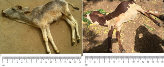
A- 2- month-old Sokoto Gudali calf weighing 30 kg was presented to our large animal clinic with complaints of reduced feed intake, recumbency, abnormal swelling in the ventral part of the abdomen, and absence of defecation (Figure 1). The calf was managed semi-intensively and suckled only with assistance. It was maintained with a dam and three other cows. On physical examination, the rectal temperature was 37.1oC, pulse rate was 69 beats per minute, and respiratory rate was 32 cycles per minute. The calf was weak and recumbent. There was enlargement of the ventrolateral abdomen, which was more to the left and hard on palpation. A bilateral serous nasal discharge was also observed.
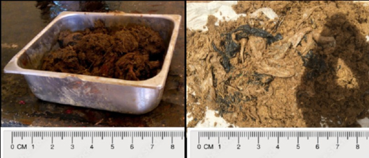
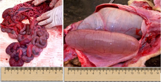
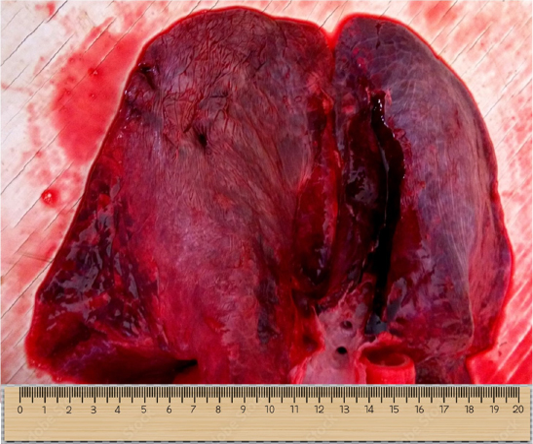
Unfortunately, the calf died before any intervention and the whole carcass was sent for necropsy examinations. During necropsy examinations, polyethylene bags, plastic materials, and other indigestible materials mixed with ruminal ingesta were found in the rumen (Figure 2). Generalized congestion of the small intestine with mucoid content and ballooned cecum with marked congestion on the mucosal surface were also observed (Figure 3). The lungs were severely congested with focal areas of emphysema and necrosis in the diaphragmatic lobe (Figure 4).
Case reports 2
A 6 months old Sokoto Gudali calf weighing 40 kg presented to the ambulatory team of our large animal clinic with the complaint of absence of regurgitation, distended abdomen, recumbency, and constipation (Figure 1). The calf was purchased three months prior to presentation and was managed semi-intensively with five (5) other cows. There was no history of medication or vaccination in the heard. On physical examination, the ocular mucous membrane was whitish, and rumen motility was low, bilateral ocular discharge, foamy salivation, and labored breathing were observed. The prescapular and submandibular lymph nodes were enlarged and a hard mass was palpated on the distended abdomen. On per rectal examinations, hard feces in rectum were found. The rectal temperature was subnormal (35.4 oC), respiratory rates were 12 cycle/min and the pulse rate was so weak and it was not detectable. Attempts to induce defecation using KC-bloat failed and the animal died on examination table.
Following necropsy examination, polyethylene bags, rugs, ropes, and other indigestible materials mixed with ruminal ingesta were observed in the rumen (Figure 2). The small and large intestines were pale and almost empty. The spleen was pale and the urinary bladder was distended with urine.
results and Discussions
Cattle in developing countries where waste disposal is not controlled are more susceptible to ruminal impaction than small ruminants because they do not use their lips for prehension . Owing to the lack of oral discrimination, cattle may ingest foreign particles that would otherwise be rejected by other species . Nutritional deficiency and poor feeding management practices can lead to the ingestion of foreign particles, which might eventually affect different organs of the animal, mainly the rumen and reticulum . When ingested, these foreign particles remain in the rumen of cattle, majorly compromising ruminal space and interfering with normal physiological functions of the rumen and to some extent cardio-pulmonary functions, thereby leading to weight loss and eventually the death of the animal ; . Ruminal impaction as a result of indigestible foreign bodies, especially plastics, also leads to ruminal distension, passing of scanty or no feces, high mortality rates, and premature calling of sick cattle ; . Rumen impaction is a very rare occurrence generally in young animals, hence this warrant the report of this case. To the best of our knowledge, there was no reported case of rumen impaction in calves less than 6-month old. However, there was reported case of rumen impaction in Iranian goat-kid with successful diagnosis and surgical intervention to recovery as reported by Mozaffari et al. (2009). It is also worth noting that abomasal impaction was also recorded in a six-month old Turkish calf by Belge at al. (2017).
In both cases, the rumen was quite solid on palpation and the content consisted of polyethene materials, rugs, and other indigestible materials. The history and clinical findings of our cases were similar to and compatible with those of previous reports in matured cattle above one year-old ; ; ; ; Vanithal et al., 2010). Regarding the findings of the postmortem examinations, the intestines in both cases were empty and congested with little or no feed ingester, which was probably due to obstruction or blockage of the distal orifices (pylorus). Akraiem and Abd Al-Galil (2016) suggested that the presence of impacted material may partially or completely block rumino-reticular orifices. Mozaffari et al. (2009) also reported that the tightly impacted rumen may stop functioning because there is no contractile force sufficient to move the contents forward along the normal digestive course nor to regurgitate them from chewing the cud. The severely congested lungs and increased respiratory rate (32c/m) observed were probably due to the pressure exerted on the lungs by the impacted rumen. The increased respiratory rate is probably due to increased pressure of the dilated rumen on the diaphragm as reported by Bakhiet (2008). Although rumen impaction is common in ruminants, especially in matured ruminant above one-year old and has been studied in detail, very little is known about its incidence in calves. Furthermore, older animals are more susceptible to the development of ruminal impaction due to plastic materials than are young animals . Cattle above 10 years of age and sheep and goats above 4-year of age were more frequently affected by indigestible materials than other age groups ; . Animals in this age group had more foreign bodies than young ones because of the gradual accumulation of these types of foreign bodies in the rumen . Therefore, this study reports the rare occurrence of rumen impaction in two calves. It is thought that the calves ingested these materials because of insufficient feeding by the owner or early weaning without sufficient milk supplementation, thus forcing them to eat foreign materials to satisfy their appetite. Additionally, it is a tradition practiced in some communities that calves are usually isolated and confined during the day, and the dams usually go out for grazing. Because of their increased metabolic demands, these practices may predispose them to ingest anything that comes their way to satisfy their energy demand.
Conclusions and Recommendations
Foreign body impaction, especially in the rumen of calves, is a serious threat to the livestock industry and can lead to the premature death of young animals if immediate intervention has not been instituted. Livestock owners should ensure the proper feeding of their young ruminants and proper disposal of plastic materials around their farms. It can be concluded that rumen impaction with non-degradable materials can also occurred in young ruminants less than six-month old, with proper diagnosis, it can be detected and surgically treated to recovery.
Acknowledgements
The authors want to thank all clinical students and technicians at the necropsy room for active participation in handing the case to conclusion. The technical assistance of animal handlers at the large animal clinic is also appreciated.
Novelty Statement
There are very limited reports of rumen and gastric impaction with foreign bodies in yonger animals less than six month old in literartures. Therefore, this make this case rare, hence the need for reporting it for clinitian to take note of.
Author’s Contribution
All the authors participated in the handling of the two clinical cases. USA and AAA drafted the initial manuscript, HAB, NA and EIO conducted the postmoterm examinatio while ZS and AMM participated in taking the clinical images and selecting the appropriate images that were included in the draft. All authors partipated in the manuscript draft and review of the manuscript.
Conflict of interest
The authors have declared no conflict of interest.
Figures

Patients on presentation (case I and II; left to right), the calves were both presented recumbent, moribund, weak and the abdomen was distended laterally and ventrally.

Constipated faeces and polyethene (non-biodegradable) materials recovered from both calves after necropsy examination, the black and whitish materials are polythene bags recovered from the rumens of the calves.

Congested, hemorrhagic and ballooned intestinal segments observed during necropsy examination.

Congested and hemorrhagic lungs observed during necropsy examination.
References
- Abdelaal AM (2014). Selected studies on foreign body impaction in goats with special reference to ultrasonography. Vet. World, 7: 522-527. https://doi.org/10.14202/vetworld.2014.522-527[CrossRef]
- Abebe F, Nuru M (2011). Prevalence of indigestible foreign body ingestion in small ruminants slaughtered at luna export Abattoir, East Shoa, Ethopia. Asian J. Anim. Vet.Adv. 10(12): 1598-1602. https://doi.org/10.3923/javaa.2011.1598.1602[CrossRef]
- Akinrinmade JF, Akinrinde AS (2012). Hematological, serum biochemical and trace mineral indices of cattle with foreign body rumen impaction. Int. J. Anim. Vet. Adv., 4(6): 344-350.
- Akraiem A, Abd Al-Galil ASA (2016). Rumen impaction in cattle due to plastic materials. J. Vet. Med. Res., 23(1): 65-70. https://doi.org/10.21608/jvmr.2016.43189[CrossRef]
- Athar H, Mohindroo J, Singh K, Singh T (2010). Clinical, Heamatobiochemical, radiographic and ultrasonographic findings in bovines with rumen impaction. Intas Polivet., 11(2): 180-183. https://doi.org/10.4061/2010/939870[CrossRef]
- Bakhiet AO (2008). Studies on the rumen pathology of Sudanese desert sheep in slaughter house. Sci. Res. Essay, 3(7): 294-298.
- Belge A. Koklu S, Atasoy A, Derincegoz OO, Akin I, Kilic N (2017). A case of abomasal impaction and ruminal trichobesoar in a calf. J. Fac. Vet. Med. Erciyes Univ., 14(2): 145-158.
- Berrie K, Tadesse E, Mossie B (2015). Study on rumen and reticulum foreign body in slaughtered cattle at Gondar Elfora Abattoir. World J. Biol. Med. Sci., 2: 133-150.
- Boodur P, Sivaprakash BV, Kasaralivar V, Rand DD (2010). Methylene blue reduction test in cattle affected with rumen impaction due to plastics. J. Ind. Poly Vet., 11(2): 184-188.
- Desiye T, Mersha C (2012). Study on rumen and reticulum foreign bodies in cattle slaughtered at Jimma Municipal Abattoir, South West Ethiopia. Am-Euras. J. Sci. Res. 7: 160-167.
- Fasil N (2016). Assessment of sheep and goat foreign bodies in rumen and reticulum in the Jigjiga municipal Abattiar. J. Adv. Dairy Res., 4: 1-6. https://doi.org/10.4172/2329-888X.1000157[CrossRef]
- Igbokwe IO, Koloand MY, Egwu GO (2003). Rumen impaction in sheep with indigestible foreign body in the semi-arid region of Nigeria. Small Rumin. Res., 49: 141-147. https://doi.org/10.1016/S0921-4488(03)00074-9[CrossRef]
- Ismail ZB, Al-Majali A., Al-Qudah K (2007). Clinical and surgical findings and outcome following rumenotomy in adult dairy cattle affected with recurrent rumen tympany associated with non-metallic foreign bodies. Am. J. Anim. Vet. Sci., 2(3): 66-71. https://doi.org/10.3844/ajavsp.2007.66.71[CrossRef]
- Mozaffari AA, Olomi MM, Vosough D (2009). Unusual and severe ruminal impaction in a goat-kid: Clinical and radiological findings. Iran. J. Vet. Surg., 4(1-2): 115-119.
- Radostits OM, Gay CC, Hinchcliff KW, Constable PD (2006). Veterinary medicine: A textbook of the diseases of cattle, horses, sheep, pigs and goats; Saunders: Philadephia, PA, USA.
- Ravindra RY, Asha LP, Sandeep RS (2014). Review on metallic and non-metallic foreign bodies a threat to livestock and environment. Int. J. Food Agric. Vet. Sci., 4: 6-14.
- Remi-Adewunmi BD, Gyang EO, Osinowo AO (2004). Abattoir survey of foreign body rumen impaction in small ruminants. Niger. Vet. J., 25: 32-38. https://doi.org/10.4314/nvj.v25i2.3472[CrossRef]
- Roman T, Hiwot Y (2010). Occurrence of rumen foreign bodies in sheep and goats slaughtered at the Addis Ababa Municipality Abattoir. Ethiop. Vet. J., 14(1): 91-100.
- Saulawa MA, Ukashatu S, Garba MG, Magaji AA, Bello MB, Magaji AS (2012). Prevalence of indigestible substances in the rumen and reticulum of small ruminants slaughtered at Katsina central abattoir, Katsina state, northwestern Nigeria. Sci. J. Pure. Appl. Sci., 1: 17-21.
- Vanitha V, Nambi AP, Gowri B, Kavitha S (2010). Rumen impaction in cattle with indigestible foreign bodies. Vet. Anim. Sci., 6(3): 138-140.
Services
Article usage statistics
Loading…