Research Article · Volume 12, Issue 12, 29 October 2024 · Open Access
Radiological Evaluation Of Acellular Fish Swim Bladder and Autologous Bone Marrow Clot on Induced Radial Bone Defects in Rabbits
·
Affiliations & Notes ⌄
1Department of Veterinary Surgery and Obstetrics, College of Veterinary Medicine, University of Baghdad, Baghdad, Iraq. *Correspondence | Nadia H.R. AL-Falahi, Department of Veterinary Surgery and Obstetrics, College of Veterinary Medicine, University of Baghdad, Baghdad, Iraq
A radiolucent segmental defect area was appeared at the mid shaft of radius bone in control group immediately post –operation as in (Figure 4A).
While, the proximal and distal ends of the radius bone segments were showed a mild periosteal reaction in the second week following the operation as in (Figure 4B).
While, the radius defect’s new bone growth was increased and extended from both ends in the sixth week following the procedure (Figure 5J).
The periosteal reaction at both ends of the segmental radius defect and the opacity of the foci in the center of the defect were increased at 4th week post –operation (Figure 6O).
Selected from the article's own text.
Abstract
The defect in segmental bone was commonly required a surgical intervention for restoration bone function. Fifteen healthy adult rabbits with 9-12 months old and weighted 1.5-1.7 Kg. were divided equally into three groups (n=5), in all animals the segmental defects of 5mm were induced in the midshaft of the radius, the first group was induced segmental defect and untreated (control group) , the second group was created defects, wrapped using the previously prepared a cellular fish swim bladder (FSB) and sutured around the bone defects (FSB group), while the third group was created defects, wrapped using FSB as previously described after filling the bone defect with clotted autologous bone marrow (FSB-BMC group). The segmental defects in all groups were evaluated radiographically immediately after operation then every two weeks till 12 weeks post operation to observe the bone formation and bridging of segmental bone defects. The results indicated a new bone formation and mineralization on radiographs in both treated groups at 4th week post operation, a partially bridging of the segmental defect in a straight direction in the FSB group was seen at 12th week post operation , while a complete bridging in a straight direction in the FSB-BMC group was observed radiographically from 8th week and continued to 12th week post operation when compared to control group in which the new bone formation was sloped to the center of the defect at 6th week post operation with higher bony connection between radius and ulna at 12th week post operation. From this study concluded the combination between FSB membrane as guided bone regeneration and bone marrow clot had a good osteoinduction properties in segmental bone defects regeneration.
Keywords | Segmental bone defect, Fish swim bladder, Bone marrow clot, Guided bone regeneration, Rabbits
INTRODUCTION
The repairing of segmental bone defects was stilled an important challenging for orthopedic surgeons and usually the segmental bone defects were resulted from trauma, infection, bone tumors resection and congenital malformation ; . The preferred treatment option for the healing of bone defects is bone grafting which it was useful in the management of delayed union, nonunion and arthrodesis . Clinically autograft, allograft and xenograft had been utilized as graft substitutes . Autograft is the gold standard for bone grafting . A variety of treatment options were developed for segmental bone defects in rabbits’ radius bone such as Masquelet technique, which it was involved the first creating of a cement spacer to induce a membrane, then it was filled with bone graft material in a second stage; alternatively, bone substitutes like avian eggshell hydroxyapatite powder, α-calcium sulfate hemihydrate (α-CSH), and porous tantalum scaffolds were showed a promising in treatment rabbits’ bone defects ; ; . Furthermore, in rabbits with critical-sized radial lesions and decellularized bone matrix (DBM) scaffolds filling with platelet-rich plasma (PRP) were also utilized to enhance bone the integration and regeneration . Critical advances were conducted in the bone tissue engineering field and they were fully determined during supplanting the utilization of autogenous bone grafts to keep away from the adverse consequences lead toimproving the regeneration of bone .
Extracellular matrix (ECM) derived biomaterials had been clinically utilized in the surgery lead to repairing different tissues and organs ; ; ; ; ; ; ; . Different types of whole ECM were utilized as biologic scaffolds to encourage the remodeling of organs and tissues; these ECM scaffolds were commonly gathered from the small intestine, urinary bladder, skin, pancreas and liver .
Other studies were demonstrated that scaffolds mimicking the mechanical properties of natural bone can be enhanced the integration and stability of the newly formed bone . The extracellular matrix was essential in the repairing of segmental bone defects in rabbits, facilitating cellular activities and providing mechanical supporting and stability to the defect site, which was a vital for load-bearing during the healing process lead to an enhancement of the biological processes such as healing. Osteoblasts and mesenchymal stem cells (MSCs) were two examples of the bone cells that may be proliferated more easily when they are supported by the extracellular matrix (ECM). While the ECM was stimulated the growth factors and signaling molecules lead to control on cellular function, this was necessary to start the mending process in bone abnormalities. These elements were affected on MSCs’ ability to differentiate into osteoblasts, which they were essential for the production of new bone. An improvement of repairing bone can be resulted from ECM components by increasing osteogenic capacity. In addition the ECM components were also contributed to bone remodeling. The ECM altered as new bone forms, making it easier to shift from a temporary scaffold to a more permanent one. Restoring the mechanical integrity of the bone depended on this remodeling. Several biocompatible scaffolds such as collagen and peptide-modified scaffolds were utilized to improve the bone healing in experimental animals. Scaffolds were intended to an enhancement the overall healing of segmental bone defects. (Lin and colleagues, 2020).
Fish swim bladder (FSB) collagen was considered an essential constituents of the ECM, it had the advantages of low immunity, significant biocompatibility and biodegradability, easily available, low cost material and easily decellularized, it was mainly composed from collagen I with low cellularity compared to other tissues with extremely strong mechanical strength because of its inflation function, which it was a crucial during tissue regeneration . Collagen membranes from fish swim bladders were used as a strong and acid-resistant suture for pH-regulated stomach perforation and tendon rupture and research on biomaterials for tissue engineering by using appropriate scaffold from FSB as an application of vascular graft (Bai et al., 2022; . Cell-based therapies were one way to enhance the healing process of injured tissues. The three primary approaches were developed and utilized for tissues regeneration, these an important approaches were included bone marrow aspirate (BMA), bone marrow concentrate and cultured mesenchymal stem cells (MSCs) . Bone marrow aspirating was founded to be a great source of endogenous reparative cells and growth factors for the successful repairing of bone, cartilage and soft tissues ; ; ; . Bone Marrow Aspiration BMA was a direct aspiration of autologous bone marrow from iliac crest and long bones such as humerus or femur. BMA tended to coagulate due to the presence of megakaryocytes, platelets and coagulation factors. There was a strong reasoning for the use of BMA clot for strategies of tissue regeneration .
Due to the lack of studies on using extracellular matrix from natural sources as scaffolds for bridging segmental bone defects in Iraq; this study focused on converting waste fish swim bladder into valuable scaffold by combining waste fish swim bladder with autologous bone marrow aspirating clot for reconstructive of segmental bone defect.
MATERIALS AND METHODS
Ethical Approval
All procedures in present study were provided ethical approval, from the local committee of animal care and use at the College of Veterinary Medicine - University of Baghdad (No: P.G/1222 at June-26th 2024).
Experimental Animals
Fifteen healthy adult rabbits weighing 1.5–1.7 kg and aged 9–12 months were used in the current study. They were housed in special cages (60 × 50× 50) centimeters metal crates in the College of Veterinary Medicine. Throughout the trial, these animals were maintained at ordinary room temperature, which it was 22 ± 3°C for 15 days to acclimatization before surgery, they were fed green grass and commercial pellets and all animals were given 0.2 mg/kg. B.W. (S/C) Ivermectin (AVICO, Jordan) .
Experimental Design
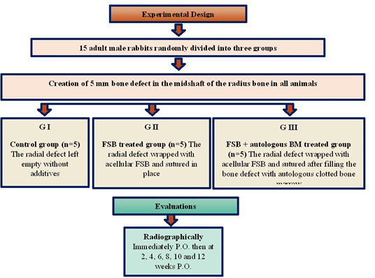
The fifteen experimental animals were divided randomly into three groups (n=5), the induction of segmental defects (5 mm) were done in the mid shaft of the radius bone in each animal by using a low-speed electrical saw and continuous irrigation with a 0.9% sterile saline solution, the first group was induced defects and left without any treatment (control group ). The second group was induced defects and treated with fish swim bladder treated group (FSB group). While the third group was induced defects and treated with fish swim bladder with autologous bone marrow clot (FSB-BMC group). The segmental defects in all groups were evaluated radiographically immediately after operation, then every two weeks till 12 weeks post-operation on a medio-lateral view with X-Ray machine (ENIE Radiologie®) at 45 kv and 5 mAs, to observe the bridging of the segmental bone defects and the development of new bone formation as in (Figure 1).
Preparation of Acellular Fish Swim Bladder (FSB)
Fresh swim bladders of carp fish were obtained from the local market and transported to the lab in a container with phosphate buffer solution (PBS). After removing the outer fat and blood capillaries. The swim bladders were rinsed and washed with PBS at 37 °C for 15 minutes. Next, the FSB scaffolds were decellularized using chemical decellularization method, which it was lysed the cell membrane and other cellular components with 0.5% sodium dodecyl sulphate (SDS) for 24 hours at room temperature and after that it was gently shaking the container three times ; . The decellularized scaffolds were cutting into approximately (1×1.5cm pieces) and terminally sterilized for 2 hours using 0.1% Peracetic acid (PAA) and 4% ethanol solution . Then the scaffolds were washed for 15 minutes at 37 °C using phosphate buffer solution (PBS) and stored in sterile PBS containing streptomycin (100μg/ml), penicillin (100 IU/ml) and Amphotericin (100μg/ml) and finally the scaffolds were preserved at 4 °C. Samples of FSB after decellularization were obtained and fixed in 10% buffer formalin solution for sectioning and staining using hematoxylin-eosin stain, then the samples were examined histologically after completing removal of cells with wavy dense staining acellular collagen fiber and loose delicate light-stained elastic fibers.
Surgical Procedure
Anesthetic protocol: Acepromazine (1 mg/kg) was injected intramuscularly into the experimental animals to sedate them (Prozil fort, Vietnam). 10% Ketamine (40 mg/kg of ) and 5 mg/kg of 2% Xylazine Hydrochloride (VMD-Belgium) were given I/M and as a general anesthesia after 10 minutes of Acepromazine injection . .
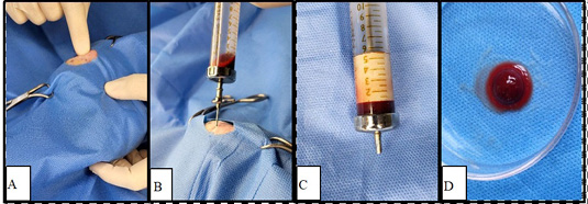
Bone marrow aspiration: After giving the general anesthesia and undergoing aseptic surgical preparation of the aspiration filed, the animals were placed into a lateral recumbency. A tiny stab skin incision was made for each animal to facilitate entry of the aspiration needle gauge 16 in the mid-way between the head of femur and the greater trochanter. The location of the trochanter fossa was determined by palpating the greater trochanter. The sterile needle of syringe was inserted toward the trochanteric fossa and carefully rotated until reaching to the medullary cavity to obtain 1ml of BM sample (Awid et al., 2014) as in (Figure 2A, B). The aspirating BM was allowed to clot for fifteen minutes before implantation and cut to fit the creating of radial defect in the same animal as in (Figure 2C, D).
Surgical Operation
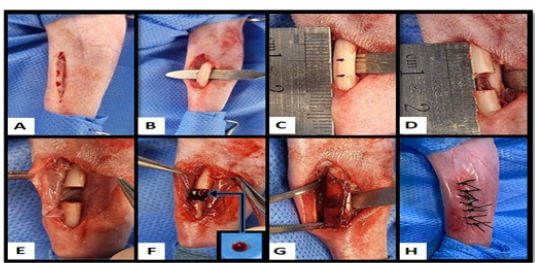
Following aseptic preparation of the surgical site in the right forelimb from the mid-shift of the humerus to the metacarpal bones, the anesthitized experimental animals were placed in a right lateral recumbency position. A 3–4 cm and skin incision was performed on the right forelimb’s cranio-medial aspect in equivalent distance between the elbow and carpal joint as in (Figure 3A). The pronator teres and flexor carpi radialis muscles were carefully separated with fine blunt scissors to expose the radius bone as in (Figure 3B). Distance (5mm) was marked at the radius mid shaft for osteotomy as in (Figure 3C), the marked segment was removed using electrical saw irrigated and (0.9%) sterile saline solution to create a defect in the radius mid shaft as in (Figure 3D). In the control group the segmental defect was left empty, whereas in the FSB group the defect was wrapped using the previously preparation of acellular FSB and it was sutured as a tube around the radius bone defect as in (Figure 3E), while in the FSB-BMC group the radial defect was wrapped using acellular FSB and filled with autologous bone marrow clot as in (Figure 3F), Subsequently, a simple continuous pattern using 5/0 polydioxanone (PDS) was used to suture the FSB membrane (Figure 3G). The muscles were approximated by simple continuous suture pattern using 3/0 polydioxanone (PDS) and an interrupted suture pattern with 3/0 silk. After that, the skin was closed (Figure 3H). Intramuscular injection of meloxicam at a dose of 1.5 mg/ kg. B.W. was given as analgesia (Ahmed et al., 2017), penicillin-streptomycin antibiotics were administered at doses of 10,000 IU/Kg B.W. and 5 mg/Kg B.W., respectively for 3 days post operation.
RESULTS AND DISCUSSION
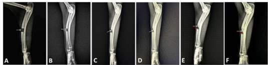
Numerous studies were recorded about bone repairing of animal models. Study done by Bigham-Sadegh and Oryan (2015), they mentioned rabbits a good animal to examine the segmental defects and grafts with structural qualities. As well as, previous reports of this model’s suitability had been mentioned rabbits did not required internal or external fixation lead to impact the healing process (Atiyah et al., 2020). Therefore, in current study a radius defect of approximately (0.5) cm was created to evaluate acellular FSB effect as guided bone regeneration to bridging the radius defect associated with autologous BMC in rabbits without needing for any fixation. Numerous research were reported on the application of tissue engineering techniques to repair radial abnormalities in New Zealand white rabbits ; ; . After middle radial defects, the intact ulna supported the rabbit’s forelimbs without internal fixation. For bone defect models, the mid-diaphyseal radius can frequently be the exacted site. A radiolucent segmental defect area was appeared at the mid shaft of radius bone in control group immediately post –operation as in (Figure 4A). While, the proximal and distal ends of the radius bone segments were showed a mild periosteal reaction in the second week following the operation as in (Figure 4B). Whereas, the proximal and distal ends of the radius bone segments were appeared an increase in thickness of both ends with callus formation and extending from the proximal end of the radius defect towards the defect center and it was attached with the ulna at ٤th week post –operation as in (Figure 4C). While the callus formation was increased from the proximal end of the radius defect, as well as, it was extended to the center of the defect and fused with ulna at 6th week post –operation as in (Figure 4D). A mild periosteal reaction in the radius defect’s distal end was appeared in the eighth week following the procedure as in (Figure 4E). In addition, the proximal end of callus from the radius defects to its center was continuously grew in along week after the surgery and the periosteal reaction in the distal end of radius defect was increased until 12th week post –operation with obvious radio-ulnar synostosis (Figure 4F). Some researchers hypothesized this reaction could be caused by scratching the surface of the bone and it was perfected for starting a biologic reaction lead to ulnar hypertrophy. Other studies were showed this reaction of the activation in periosteal cambium cells leads to the synthesis of callus matrix ; . This result may be attributed to a biologic reaction from cells in the surrounding tissues and this explaining of current study was agreed with other studies demonstrated a distinct fusing of the radius and ulna at the sites of defect creation by biological reaction . Other study reported by Bodde et al. (2008), who mentioned that the membrane interossea between the two bones and the periosteal were remained from the proximal and distal ends of the defect and this result perhaps due to the charge of the synostosis or fusion between the rabbit’s ulna and radius. According to study mentioned by Torbjörn et al. (2021), they showed the assessments of the osseoinductive and osseoconductive qualities in the investigation were made more difficult by the osseous growth of the ulnar bone and its union with the radius.
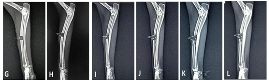
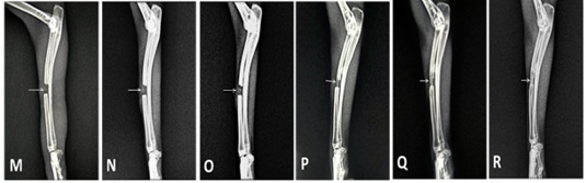
In present study A radiolucent segmental defect area of FSB treated group immediately post –operation was seen in (Figure 5G), Furthermore, a little periosteal response at both ends of the radius bone defect was appeared in the second week following surgery as in (Figure 5H). There was an increase in the periosteal reaction and radiopaque foci of newly formed bone was showed in the fourth week following surgery as in (Figure 5I). While, the radius defect’s new bone growth was increased and extended from both ends in the sixth week following the procedure (Figure 5J). As well as, an increase in newly formed bone and extended from both ends of the radius defect towards the central new bone formation foci was appeared in eight weeks after the procedure (Figure 5K). The opacity of new bone formation was increased and extended from both ends of the radius defect towards the central new bone formation foci at 10th week post –operation and 12th week post –operation. Furthermore, The opacity of new bone formation was extended from both ends of the radius defect in a straight direction with partially bridging the radius defect as in (Figure 5L), these results might be related to the heamatoma formation which it was considered as the first an important step for the healing of fractures and it was produced in the acellular FSB tube that it was encircled the defect lead to the production of new bone at the defect’s center and a clear extension of the fracture ends. These outcomes were consistent with those recorded by Gu et al. (2023) and Hwang et al. (2016). According to other research, a collagen membrane’s ability was resisted the proteolytic attacking from the surrounding tissue lead to maintaining the space beneath and preventing the growth of fibrous connective tissues as its initial mechanical strength . Furthermore, in contrast to the control group, the using of an acellular FSB scaffold in this investigation might be prevented ulnar osseous hypertrophy from influencing bone growth in the radius defect, this result agreed with findings of Bodde et al. (2008) and Zhao et al. (2016). A radiolucent segmental defect area of FSB-BMC treated group post –operation was observed in (Figure 6M). A slight periosteal reaction at both ends of the segmental radius defect with slight radiopaque foci in the center of the defect were seen at 2nd week post –operation (Figure 6N). The periosteal reaction at both ends of the segmental radius defect and the opacity of the foci in the center of the defect were increased at 4th week post –operation (Figure 6O). An increase in a new bone growth was observed at the six-week following surgery, and it was extended as a straight line from the proximal to the distal end of the bone defect as in (Figure 6P). The segmental bone defect was bridged by an increase in callus formation opacity in the eighth week after surgery, (Figure 6Q). The segmental bone defect was constantly continued to complete bridging of the segmental defect of radius bone by new bone formation at10th and 12th week post-operation (Figure 6R), these results might be attributed to action of acellular FSB scaffold which it was considered an essential to the guiding bone regeneration technique. . In addition to the action of autologous BMC that it was played an important role in healing the site of the defect. The present study showed allowing the aspirated autologous bone marrow to clot lead to producing implantable material with good handling properties to fit the site of the bone defect, this clarification was in agreement with the findings reported by Healey et al. (1990). According to Travlos (2006) and Korf-klingebiel (2008), they mentioned that bone marrow contained a variety of blood cell types, growth factors, cytokines, and stem cells. All of these cell types lead to an enhancemen of bone defect healing process. Eesa et al., (2006) and Thanoon et al., (2019) they also discovered that improving of bone marrow by these cells had a critical role in promoting and enhancing the fracture healing.
The result of radiographic results at six weeks might be related to the activation of platelets in the BMC lead to degranulation and releasing of pro-osteogenic, pro-angiogenic and pro-mitogenic growth factors such as BMP-2, vascular endothelial growth factor (VEGF), platelet-derived growth factor (PDGF) and fibroblast growth factor-2 (FGF-2) during BMA clotting ; ; ; . Other researchers found higher levels of growth factors in BMA clot compared with peripheral blood clot. In consequently, the a raising in chemotactic response of reparative cells such as MSCs lead to enhancement the ability of various kinds of tissue cells to differentiate and multiply such as chondroblast, osteoblast and osteocyte which they were played an important role in enhancing the healing process of bone defect and this result of current study was agreed with findings reported by Fiedler et al. (2002); Crovace et al., (2008); Shoji et al. (2017); Harwood et al. (2010), Claes et al. (2012); Thanoon et al. (2019).
In general, the procedure succeed in using barrier membrane as guiding bone regeneration depended on the defect size lead to allow in a new bone formation ; . Insufficient mechanical strength or early degradation of the barrier membrane might be caused soft tissue invasion. For this reason, a crosslinking agent was incorporated during preparation of some collagen membrane to improve its mechanical and degradation properties .
CONCLUSIONS AND RECOMMENDATIONS
Fish swim bladder (FSB) scaffolds were easily prepared from the waste product of carp fish with mild chemicals, and it was acted as a good guided for bone regeneration. In addition, the combination between FSB and autologous bone marrow clot gave a success results in bone regeneration and bridging segmental bone defect without any clinical complications related to infection or rejection. The study recommended to an additional efforts were required to optimizing the prepared FSB and determining the effectiveness of FSB in conjunction with other materials like hydrogel, platelet-rich fibrin matrix, hydroxyapatite or other sources of autologous cells in regeneration of a critical segmental bone defect.
ACKNOWLEDGEMENTS
The authors express their gratitude to the members of the surgery department at the University of Baghdad’s College of Veterinary Medicine for their assistance in this work.
NOVELTY STATEMENT
The distinctive feature of this work is that it is the first to prepare fish swim bladders for local application as guided bone regeneration using autologous bone marrow clots to treat segmental bone defects. The project also aims to explore the potential of these treatments for tissue regeneration after injury.
AUTHOR’S CONTRIBUTIONS
The authors’ combined efforts have resulted in the completion of this article.
Data Availability
All data will be provided by the authors upon reasonable request.
Conflict of Interest
The authors of this article worked on it and declared that they had no conflicts of interest.
Figures
Figure 1:.
Experimental design.
Figure 2:.
Steps of bone marrow aspiration; A: Palpating the greater trochanter; B: The aspiration needle is inserted toward the trochanteric fossa; C: The aspirated BM is left to clot before implantation; D: Bone marrow clot sample.
Figure 3:.
Steps of surgical procedure; A: 3–4 cm skin incision is performed on the right forelimb’s cranio-medial aspect; B: Expose the radius bone after separating the muscles; C: 5mm distance at the radius mid shaft is marked for osteotomy; D: Creation of 5mm segmental bone defect; E: The radial defect is wrapped with acellular FSB; F: The radial defect is filled with bone marrow clot after wrapping with acellular FSB; G: The FSB membrane is sutured; H: Sutured skin.
Figure 4:.
A mediolateral view of radiological image in control group (A) Immediately post –operation, shows a radiolucent segmental defect area appeared at the mid shaft of radius bone. (B) At 2nd week post –operation, shows the proximal and distal ends of the radius bone segments with a mild periosteal reaction. (C) At 4th week post –operation, shows thickening of both ends of the defect with callus formation directed from the proximal end of the radius defect towards the defect center and attached with the ulna. (D) At 6th week post –operation, callus formation was increased from the proximal end of the radius defect sloped to the center of the defect and fused with ulna. (E) At 8th week post –operation, mild periosteal reaction in the radius defect’s distal end. (F) At 12th week post –operation, the sloping callus from the radius defect’s proximal end to its center was continuously growing with radio-ulnar synostosis.
Figure 5:.
A mediolateral view of radiological image in FSB treated group. (G) Immediately post –operation, shows a radiolucent segmental defect area in the mid shaft of radius bone. (H) At 2nd week post –operation, shows a little periosteal response at both ends of the radius bone defect. (I) At 4th week post –operation, there was an increase in the periosteal reaction, and a radiopaque foci of newly formed bone is visible in the defect’s center. (J) At 6th week post –operation, the radius defect’s new bone growth was increased and extended from both ends. (K) At 8th week post –operation. there was an increase in newly formed bone and extended from both ends of the radius defect towards the central new bone formation foci. (L) At 12th week post –operation, shows increased the opacity of new bone formation and extended in a straight direction from both ends of the radius defect towards the central new bone formation foci, with partially bridging the radius defect.
Figure 6:.
A mediolateral view of radiological image in FSB-BMC treated group. (M) Immediately post –operation, shows a radiolucent segmental defect area in mid shaft of radius bone (N) At 2nd week post –operation, shows a slight periosteal reaction at both ends of the segmental radius defect with slight radiopaque foci in the center of the defect (O) At 4th week post –operation, shows increased the periosteal reaction at both ends of the segmental radius defect with increased the opacity of the foci in the center of the defect (P) At 6th week post –operation, shows increased new bone growth and it extended in a straight direction from the proximal to the distal end of the bone defect. (Q) At 8th week post –operation, the segmental bone defect had been bridged by an increase in callus formation opacity. (R) At 12th week post –operation, shows a complete bridging of the segmental defect of radius bone by new bone formation.
References
Ahmad A, Shah Nawaz R, Soomro SA, Rao N, Qasim M (٢٠١٧). The effects of meloxicam, a non-steroidal anti-inflammatory drug on the biochemical parameters of rabbits. Int. J. Biosci., 11(2): 58-67. https://doi.org/10.12692/ijb/11.2.58-67[CrossRef]
Al-Bayati AH, Al-Tememe HA, Al-Mudallal NH (2016). Role of acellular bovine urinary bladder submucosa on skin wound healing in Iraqi goats. The Iraqi J. Vet. Med., 40(1): 53-60. https://doi.org/10.30539/iraqijvm.v40i1.138[CrossRef]
Al-Ebadi AK, Al-Bayati A H (2019). Effect of acellular bovine pericardium and urinary bladder submucosa matrixes in reconstruction of ventro-lateral hernias in bucks; molecular evaluation. The Iraqi J. Vet. Med., 43(1): 67-74. https://doi.org/10.30539/iraqijvm.v43i1.474[CrossRef]
Al-Falahi NH (2009). Comparative study for using of sub mucosa of small intestine in healing of clean and infected avulsion wounds. The Iraqi J. Vet. Med., 33(1): 90-97. https://doi.org/10.30539/iraqijvm.v33i1.720[CrossRef]
AL-Falahi NH, Salih SI, Obaid A (2016). A comparative biomechanical study of repaired tendons wrapped with two biological matrices in Bucks. The Iraqi J. Vet. Med., 40(1): 73-78. https://doi.org/10.30539/iraqijvm.v40i1.141[CrossRef]
Al-Falahi NH, Ab Abood D, Dauood MS (2017). Comparative evaluation of bovine pericardial membrane and amniotic membrane in wounds skin healing in rabbits. The Iraqi J. Vet. Med., 41(2):137-145. https://doi.org/10.30539/iraqijvm.v41i2.63[CrossRef]
Atiyah AG, AL-Falahi NHR, Hasan MS , Owain MS (2020). Study the effect of avian eggshell hydroxyapatite powder on bone gaps healing in rabbits. Veterinary Practitioner, 21(2):429-434.
Awid AC, Alfaris AA, Sawad AA (2014). Effects of the cyclosporine on some hematological indices in bone marrow transplanted-rabbit. Bas. J. Vet. Res., 1(2):23-35. https://doi.org/10.33762/bvetr.2014.98790[CrossRef]
Badylak SF (2007). The extracellular matrix as a biologic scaffold material. Biomaterials., 28(25): 3587-3593. https://doi.org/10.1016/j.biomaterials.2007.04.043[CrossRef]
Bai H, Sun P, Wu H, Wei S, Xie B, Wang W, Li Z (2021). The application of tissue-engineered fish swim bladder vascular graft. Commun. Biol., 4(1): 1153. https://doi.org/10.1038/s42003-021-02696-9[CrossRef]
Bigham -Sadegh A, Oryan A (2015). Selection of animal models for pre-clinical strategies in evaluating the fracture healing, bone graft substitutes and bone tissue regeneration and engineering Connect. Tissue Res., 56: 175-94. https://doi.org/10.3109/03008207.2015.1027341[CrossRef]
Bodde EW, Spauwen PH, Mikos AG, Jansen JA (2008). Closing capacity of segmental radius defects in rabbits. J. Biomed. Mater Res., 85: 206-217. https://doi.org/10.1002/jbm.a.31549[CrossRef]
Cho EH, Park JC, Cha JK, Kim YT, Jung UW, Kim CS, Kim CK (2011). Dimensional change of the healed periosteum on surgically created defects. J. Periodontal Implant Sci., 41(4): 176-184. https://doi.org/10.5051/jpis.2011.41.4.176[CrossRef]
Claes L, Recknagel S, Ignatius A (2012). Fracture healing under healthy and inflammatory conditions. Natur. Revi. Rheumat., 8: 133-143. https://doi.org/10.1038/nrrheum.2012.1[CrossRef]
Crapo PM, Gilbert TW, Badylak SF (2011). An overview of tissue and whole organ de-cellularization processes. Biomaterials, 32(12):32. https://doi.org/10.1016/j.biomaterials.2011.01.057[CrossRef]
Crovace A, Favia A, Lacitignola LD , Comite MS , Staffieri F, Francioso E (2008). Use of autologous bone marrow mononuclear cells and cultured bone marrow stromal cells in dogs with orthopaedic lesions. Vet. Res. Commun., 32: 39-44. https://doi.org/10.1007/s11259-008-9095-1[CrossRef]
Eesa MJ, Thanoon MG, Ibrahim SM (2006). The Effect of bone marrow autograft on fracture healing with destruction of periosteum and endosteum in rabbits. Iraqi J. Vet. Sci., 20(2): 163-172. https://doi.org/10.33899/ijvs.2006.62494[CrossRef]
Fiedler J, Röderer G, Günther KP, Brenner RE (2002). BMP-2, BMP-4, and PDGF-bb stimulate chemotactic migration of primary human mesenchymal progenitor cells. J. Cell Biochem., 87:305-312. https://doi.org/10.1002/jcb.10309[CrossRef]
Flecknell P (2015). Laboratory animal anaesthesia. (4th edition). United States of America Academic press., 220-222.
Gangji V, De Maertelaer V, Hauzeur JP (2011). Autologous bone marrow cell implantation in the treatment of non-traumatic osteonecrosis of the femoral head: Five year follow-up of a prospective controlled study. Bone, 49(5): 1005–1009. https://doi.org/10.1016/j.bone.2011.07.032[CrossRef]
Gu F, Zhang K, Zhu W, Sui Z, Li J, Xie X, Yu T (2023). Silicon rubber sealed channel induced self-healing of large bone defects: Where is the limit of self-healing of bone. J. Orthop. Transl., 43: 21- 35. https://doi.org/10.1016/j.jot.2023.09.001[CrossRef]
Gumaa BH, AL-Bayati AH (2021). Molecular evaluation of efficacy of freshwater and marine acellular fish skin matrixes in reconstruction of ventro-lateral hernia in bucks. Indian J. Forensic Med. Toxicol., 15(4): 880-886. https://doi.org/10.37506/ijfmt.v15i4.16814[CrossRef]
Hämmerle CHF, Karring T (1998). Guided bone regeneration at oral implant sites. Periodontol, 17(1):151-75. https://doi.org/10.1111/j.1600-0757.1998.tb00132.x[CrossRef]
Harwood PJ, Newman JB, Michael AL (2010). (ii) An update on fracture healing and non-union. Orthop. Trauma., 24(1): 9-23. https://doi.org/10.1016/j.mporth.2009.12.004[CrossRef]
Healey JH, Zimmerman PA, McDonnell JM, Lane JM (1990). Percutaneous bone marrow grafting of delayed union and nonunion in cancer patients. Clin. Orthop. Relat. Res., 256: 280-285. https://doi.org/10.1097/00003086-199007000-00039[CrossRef]
Huang Q, Liu Y, Ouyang Z, Feng Q (2020). Comparing the regeneration potential between PLLA/Aragonite and PLLA/Vaterite pearl composite scaffolds in rabbit radius segmental bone defects. Bioact. Mater., 5(4): 980989. https://doi.org/10.1016/j.bioactmat.2020.06.018[CrossRef]
Hwang JW, Kim S, Kim SW, Lee JH (2016). Effect of Extracellular Matrix Membrane on Bone Formation in a Rabbit Tibial Defect Model. BioMed. Res. Int., 1-8. https://doi.org/10.1155/2016/6715295[CrossRef]
Italiano JE, Richardson JL, Patel-Hett S (2008). Angiogenesis is regulated by a novel mechanism: pro- and antiangiogenic proteins are organized into separate platelet alpha granules and differentially released. Blood, 111:1227-1233 . https://doi.org/10.1182/blood-2007-09-113837[CrossRef]
Jonitz A, Lochner K, Lindner T, Hansmann D, Marrot A, Bader R (2011). Oxygen consumption, acidification and migration capacity of human primary osteoblasts within a three-dimensional tantalum scaffold. J. Mater. Sci. Mater. Med., 22: 2089-2095. https://doi.org/10.1007/s10856-011-4384-6[CrossRef]
Kalén A, Wahlström O, Linder CH, Magnusson P (2008). The content of bone morphogenetic proteins in platelets varies greatly between different platelet donors. Biochem. Biophys. Res. Commun., 375:261-264. https://doi.org/10.1016/j.bbrc.2008.08.014[CrossRef]
Kasten P, Vogel J, Geiger F, Niemeyer P, Luginbuhl R, Szalay K (2008). The effect of platelet-rich plasma on healing in critical-size long-bone defects. Biomaterials., 29: 3983–3992. PMID: 18614227. https://doi.org/10.1016/j.biomaterials.2008.06.014[CrossRef]
Khoshkhoonejad AA, Miremadi A, Rokn AR, Eslami B, Dehghan M, Kalbassi H (2006). Effect of GBR in Combination with Deproteinized Bovine Bone Mineral on the Healing of Calvarial Defects in Rabbits. J. Dent., (3)2: 77-86.
Korf-Klingebiel M, Kempf T, Sauer T,Brinkmann E, Fischer P, Meyer GP , Wollert KC (2008). Bone marrow cells are a rich source of growth factors and cytokines: implications for cell therapy trials after myocardial infarction. Eur. heart J., 29(23): 2851-2858. https://doi.org/10.1093/eurheartj/ehn456[CrossRef]
Kumar V, Kumar N, Gangwar AK, Singh H, Singh R (2015). Comparative histologic and immunologic evaluation of 1, 4-butanediol diglycidyl ether crosslinked versus noncrosslinked acellular swim bladder matrix for healing of full-thickness skin wounds inrabbits. J. Surg. Res., 197 (2): 436446. https://doi.org/10.1016/j.jss.2015.04.080[CrossRef]
Lim ZXH, Rai B, Tan TC, Ramruttun AK, Hui JH, Nurcombe V, Teoh SH, Cool SM (2019). Autologous bone marrow clot as an alternative to autograft for bone defect healing. Bone Joint Res., 8: 107-117. https://doi.org/10.1302/2046-3758.83.BJR-2018-0096.R1[CrossRef]
Luan LS, Wang W, Xu K, Ye S, Dan R, Zhang H, Shu Z, Wang T, Fan C (2022). Aligned nanofibrous collagen membranes from fish swim bladder as a tough and acid-resistant suture for pH-regulated stomach perforation and tendon rupture. Biomater. Res., 26(1): 60. https://doi.org/10.1186/s40824-022-00306-1[CrossRef]
Leng Y, Ren G, Cui Y, Peng C, Wang J, Wu D, Liu H (2020). Platelet-rich plasma- enhanced osseointegration of decellularized bone matrix in critical-size radial defects in rabbits. Ann. Transl. Med., 8(5). https://doi.org/10.21037/atm.2020.01.53[CrossRef]
Lin X, Patil S, Gao YG, Qian A (2020). The bone extracellular matrix in bone formation and regeneration. Front. Pharmacol., 11: 757. https://doi.org/10.3389/fphar.2020.00757[CrossRef]
Mohammad FA, Al-Ebadi AK (2023). Macroscopically and histopathological study of Using Fenestrated and Non-Fenestrated Catfish Acellular Dermal Matrix on Healing Ventro-Lateral Hernia in Bucks. Journal of Survey in Fish. Sci., 10(3S): 844-859.
Mohammed MS, Salih SI (2022). Histopathological Study of Using Fetal Caprine Acellular Dermal Matrix Alone and in Combination with Non-thermal Plasma in Healing of Full-thickness Acute Skin Wounds in Bucks. Int. J. Health Sci., 5 (3): 8784-8806. https://doi.org/10.53730/ijhs.v6nS3.8105[CrossRef]
Mollon B, Kandel R, Chahal J, Theodoropoulos J (2013). The clinical status of cartilage tissue regeneration in humans. Osteoarthritis and cartilage, 21(12): 1824-1833. https://doi.org/10.1016/j.joca.2013.08.024[CrossRef]
Mredha MTI, Kitamura N, Nonoyama T, Wada S, Goto K, Zhang X, Gong JP (2017). Anisotropic tough double network hydrogel from fish collagen and its spontaneous in vivo bonding to bone. Biomaterials, 132: 85-95. https://doi.org/10.1016/j.biomaterials.2017.04.005[CrossRef]
Meng ZL, Wu ZQ, Shen BX, Li HB, Bian YY, Zeng DL, Peng L (2019). Reconstruction of large segmental bone defects in rabbit using the Masquelet technique with α-calcium sulfate hemihydrate. J. Orthop. Surg. Res., 14:192 https://doi.org/10.1186/s13018-019-1235-5[CrossRef]
Olaechea A, Mendoza-Azpur G, Valdivia E, Rasperini G (2016). Biodegradation of three different collagen membranes: A histological study. J. Osseointegration, 8(2):15-9.
Peng F, Zhang X, Wang Y, Zhao R, Cao Z, Chen S, Zhang X (2023). Guided bone regeneration in long-bone defect with a bilayer mineralized collagen membrane. Coll. Leather, 5(1): 36. https://doi.org/10.1186/s42825-023-00144-4[CrossRef]
Pereira HF, Cengiz IF, Silva FS , Reis RL , Oliveira JM (2020). Scaffolds and coatings for bone regeneration. J. Mater. Sci. Mater. Med., 31:27. https://doi.org/10.1007/s10856-020-06364-y[CrossRef]
Peric M, Dumic-Cule I, Grcevic D, Matijasic M, Verbanac D, Paul R, Grgurevic L, Trkulja V, Bagi CM, Vukicevic S (2015). The rational use of animal models in the evaluation of novel bone regenerative therapies. Bone, 70:7386. https://doi.org/10.1016/j.bone.2014.07.010[CrossRef]
Salamanna F, Contartese D, Nicoli Aldini N, Barbanti Brodano G, Griffoni C, Gasbarrini A, Fini M (2018). Bone marrow aspirate clot: A technical complication or a smart approach for musculoskeletal tissue regeneration. J. Cell. Physiol., 233(4): 2723-2732. https://doi.org/10.1002/jcp.26065[CrossRef]
Shoji T, Nakasa T, Yoshizuka M (2017). Comparison of fibrin clots derived from peripheral blood and bone marrow. Connect Tissue Res., 58:208-214. https://doi.org/10.1080/03008207.2016.1215443[CrossRef]
Sipe JB, Zhang J, Waits C (2004). Localization of bone morphogenetic proteins (BMPs)-2, -4, and -6 within megakaryocytes and platelets. Bone, 35:1316- 1322. https://doi.org/10.1016/j.bone.2004.08.020[CrossRef]
Sivajothi S, Reddy SB, Rayulu VC (2014). Effect of Ivermectin against Psoroptic mange in rabbits. Inter., J. of Sci. World, 2 (1): 10- 12. https://doi.org/10.14419/ijsw.v2i1.1893[CrossRef]
Tao M, Ao T, Mao X, Yan X, Javed R, Hou W, Yu T (2021). Sterilization and disinfection methods for decellularized matrix materials., Review, consideration and proposal. Bioact. Mater., (9): 29272945. https://doi.org/10.1016/j.bioactmat.2021.02.010[CrossRef]
Thanoon M, Eesa MJ, Abed ER (2019). Effects of platelets rich fibrin and bone marrow on the healing of distal radial fracture in local dogs: Comparative study. Iraqi J. Vet. Sci., 33(2): 419-425. https://doi.org/10.33899/ijvs.2019.163169[CrossRef]
Torbjörn M, Amela T, Andreas T, Ekman Stina E, Ley Cecilia L, Caroline O, Petra HJ, Jensen-Waern Marianne JW, Patricia H (2021). Guided bone tissue regeneration using a hollow calcium phosphate based implant in a critical size rabbit radius defect. Biomed. Mater., 16:1-14. https://doi.org/10.1088/1748-605X/abde6f[CrossRef]
Travlos GS (2006). Normal structure, function, and histology of the bone marrow. Toxicol. Pathol., 34 (5): 548-565. https://doi.org/10.1080/01926230600939856[CrossRef]
Veronesi F, Salamanna F, Tschon M, Maglio M, Nicoli Aldini N, Fini M (2017). Mesenchymal stem cells for tendon healing: what is on the horizon. J. Tissue Eng. Regenerative Med., 11(11):3202-3219. https://doi.org/10.1002/term.2209[CrossRef]
Veronesi F, Giavaresi G, Tschon M, Borsari V, Nicoli Aldini N, Fini M (2013). Clinical use of bone marrow, bone marrow concentrate, and expanded bone marrow mesenchymal stem cells in cartilage disease Stem Cells Dev., 22(2):181–192. https://doi.org/10.1089/scd.2012.0373[CrossRef]
Wubneh A, Tsekoura EK, Ayranci C, Uluda˘g H (2018). Current state of fabrication technologies and materials for bone tissue engineering. Acta Biomater, 80: 1 https://doi.org/10.1016/j.actbio.2018.09.031[CrossRef]
Wang H, Li Q, Wang Q, Zhang H, Shi W, Gan H, Wang Z (2017). Enhanced repair of segmental bone defects in rabbit radius by porous tantalum scaffolds modified with the RGD peptide. J. Mater. Sci. Mater. Med., 28:1-10. https://doi.org/10.1007/s10856-017-5860-4[CrossRef]
Weadock KS, Miller EJ, Keuffel EL, Dunn MG. (1996). Effect of physical crosslinking methods on collagen‐fiber durability in proteolytic solutions. J. Biomed. Materi. Res., 32(2):221-6. https://doi.org/10.1002/(SICI)1097-4636(199610)32:2<221::AID-JBM11>3.0.CO;2-M[CrossRef]
Zhao MD, Huang JS , Zhang XC, Gui KK , Xiong M, Yin WP, Yuan FL , Cai GP (2016). Construction of radial defect models in rabbits to determine the critical size defects. PLoS One, 11 -463. https://doi.org/10.1371/journal.pone.0146301[CrossRef]
Zhao M, Zhou J, Li X, Fang T, Dai W, Yin W (2011). Repair of bone defect with vascularized tissue engineered bone graft seeded with mesenchymal stem cells in rabbits. Microsurgery, 31: 130–137. https://doi.org/10.1002/micr.20854[CrossRef]