
Rab GTPases are important regulators of intracellular trafficking, having distinct intracellular localisation for controlling endocytic, exocytic, and recycling pathways. The impairments in Rab pathways have been linked with drug resistance in various cancers. However, the role of Rab21, an endocytic Rab GTPase, in the emergence of drug resistance is not known. Therefore, we aimed to investigate the role of Rab21 in MRP-1, drug efflux pump, mediated resistance in prostate cancer cells. Drug sensitive (PC-3/Wt) and epirubicin resistant (PC-3/Res) prostate cancer cell lines were used in this study. Our data revealed that PC-3/Res cells had high expression of MRP-1 mRNA and protein in comparison with PC-3/Wt cells. MRP-1 was found distributed between intracellular and cell surface pools in PC-3/Res cells and was capable of drug efflux as shown by doxorubicin and epirubicin efflux assays. Moreover, qRT-PCR and Western blot analysis showed that PC-3/Res cells also had significantly up-regulated expression of Rab21. To study the effect of Rab21 on MRP-1 mediated multidrug resistance, siRNA mediated knockdown of Rab21 was performed. Results showed that the knocking down of Rab21 decreased the drug efflux ability of PC-3/Res cells, possibly by altering the surface localisation of MRP-1. These results suggest that interruption in MRP-1 trafficking to the plasma membrane by Rab21 is a potential strategy to overcome multi drug resistance in cancer cells.
Research Article · Volume 52, Issue 6, 5 September 2020 · Open Access
Rab21, an Endocytic Rab GTPase, is Involved in Drug Resistance in Prostate Cancer Cells
· · ·
Affiliations & Notes ⌄
- 1Institute of Biochemistry and Biotechnology, Quaid e Azam Campus, University of the Punjab, Lahore, Pakistan 2School of Biological Sciences, University of the Punjab, Lahore, Pakistan , * Corresponding author: moazzam.ibb@pu.edu.pk
Article Info ⌄
- Journal:
- Pakistan Journal of Zoology
- Published:
- 5 September 2020

From the article
- In the present study, we found that MRP-1 expression was significantly increased in epirubicin resistant PC-3 cells, suggesting the role of MRP-1 in mediating MDR in prostate cancer cells.
- Our findings suggested that up-regulation of endogenous Rab21 level by epirubicin resistant cells evolved as a contributing factor to favour cell surface localisation of MRP-1 and hence drug resistance.
- Our findings reveal the importance of Rab21 in MRP-1 mediated multidrug resistance.
- Interruption in membrane trafficking of MRP-1 using Rab21 siRNA resulted in increased intracellular drug retention, providing a potential strategy to overcome MDR.
Selected from the article's own text.
Abstract
Introduction
Multidrug resistance associated protein 1 (MRP-1), is a drug efflux pump encoded by ABCC1 gene ; . MRP-1 was first identified in chemoresistant lung cancer cell line, as over-expressed transporter . Later on, variable expression of MRP-1 was also reported in various human tissues, for instance; high expression levels of MRP-1 were found in heart, kidney, lung, placenta, testis, skeletal muscles and adrenal glands. Whereas, the lower expression levels were reported in colon, brain, intestine, and liver ; ; . MRP-1 is expressed in basolateral membrane of the polarised epithelial cells, where it plays an important role in protecting cells against toxic metabolites and xenobiotics . Unlike other ABC transporters, MRP-1 or ABCC1 is composed of three transmembrane domains (TMDs) and the two nucleotide binding domains (NBDs) . The additional atypical TMD0 in MRP1 is believed to play an important role in sub-cellular localisation , 2005) as truncated MRP1, lacking atypical TMD accumulates in endosomes ; . TMD0 is needed for cell surface expression of MRP1 and its loss accelerates the internalisation of MRP1 . However, the exact mechanisms of MRP-1 trafficking and degradation still remain unclear.
The drug efflux ability of MRP-1 reduces the treatment efficacy against different diseases including epilepsy, depression, and cancer ; ; . In fact, MRP-1 over-expression is reported to play a key role in emergence of multidrug resistance in various cancers during chemotherapy . Moreover, the over-expression of MRP-1 along with MDR1 in various cancers has been found to be associated with relapse and considerable reduction in survival of cancer patients ; ; ; ; . MRP-1 is also a major factor in developing chemoresistance in prostate cancer . Over-expression of MRP1 was found in drug resistant prostate cancer cell lines ; Its ability to extrude antiandrogen compounds also suggests its role in antiandrogen therapeutic failure .
Rab GTPases are the key regulators of membrane trafficking and are involved in endocytic, exocytic, and recycling pathways. Human genome encodes more than 70 Rab proteins that function as molecular switches, by cycling between the active and the inactive states . Rabs are localised at different intracellular compartments to perform specific functions. Together with their effector proteins Rab GTPases regulate trafficking pathways and determine the destinations of cargo proteins. Dysregulation of Rab proteins have been implicated in cancer progression by disrupting the regulatory mechanisms of membrane trafficking . For instance, over-expression of Rab5 in colon cancer cells resulted in translocation of ABCB1 from plasma membrane to intracellular compartments, suggesting the role of Rab5 in endocytosis of ABCB1 . Studies have shown that Rab21, a homolog of Rab5, is also involved in endocytic pathways . Rab21 is known for its role of protein cargo sorting from early to late endosomes and lysosomes (Egami and Araki, 2008, 2009). However, the role of Rab21 in regulating the membrane trafficking of MRP-1 and its consequence on multi drug resistance (MDR) remains unclear.
Over-expression of MRP-1 in epirubicin resistant prostate cancer cell (PC3/Res) led us to investigate its involvement in drug extrusion. Here, we hypothesised that altered expression of Rab21 might be involved in MRP-1 mediated epirubicin resistance in PC3 cells.
MATERIALs AND METHODS
Materials
The monoclonal anti-MRP-1 antibody (clone, QCRL-1 Cat No sc-18835) and the anti-Rab21 antibody (clone B16K, Cat No sc-81917) were obtained from Santa Cruz Biotechnology. Alexa Fluor 568 conjugated secondary antibody (Cat No A11031), goat anti mouse HRP conjugated secondary antibody (Cat No G21040) and SlowFade® Gold Antifade mounting media with DAPI were from Invitrogen. Rab21 siRNA (Cat No sc-76322) was obtained from Santa Cruz Biotechnology. Doxorubicin hydrochloride and epirubicin hydrochloride were obtained from Sigma Aldrich. Dulbecco’s modified eagle media and all other cell culture reagents were obtained from Thermofisher scientific.
Cell culture and transfection
Both epirubicin sensitive (PC-3/Wt) and resistant (PC-3/Res) prostate cancer cell lines were maintained in DMEM, with 10% fetal bovine serum, penicillin 100 units/ml and streptomycin, 100 ug/ml. Cells were maintained at 37°C with 5% CO2 in a humidified incubator. The epirubicin resistant PC-3 cells were maintained under 0.5 micro molar epirubicin. Cells were transfected with siRNA against Rab21 using LipofectamineTM 3000 reagent (Invitrogen) according to manufacturerʼs instructions and analysed 72 h post transfection.
Quantitative Real-time PCR
RNA was isolated using GeneJET RNA Purification Kit and DNA contamination was removed by treating total RNA samples with DNase I (both from Thermo Scientific). cDNA was synthesised using RevertAid™ First Strand cDNA Synthesis Kit (Thermo Scientific) following manufacturer’s instructions. The quantitative RT-PCR was performed using Thermo Scientific’s Maxima SYBR Green qPCR Master Mix (2X). Primers used were
MRP-1F: 5’-AGACAGAGATGCGAACCACC -3’,
MRP-1R: 5’-GGAAGCACCAGGAAACCACT -3’, Rab21F: 5’-GGGAAAAGAGTAAACCTTGCCA-3’, Rab21R: 5’-TAAACTAAAATCGCTCCATTTGAAT-3’ B2MF: 5’-TGCTGTCTCCATGTTTGATGTATCT-3’, and B2MR: 5’-TCTCTGCTCCCCACCTCTAA
GT-3’. The relative mRNA level was calculated by delta delta Ct method, normalising against B2M reference mRNA using CFX manager software (Bio-Rad).
Western blotting
Cells were lysed with SDS sample buffer by heating at 95°C for 10min. Cell lysates were resolved using 12% SDS-PAGE at 0.1 × 10⁶ cell equivalents per lane, and then transferred to PVDF membranes (Immobilon; Millipore). Immunoblotting was performed using mouse anti MRP-1 and anti Rab21 monoclonal antibodies along with goat anti-mouse horseradish peroxidase conjugated secondary antibody. LuminataTM Forte Western HRP Substrate (Millipore) was used for detection. Photographic films were exposed for appropriate time and developed using automated film developer by Konica Minolta. Films were scanned using backlit film scanner and protein band intensities were quantified by normalising against Ponceau S staining as a loading control using ImageJ software.
Immunocytochemistry
PC-3 wild type and resistant cells were grown on coverslips. For immunofluorescent detection of MRP-1, cells were washed with dPBS and fixed using 4% formaldehyde solution (made freshly using paraformaldehyde) for 20 min. Following fixation, cells were permeabilised using 0.1% Triton X-100 and blocked with 5 % FBS for one hour. Cells were treated with 1: 50 dilution of anti-MRP-1 antibody for 1 h, washed thrice with dPBS and incubated with 1: 1000 dilution of Alexa Fluor 568 conjugated goat anti-mouse antibody for 1 hr. Cells were washed again, air dried and mounted using DAPI containing mountant. Labelled cells were observed using Olympus BXP-51 fluorescence microscope.
Drug efflux assays
For drug efflux assays; 0.5×10⁶ cells/well were seeded on cover slips and allowed to grow. Cells were washed with dPBS and incubated with 5uM epirubicin/ doxorubicin at 37°C for 2 h. After incubation, cells were allowed to efflux in drug free media for 1 h at 37°C. Cells were washed again and fixed with 4% formaldehyde for 20 min. DAPI Slowfade mounting media was used to counter satin nuclei. Non saturating microscopic images were obtained and mean fluorescence intensities of drugs were analysed and quantified using ImageJ Software.
Statistical analysis
GraphPad Prism software was used to analyse data. An unpaired t-test was used to calculate the p-value. Results were confirmed by performing at least two independent experiments with three technical replicates each. All results are presented as mean ± standard error of mean.
RESULTS
MRP-1 over expression and localisation in epirubicin resistant PC-3 cells
To study the effects of Rab21 on MRP-1 mediated MDR in prostate cancer PC-3 cells, the MRP-1 expression was compared in epirubicin (sensitive PC-3/Wt) resistant (PC-3/Res) cell lines. Our data revealed that MRP-1 mRNA was highly over expressed in PC-3/Res cells as compared to PC-3/Wt cells (Fig. 1A), Similarly, protein level of MRP-1 was also found elevated in PC-3/Res cells when compared with PC-3/Wt cells (Fig. 1B). Increased expression of MRP-1 in PC-3/Res cells suggest the possible role of MRP-1 in the development of resistance against epirubicin in prostate cancer. To investigate the sub cellular localisation of MRP-1 in PC-3/Res cells, immunofluorescence analysis was performed using specific anti-MRP-1 antibody. In drug sensitive PC-3/Wt cells, expression of MRP-1 was not observed. While, in epirubicin resistant PC-3/Res cell line, MRP-1 was predominantly found in plasma membrane in addition to its cytoplasmic localisation (Fig. 1C). Increased expression and cell surface localisation of MRP-1 in resistant PC-3/Res cells compared to drug sensitive PC-3/Wt cells indicated the role of MRP-1 in mediating MDR.
Functional analysis of MRP-1 in PC-3/Res cells
To substantiate the role of MRP-1 in mediating MDR in PC-3/Res cell line, we performed drug efflux assays. Doxorubicin and epirubicin were used to quantify drug retention in PC-3/Wt and PC-3/Res cells. These drugs are fluorescent in nature and their uptake or efflux by cells can be quantified by observing cells under a fluorescent microscope. Post efflux images were used to quantify residual intracellular doxorubicin. As shown in Figure 2A, the PC-3/Res cells showed decreased retention of doxorubicin than PC-3/Wt cells. Quantification of fluorescent intensity of doxorubicin showed a significant reduction in doxorubicin in PC-3/Res cells (Fig. 2B). Similar results were observed using epirubicin as a substrate of MRP-1. Decreased intracellular retention of epirubicin was observed in PC-3/ Res cells than PC-3/Wt cells (Fig. 3A and B). These findings indicate that the over expression of MRP-1 confers MDR in prostate cancer cells through drug efflux mechanism.
High expression level of Rab21 in PC-3/Res cells
To examine whether expression of Rab21 is a contributing factor in MRP-1 mediated epirubicin resistance in PC-3 cells, we performed quantitative RT-PCR and Western blot analysis to measure mRNA and protein expression levels of Rab21 in PC-3/Wt and PC-3/Res cells. As shown in Figure 4A, the mRNA expression level of Rab21 was higher in PC-3/ Res cells as compared to PC-3/Wt cells. Similar results were obtained after Western blotting using anti-Rab21 antibody (Fig. 4B). The protein band intensity was also quantified and normalised against total protein loading control using Image J Software. Results showed a significantly increased expression of Rab21 in PC-3 epirubicin resistant cells (Fig. 4C).
Knock down of Rab21 decreases epirubicin resistance in PC-3/Res cells
Rab21 was up-regulated in PC-3/Res cells as compared to PC-3/Wt cells, suggesting that increased expression of Rab21 might contribute to MDR. We next explored whether epirubicin resistance could be attenuated by knocking down of Rab21 using Rab21 siRNA. Western blotting was performed post siRNA transfection and expression of Rab21 was found significantly reduced in PC-3/Res (Fig. 4D). We further examined whether down regulation of Rab21 had any effect on total MRP-1 protein expression level. Western blotting performed using anti-MRP-1 antibody showed that total MRP-1 protein remained unchanged after Rab21 knockdown as compared to control untransfected PC-3/Res cells (Fig. 4D). To see the effect of Rab21 knockdown on MRP-1 mediated drug resistance, epirubicin efflux assay was performed. Results showed that down regulation of Rab21 decreased the epirubicin efflux in PC-3/Res cells (Fig. 5A). The fluorescence intensity of epirubicin was also quantified using ImageJ Software and results showed a significant increase in the residual epirubicin in Rab21 knockdown PC-3/Res cells after efflux (Fig. 5B). The loss of drug efflux ability after Rab21 knockdown despite of unchanged total MRP-1 level, suggests that Rab21 down regulation may reduce MRP-1 surface localisation in PC-3/Res cells.
DISCUSSION
MRP-1 was first identified in drug resistant lung cancer cells and later in drug resistant acute lymphoblastic leukaemia, neuroblastoma, breast cancer, non-small cell lung cancer, and prostate cancer ; . MRP-1 is an important drug efflux pump that transport various anticancer drugs including anthracyclins, vincristine, methotrexate etc. In addition to its role in drug extrusion, MRP-1 also exports a variety of physiological important molecules. Unlike P-gp, MRP-1 recognises different substrates due to bipartite nature and the plasticity of the single substrate binding site of MRP-1 .
In the present study, we found that MRP-1 expression was significantly increased in epirubicin resistant PC-3 cells, suggesting the role of MRP-1 in mediating MDR in prostate cancer cells. This role of MRP-1 is in agreement with previous studies where expression levels of MRP-1 are known to increase with the progression of disease and invasiveness in prostate cancer . Moreover, over-expression of MRP-1 is known to confer chemoresistance in doxorubicin exposed prostate cancer cell lines . The sub cellular localisation of MRP-1 in PC-3/Res cells is also consistent with previous studies where MRP-1 showed both plasma membrane and internal pools. MRP-1 is known to localise in prostasomes of drug resistant prostate cancer cells which serve as reservoir of MRP-1 for translocation to the plasma membrane .
Rab proteins are well known regulators of membrane trafficking . We have observed the differential expression of Rab21 between drug resistant and drug sensitive PC-3 cells. Rab21 is ubiquitously expressed protein predominantly localised to endosomes and known to play a key role in early endocytic pathways. Rab21 has been reported to regulate endosomal trafficking of integrins from where they can be recycled back to plasma membrane through exocytic or recycling Rabs ; . Down regulation of Rab21 in drug resistant PC-3 cells using Rab21 specific siRNA, resulted in reduction of MDR phenotype in epirubicin resistant PC-3 cells. Our findings suggested that up-regulation of endogenous Rab21 level by epirubicin resistant cells evolved as a contributing factor to favour cell surface localisation of MRP-1 and hence drug resistance. Further studies are needed to better understand molecular link between Rab21 and MRP-1 and to find out if Rab21 has a role in other types of MDR cancer cells.
CONCLUSION
Our findings reveal the importance of Rab21 in MRP-1 mediated multidrug resistance. Interruption in membrane trafficking of MRP-1 using Rab21 siRNA resulted in increased intracellular drug retention, providing a potential strategy to overcome MDR.
ACKNOWLEDGEMENTS
This study was supported by grant from Higher Education Commission Pakistan (NRPU/2014/4506) to Dr. Moazzam Ali.
Statement of conflict of interest
The authors have declared no conflict of interest.
Figures
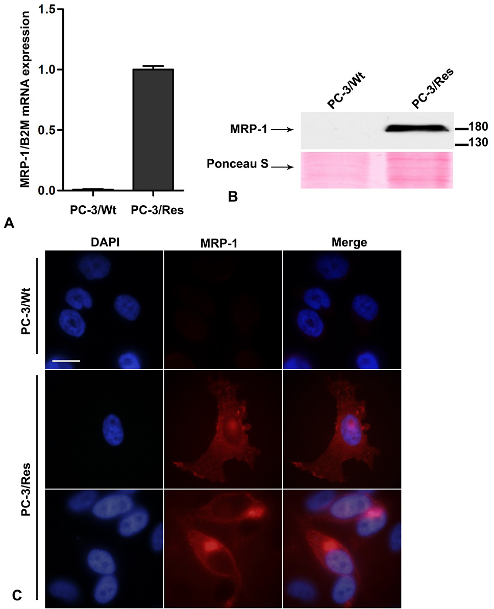
Expression and localisation of MRP-1 in prostate cancer cells. (A) The mRNA levels of ABCC1 in PC-3/Wt and PC-3/Res cells were obtained by real time RT-PCR and normalised to beta 2 microglobulin (B2M) expression (B) ABCC1 protein expression was analysed by Western blot using anti-MRP-1 antibody. Ponceau S staining is shown as loading control (C) Immunofluorescence analysis of ABCC1 protein in PC-3/Wt (upper panel) and PC-3/Res cells (lower panels). Cells were fixed and immunolabelled for ABCC1 (red) using anti-MRP-1 antibody. Merged images, including nuclei (stained with DAPI), were assembled using Adobe Photoshop CS6 Software. Bar, 20µm.
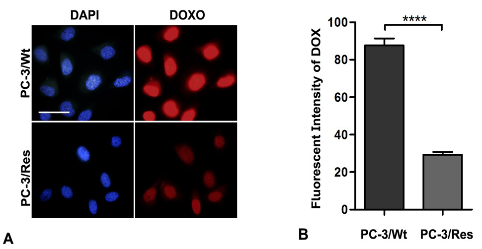
Functional analysis of MRP-1 over expression in PC-3/Res cells. (A) Efflux of doxorubicin in PC-3/Wt and PC-3/Res cells monitored using Olympus BX-51 fluorescent microscope. Cells were incubated with 5μM doxorubicin for 2 h and then allowed to efflux for 1h in drug free media. Bar, 50µm. (B) The intracellular fluorescent intensity of doxorubicin post efflux was measured and expressed as mean fluorescent intensity. Error bars represent standard error of mean, n=70; ****P Ë‚ 0.0001.
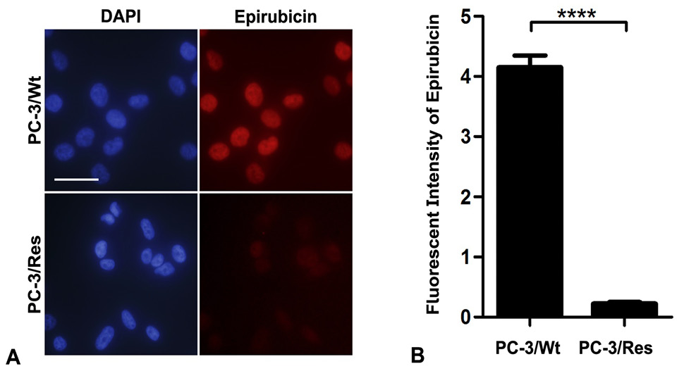
Decreased intracellular retention of epirubicin in PC-3/ Res cells. (A) Fluorescent microscope images of PC-3/Wt and PC-3/Res cells. Cells were incubated with 5μM epirubicin for 2 h and then allowed to efflux for 1 h in drug free media. Bar, 50µm. (B) Intracellular fluorescent intensity of epirubicin was quantified using Image J Software. Error bars represent standard error of mean, n=30; ****P Ë‚ 0.0001.
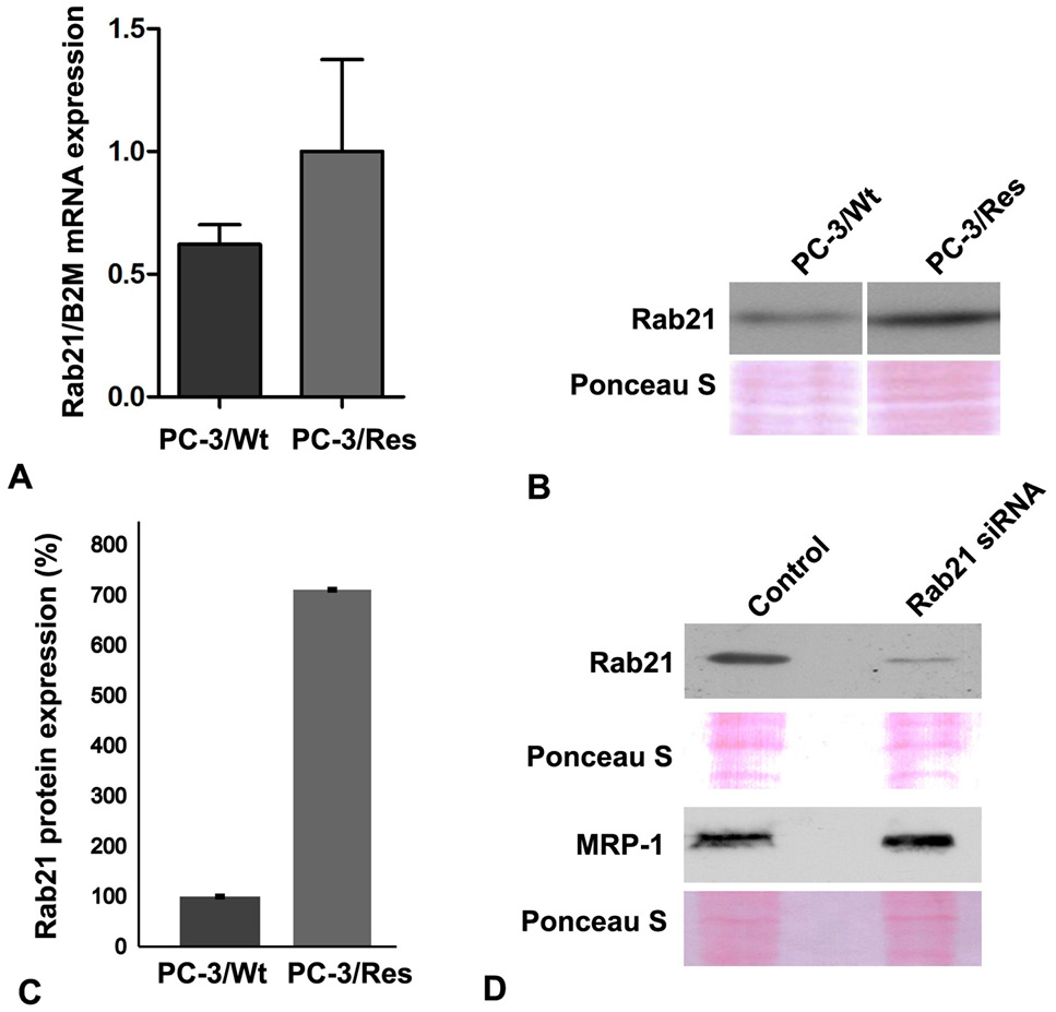
Increased expression of Rab21 in epirubicin resistant PC-3 cells. (A) The mRNA expression level of Rab21 was quantified by RT-PCR and normalised against B2M expression level. (B) Rab21 protein expression was determined by Western immunoblotting using Rab21 specific antibody. Ponceau S staining was used as a loading control. (C) Normalised expression of Rab21 protein in PC-3/Res cells represented as percentage of PC-3/Wt level. (D) Protein expression levels of Rab21 and ABCC1 in control and Rab21 siRNA transfected PC-3/Res cells.
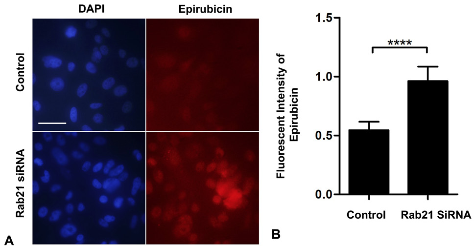
Down regulation of Rab21 decreases epirubicin efflux. (A) Fluorescent microscope images showing intracellular retention of epirubicin after efflux in control and Rab21 siRNA transfected PC-3/Res cells. All images were captured at the same magnification and exposure settings using an Olympus BX-51 fluorescence microscope. Bar, 50µm. (B) Fluorescent intensity of epirubicin was quantified in control and Rab21 siRNA transfected PC-3/Res cells using ImageJ Software. Error bars represent standard error of mean, n=30; ****P Ë‚ 0.0001.
References
- Alisi, A., Cho, W.C., Locatelli, F., and Fruci, D., 2013. Multidrug resistance and cancer stem cells in neuroblastoma and hepatoblastoma. Int. J. mol. Sci., 14: 24706–24725. https://doi.org/10.3390/ijms141224706[CrossRef]
- Bakos, E., and Homolya, L., 2007. Portrait of multifaceted transporter, the multidrug resistance-associated protein 1 (MRP1/ABCC1). Pflugers Arch., 453: 621–641. https://doi.org/10.1007/s00424-006-0160-8[CrossRef]
- Bao, G.S., Wang, W.A., Wang, T.Z., Huang, J.K., He, H., Liu, Z. and Huang, F.D., 2011. Overexpression of human MRP1 in neurons causes resistance to antiepileptic drugs in Drosophila seizure mutants. J. Neurogenet., 25: 201–206. https://doi.org/10.3109/01677063.2011.620662[CrossRef]
- Chang, X.B., 2007. A molecular understanding of ATP-dependent solute transport by multidrug resistance-associated protein MRP1. Cancer Metast. Rev., 26: 15–37. https://doi.org/10.1007/s10555-007-9041-7[CrossRef]
- Chen, Y.H., Wang, C.C., Xiao, X., Wei, L., and Xu, G., 2013. Multidrug resistance associated protein 1 decreases the concentrations of antiepileptic drugs in cortical extracellular fluid in amygdale kindling rats. Acta Pharmacol. Sin., 34: 473–479. https://doi.org/10.1038/aps.2012.183[CrossRef]
- Cole, S.P., 2014. Targeting multidrug resistance protein 1 (MRP1, ABCC1): Past, present, and future. Annu. Rev. Pharmacol. Toxicol., 54: 95–117. https://doi.org/10.1146/annurev-pharmtox-011613-135959[CrossRef]
- Cole, S.P., Bhardwaj, G., Gerlach, J.H., Mackie, J.E., Grant, C.E., Almquist, K.C., Stewart, A.J., Kurz, E.U., Duncan, A.M., and Deeley, R.G., 1992. Overexpression of a transporter gene in a multidrug-resistant human lung cancer cell line. Science, 258: 1650–1654. https://doi.org/10.1126/science.1360704[CrossRef]
- Diestra, J.E., Condom, E., Del Muro, X.G., Scheffer, G.L., Perez, J., Zurita, A.J., Munoz-Segui, J., Vigues, F., Scheper, R.J., Capella, G., Germa-Lluch, J.R. and Izquierdo, M.A., 2003. Expression of multidrug resistance proteins P-glycoprotein, multidrug resistance protein 1, breast cancer resistance protein and lung resistance related protein in locally advanced bladder cancer treated with neoadjuvant chemotherapy: Biological and clinical implications. J. Urol., 170: 1383–1387. https://doi.org/10.1097/01.ju.0000074710.96154.c9[CrossRef]
- Egami, Y. and Araki, N., 2008. Characterization of Rab21-positive tubular endosomes induced by PI3K inhibitors. Exp. Cell. Res., 314: 729-737. https://doi:10.1016/j.yexcr.2007.11.018[CrossRef]
- Egami, Y. and Araki, N., 2009. Dynamic changes in the spatiotemporal localization of Rab21 in live RAW264 cells during macropinocytosis. PLoS One, 4: e6689. https://doi:10.1371/journal.pone.0006689[CrossRef]
- Eva, B., Raymond, E., Gergely, S., Gabor, E., Tusna, D., Ervin, W., Katalin, S., Marcel, D.H., Liesbeth, V.D., Piet, B., Andra, V.R, and Bala, S., 1998. Functional multidrug resistance protein (MRP1) lacking the N-terminal transmembrane domain. J. biol. Chem., 273: 32167–32175. https://doi.org/10.1074/jbc.273.48.32167[CrossRef]
- Flens, M.J., Zaman, G.J., van der Valk, P., Izquierdo, M.A., Schroeijers, A.B., Scheffer, G.L., van der Groep, P., Haas, M., Meijer, C.J., and Scheper, R.J., 1996. Tissue distribution of the multidrug resistance protein. Am. J. Pathol., 48: 1237–1247.
- Goma, A., Mir, R., Martinez-Soler, F., Tortosa, A., Vidal, A., Condom, E., Perez–Tomas, R., and Gimenez-Bonafe, P., 2014. Multidrug resistance protein 1 localization in lipid raft domains and prostasomes in prostate cancer cell lines. Onco Targets Ther., 7: 2215-2225. https://doi.org/10.2147/OTT.S69530[CrossRef]
- Gregory, F.S., Peter, S.A, Jocelyn, D.V., Carmelita, J.A., Jin-Ming, Y., and William, N.H., 1998. The expression of drug resistance gene products during the progression of human prostate cancer. Clin. Cancer Res., 4: 1393-1403.
- Haber, M., Smith, J., Bordow, S.B., Flemming, C., Cohn, S.L., London, W.B., Marshall, G.M. and Norris, M.D., 2006. Association of high-level MRP1 expression with poor clinical outcome in a large prospective study of primary neuroblastoma. J. clin. Oncol., 24: 1546–1553. https://doi.org/10.1200/JCO.2005.01.6196[CrossRef]
- John, T.L., Brian, D.L., David, M. T., William, H.C., Franca, S., Massimo, L., Alberto, M.M., Linda, S.S., and James, A.M., 2008. Targeting prostate cancer based on signal transduction and cell cycle pathways. Cell Cycle, 7: 1745–1762. https://doi.org/10.4161/cc.7.12.6166[CrossRef]
- Johnson, Z.L. and J. Chen, 2017. Structural basis of substrate recognition by the multidrug resistance protein MRP1. Cell, 168: 1075-1085. https://doi.org/10.1016/j.cell.2017.01.041[CrossRef]
- Jones, M.C., Caswell, P.T., and Norman, J.C., 2006. Endocytic recycling pathways: Emerging regulators of cell migration. Curr. Opin. Cell Biol., 18: 549-557. https://doi.org/10.1016/j.ceb.2006.08.003[CrossRef]
- Kim, H., Barroso, M., Samanta, R., Greenberger, L. and Sztul, E., 1997. Experimentally induced changes in the endocytic traffic of P-glycoprotein alter drug resistance of cancer cells. Am. J. Physiol., 273: 687-702. https://doi.org/10.1152/ajpcell.1997.273.2.C687[CrossRef]
- Leslie, E.M., Deeley, R.G., and Cole, S.P., 2005. Multidrug resistance proteins: role of P-glycoprotein, MRP1, MRP2, and BCRP (ABCG2) in tissue defense. Toxicol. appl. Pharmacol., 204: 216–237. https://doi.org/10.1016/j.taap.2004.10.012[CrossRef]
- Lu, J.F., Pokharel, D., and Bebawy, M., 2015. MRP1 and its role in anticancer drug resistance. Drug Metab. Rev., 47: 406-419. https://doi.org/10.3109/03602532.2015.1105253[CrossRef]
- Matthew, J., Grzywacz, J.M.Y., and William, N.H., 2003. Effect of the multidrug resistance protein on the transport of the antiandrogen flutamide. Cancer Res., 63: 2492–2498.
- Munoz, M., Henderson, M., Haber, M., and Norris, M., 2007. Role of the MRP1/ABCC1 multidrug transporter protein in cancer. IUBMB Life, 59: 752-757. https://doi.org/10.1080/15216540701736285[CrossRef]
- Opdam, F., 2000. Expression of Rab small GTPases in epithelial Caco-2 cells: Rab21 is an apically located GTP-binding protein in polarised intestinal epithelial cells. Eur. J. Cell Biol., 79: 308-316. https://doi.org/10.1078/S0171-9335(04)70034-5[CrossRef]
- Opdam, F.J.M., Kamps, G., Croes, H., Bokhoven, H., Ginsel, L.A. and Fransen, J.A.M., 2000. Expression of Rab small GTPases in epithelial Caco-2 cells: Rab21 is an apically located GTP-binding protein in polarised intestinal epithelial cells. Eur. J. Cell Biol., 79: 308–316. https://doi.org/10.1078/S0171-9335(04)70034-5[CrossRef]
- Pellinen, T., Arjonen, A., Vuoriluoto, K., Kallio, K., Fransen, J.A. and Ivaska, J., 2006. Small GTPase Rab21 regulates cell adhesion and controls endosomal traffic of beta1-integrins. J. Cell Biol., 173: 767-780. https://doi.org/10.1083/jcb.200509019[CrossRef]
- Samantha, L.S., Canhong, C., Olena, P., Alexey, R., and Angela, W., 2008. Rab GTPases at a glance. J. Cell Sci., 121: 246. https://doi.org/10.1242/jcs.03495[CrossRef]
- Sharom, F.J., 2008. ABC multidrug transporters: structure, function and role in chemoresistance. Pharmacogenomics, 9: 105–127. https://doi.org/10.2217/14622416.9.1.105[CrossRef]
- Stenmark, H., 2009. Rab GTPases as coordinators of vesicle traffic. Nat. Rev. Mol. Cell Biol., 10: 513-525. https://doi.org/10.1038/nrm2728[CrossRef]
- Takeda, M., Mizokami, A., Mamiya, K., Li, Y.Q., Zhang, J., Keller, E.T. and Namiki, M., 2007. The establishment of two paclitaxel-resistant prostate cancer cell lines and the mechanisms of paclitaxel resistance with two cell lines. Prostate, 67: 955-967. https://doi.org/10.1002/pros.20581[CrossRef]
- Tzeng, H.T. and Wang, Y.C., 2016. Rab-mediated vesicle trafficking in cancer. J. biomed. Sci., 23: 70. https://doi.org/10.1186/s12929-016-0287-7[CrossRef]
- Vulsteke, C., Lambrechts, D., Dieudonne, A., Hatse, S., Brouwers, B., van Brussel, T., Neven, P., Belmans, A., Schoffski, P., Paridaens, R. and Wildiers, H., 2013. Genetic variability in the multidrug resistance associated protein-1 (ABCC1/ MRP1) predicts hematological toxicity in breast cancer patients receiving (neo-) adjuvant chemotherapy with 5-fluorouracil, epirubicin and cyclophosphamide (FEC). Annls Oncol., 24: 1513–1525. https://doi.org/10.1093/annonc/mdt008[CrossRef]
- Westlake, C.J., Cole, S.P., and Deeley, R.G., 2005. Role of the NH2-terminal membrane spanning domain of multidrug resistance protein 1/ABCC1 in protein processing and trafficking. Mol. Biol. Cell, 16: 2483-2492. https://doi.org/10.1091/mbc.e04-12-1113[CrossRef]
- Westlake, C.J., Payen, L., Gao, M., Cole, S.P. and Deeley, R.G., 2004. Identification and characterization of functionally important elements in the multidrug resistance protein 1 COOH-terminal region. J. biol. Chem., 279: 53571-53583. https://doi.org/10.1074/jbc.M402528200[CrossRef]
- Winter, S.S., Ricci, J., Luo, L., Lovato, D.M., Khawaja, H.M., Serna-Gallegos, T., Debassige, N. and Larson, R.S., 2013. ATP Binding Cassette C1 (ABCC1/MRP1)-mediated drug efflux contributes to disease progression in T-lineage acute lymphoblastic leukemia. Health, 5: 41–50. https://doi.org/10.4236/health.2013.55A005[CrossRef]
- Zalcberg, J., Hu, X.F., Slater, A., Parisot, J., El-Osta, S., Kantharidis, P., Chou, S.T., and Parkin, J.D., 2000. MRP1 not MDR1 gene expression is the predominant mechanism of acquired multidrug resistance in two prostate carcinoma cell lines. Prost. Cancer Prost. Dis., 3: 66-75. https://doi.org/10.1038/sj.pcan.4500394[CrossRef]
- Zhang, B., Liu, M., Tang, H.K., Ma, H.B., Wang, C., Chen, X. and Huang, H.Z., 2012. The expression and significance of MRP1, LRP, TOPOIIb, and BCL2 in tongue squamous cell carcinoma. J. Oral Pathol. Med., 41: 141–148. https://doi.org/10.1111/j.1600-0714.2011.01066.x[CrossRef]
Services
Article usage statistics
Loading…