
Next generation sequencing (NGS) provides an accurate analysis of hepatitis B virus (HBV) as compared to Sanger sequencing. In 2019, samples from HBV infected family of mother and her two sons samples were collected from Lahore, Pakistan. Sequencing of S gene, Geno2pheno (HBV) 2.0 tool and GHOST platform were used to analyze the genotype, mutations (natural and escape) and quasispecies analysis of the HBV. All samples in family were infected with genotype HBV/D1 and had high viral load (10e8 IU). An vaccine escape mutation “C137W” was identified in mother and a son. Mother sample were shown more number of haplotypes and total diversity as compared to sons. The quasispecies analysis showed the mother and sons were infected with HBV having similar genetic makeup, indicating the vertical transfer of infection. This case study provides knowledge about genetic variations that contributes to the improved diagnosis of the infection, treatment therapy, follow-up, and future treatment strategies.
Short Communication · Volume 56, Issue 5, 15 September 2024 · Open Access
Quasispecies Analysis Through NGS and Identification of Escape Mutation in a Family Infected with Hepatitis B Virus: A Study from Lahore, Pakistan
· · · · ·
Affiliations & Notes ⌄
- 1Centre of Excellence in Molecular Biology, University of the Punjab, Lahore, Pakistan. * Corresponding author: samiaraza@live.com
Article Info ⌄
- Journal:
- Pakistan Journal of Zoology
- Published:
- 15 September 2024

From the article
- Mutational analysis indicated the presence of vaccine Escape mutation C137W in Mother and one of her sons.
- The circular pattern indicated the acute infection with fewer variations in HBV genetics shown in Figure 1.
- Mother (P42) were shown more number of haplotypes and total diversity as compared to sons (P41 and 43) indicating more evolution of HBV quasispecies in mother.
- All samples indicated acute infections with a similar genetic makeup showing the vertical transfer of HBV infection.
Selected from the article's own text.
Abstract
Materials and methods
In the present study, blood samples of a family of 28 years old mother and her 2 sons (6 and 7 years old) infected with HBV were collected at Genome Centre and Medical Diagnostics (GCMD), Lahore for the detection, quantification and genotyping of hepatitis B (HBV). Well-informed written consents were taken from all the participants at the time of sampling. All the important information such as demographics and clinical data from all the participants was recorded on a data sheet. For the detection of HBeAg in the serum of HBV infected patients, an ETI-EBK PLUS kit was used. MagNA Pure LC Total Nucleic Acid Isolation Kit (Roche, Catalogue Number, 05323738001) was used for the Nucleic Acid Extraction from the serum samples. Primers used for Sanger sequencing 1st round PCR were: EXS164F ACATCACATCAGGACTCCTAGGA and EXS656R GAGGCCCACTCCCATAGGTAT whereas primers used for nested PCR second round were INS183F AGGACCCCTTCTCGTGTTACA and INS624R CCAAGATGATGGGATGGGAAT. The S gene was amplified by using Lightcycler 480 instrument (Roche Diagnostics Corporation, Indianapolis, IN) with thermal Profile consists of denaturation at 95°C for 10 min, PCR amplification: 30 cycles at 95°C for 30 sec, 50°C for 30 sec and 72°C for 30 sec and melting curve at 76°C for 10 min. For Sanger sequencing, the S gene PCR product was sequenced in both directions by using forward and reverse primers. The sequencing was performed by using Big Dye-Terminator version 3.1 cycling methodology (Applied Biosystems, CA). Sequencing was done by an automated sequencer (ABI 3130xl Genetic Analyzer, Applied Biosystems). The obtained sequences were cleaned and aligned by SeqMan (version 10.1.2, DNASTAR, Madison, WI). Further analysis was done by using Geno2pheno (HBV) 2.0 (https://hbv.geno2pheno.org). For NGS, 1st round was the same as used before in sanger sequencing. 32 specific barcode PCR primers were the nested primers with specific 32 unique 10 bps tags (barcode). 8 bps unique tags were added to the index primer sequences to make 32 unique index primers. The PCR products were purified by using Ampure XP (Agilent). Each product was quantified by using Tape station instrument (Agilent, CA) according to protocol described. The quantified products were normalized and diluted to make an NGS library pool of 10pmol. Sequencing was done by using MiSeq Reagent Kit v3 (600-cycle) in Illumina MiSeq instrument. NGS pooled Library was automatically de-multiplexed by the Miseq Instrument. Further de-multiplexing at paired reads was processed by GHOST system . The way to visualize the genetic relatedness among all haplotypes present in each HBV infected specimen is through k-step networks.
Results
High viral loads of HBV and HBeAg positive were observed in all three cases of family members. Sanger sequences of all the samples were of genotype D1/HBV, identified by using Geno2pheno (HBV) and further confirmed by GHOST software. Mutational analysis indicated the presence of vaccine Escape mutation C137W in Mother and one of her sons. Whereas another mutation T127L were also identified in family members as shown in supplementary section. NGS data indictaed that mother (P42) were shown more number of haplotypes and total diversity as compared to sons (P41 and 43) shown in Table I. K steps represented the Intra-Host variations and Inter Host variations. The circular pattern indicated the acute infection with fewer variations in HBV genetics shown in Figure 1. K step diagram of the 3 samples forming a cluster indicating the similar genetic makeup of HBV species in family indicating the vertical transmission, as shown in Figure 2.
Table I. Characteristics and GHOST analysis data of HBV infected family.
| ID/ Name | Genotype | nhap | nreads | Total div | Max. freq. | Hbe Ag | Viral load |
| P41 (Son) | d | 11456 | 19168 | 0.015004 | 3540 | Positive | > 10˄8 |
| P42 (Mother) | d | 11805 | 18965 | 0.015511 | 3133 | Positive | > 10˄8 |
| P43 (Son) | d | 10851 | 19192 | 0.014425 | 4031 | Positive | > 10˄8 |
.
Discussion
Pakistan has a high prevalence rate of HBV that leads towards a high modality due to liver cirrhosis and hepatocellular carcinoma. Replication of HBV takes place by reverse transcription of an RNA intermediate and the errors in HBV DNA replication occur at a much higher rate than for other DNA viruses due to a lack of RT proofreading function. The nucleotide
substitution estimated rate per year is approximately 1.4-3.2 × 10⁻⁵ per site . The evolution of these naturally occurring mutants occurs during the infection period under antiviral pressure of the host immune system or by other factors like specific therapy or immunization . In this case study, the mother and her sons were infected with HBV/D genotype, the most prevalent genotype in Pakistan. Sequencing identified a mother and one of her sons who share a mutation T127L whereas an escape mutation C137W also be identified in a mother and one of her sons. This vaccine mutation may be responsible for the transmission of HBV from mother to her sons. Escape mutations, C137W along with G145R, was identified by showing false negative result for HbsAg .
A complex mixture of HBV variants with the related genomic sequence that infects a certain host is known as the viral quasispecies . The sequencing technologies have been extensively employed to uncover novel endeavors of HBV and the infection that includes valuable data concerning the identification of the targets for diagnosis and the factors that play their role in the disease progression. For example, a spontaneous mutation in preS/S, pre-C/C and X was noted via Sanger sequencing and a further link was established with the progression of liver disease ; ; . However, these research studies pose a limitation that links with the sensitivity of Sanger and endorse the benefit NGS approaches may have contributed, especially with the depth and high quality ensured by the NGS technologies.
In this case study, Sanger sequences and major sequences by NGS showed the same results of all the samples but through NGS, all the minor populations were also obtained. Total diversity defines as the Measurement of the amount of heterogeneity of a sample from a range of 0 to 1, where 0 is more homogenous and 1 is very heterogenous. Mother (P42) were shown more number of haplotypes and total diversity as compared to sons (P41 and 43) indicating more evolution of HBV quasispecies in mother. Intra-host and inter-host variation were observed in the form of K steps by GHOST. All samples indicated acute infections with a similar genetic makeup showing the vertical transfer of HBV infection. In 2002, Pakistan initiated the HBV Childhood vaccination Program . Almost 72% to 86% of children are HBV vaccinated since the launch of the HBV childhood Program . As the children were supposed to be vaccinated but still, they are infected with HBV also confirms the vertical transmission.
This study contributes to genetic diversity of HBV that will helpful to improve the diagnosis of the infection, its treatment therapy, follow-up, and future treatment strategies. There is a need to analyse the frequency of specific mutations in HBV genome rather than specific to S gene that are responsible for the vaccination failure and vertical transmission.
Acknowledgement
Authors acknowledge Genome Centre for Molecular Diagnostic and Research Lab for providing facility for patients sample collection, and Centre for Disease control and Prevention (CDC), USA for providing facility for analysis of HBV samples.
Funding
The funding for this study was provided by Higher Education Commission Pakistan.
IRB approval
Institutional Review Board committee, CEMB, Lahore approved the study (CEMB Ref # 03/22, dated 06/06/2022).
Ethical statement
A written consent was obtained from all patients before sample collection for this study.
Statement of conflict of interest
The authors have declared no conflict of interest.
Figures
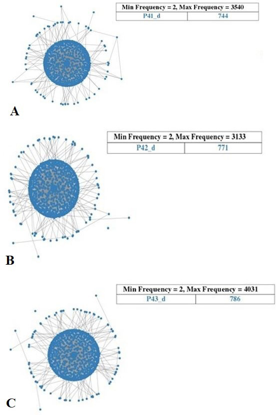
K-steps diagrams indicating the intra-host variations showing HBV quasispecies of HBV mother (B) and her sons (A and C).
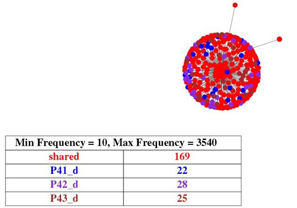
K step diagram showing the cluster formation of HBV of family, sharing the same genetic makeup. The red colour represents the same genetic sequences present in all samples.
References
- Ali, S.A., Donahue, R.M., Qureshi, H. and Vermund, S.H., 2009. Int. J. Infect. Dis., 13: 9-19. https://doi.org/10.1016/j.ijid.2008.06.019[CrossRef]
- Domingo, E., Sheldon, J. and Perales, C., 2012. Microbiol. Mol. Biol. Rev., 76: 159-216. https://doi.org/10.1128/MMBR.05023-11[CrossRef]
- Dong, Y., Liu, S.L., Zhai, X.J., Zhu, F.C., Pan, H., Yu, J.X., Chen, Y.Z., Xie, Y.R., Zhang, X.Y. and Zhang, H.M., 2009. J. med. Virol., 81: 1517-1524. https://doi.org/10.1002/jmv.21522[CrossRef]
- Forbi, J.C., Dillon, M., Purdy, M.A., Drammeh, B.S., Tejada-Strop, A., McGovern, D., Xia, G.L., Lin, Y., Ganova-Raeva, L.M. and Campo, D.S., 2017. J. Gen. Virol., 98: 1048-1057. https://doi.org/10.1099/jgv.0.000776[CrossRef]
- Gerlich, W.H., 2006. J. clin. Virol., 36: S18-S22. https://doi.org/10.1016/S1386-6532(06)80004-1[CrossRef]
- Günther, S., Fischer, L., Pult, I., Sterneck, M. and Will, H., 1999. Adv. Virus Res., 52: 25-137.
- Harris, B.J., Holzmayer, V., Qureshi, H., Khan, M.A., Khan, S.A., Salamat, N., Jafri, S. and Brennan, C.A., 2017. PLoS One, 12: e0178988. https://doi.org/10.1371/journal.pone.0178988[CrossRef]
- Jafri, W., Jafri, N., Yakoob, J., Islam, M., Tirmizi, S.F.A., Jafar, T., Akhtar, S., Hamid, S., Shah, H.A. and Nizami, S.Q., 2006. BMC Infect. Dis., 6: 1-10. https://doi.org/10.1186/1471-2334-6-101[CrossRef]
- Kyaw, Y.Y., Lwin, A.A., Aye, K.S., Thu, H.M., Htun, M.M., Soe, H.O., Aye, K.T., Thant, K.Z., Hwang, H.J., and Cheong, J., 2020. BMC Infect. Dis., 20: 1-11. https://doi.org/10.1186/s12879-020-05269-z[CrossRef]
- Lazarevic, I., Banko, A., Miljanovic, D. and Cupic, M., 2019. Viruses, 11: 778. https://doi.org/10.3390/v11090778[CrossRef]
- Longmire, A.G., Sims, S., Rytsareva, I., Campo, D.S., Skums, P., Dimitrova, Z., Ramachandran, S., Medrzycki, M., Thai, H. and Ganova-Raeva, L., 2017. BMC Genomics, 18: 21-32. https://doi.org/10.1186/s12864-017-4268-3[CrossRef]
- McNaughton, A.L., Roberts, H.E., Bonsall, D., de Cesare, M., Mokaya, J., Lumley, S.F., Golubchik, T., Piazza, P., Martin, J.B. and de Lara, C., 2019a. Sci. Rep., 9: 1-14. https://doi.org/10.1038/s41598-019-43524-9[CrossRef]
- McNaughton, A.L., D’Arienzo, V., Ansari, M.A., Lumley, S.F., Littlejohn, M., Revill, P., McKeating, J.A. and Matthews, P.C., 2019b. Gastroenterology, 156: 384-399. https://doi.org/10.1053/j.gastro.2018.07.058[CrossRef]
- Okamoto, H., Imai, M., Tsuda, F., Tanaka, T., Miyakawa, Y. and Mayumi, M. 1987. J. Virol., 61: 3030-3034.
- Purdy, M.A., 2007. Asian J. Transfus. Sci., 1: 62. https://doi.org/10.4103/0973-6247.33445[CrossRef]
- Shen, T. and Yan, X.M., 2014. World J. Gastroenterol., 20: 5435. https://doi.org/10.3748/wjg.v20.i18.5435[CrossRef]
- Tatsukawa, M., Takaki, A., Shiraha, H., Koike, K., Iwasaki, Y., Kobashi, H., Fujioka, S.I., Sakaguchi, K. and Yamamoto, K., 2011. BMC Cancer, 11: 1-9. https://doi.org/10.1186/1471-2407-11-458[CrossRef]
- Yang, J.H., Zhang, H., Chen, X.B., Chen, G. and Wang, X., 2013. World J. Gastroenterol., 19: 3861.
Services
Article usage statistics
Loading…