
Tamarindus indica L. seed extract (TSE) efficacy against γ-rays-induced nephrotoxicity in irradiated rats was investigated. Forty albino-rats were divided into four groups: Control, rats were orally administered the vehicle for 6weeks, TSE-treated (each rat received 230mg TSE; once daily, for 6weeks), irradiated (animals subjected to whole body γ-rays (8Gy), and TSE-treated and irradiated (each rat received 230mg TSE; once daily for 6week, then one-hour later after last-treatment, rats were exposed to whole body γ-rays (8Gy). The results revealed that TSE before-irradiation significantly abolish γ-rays-induced alleviation in renal catalase (CAT) and glutathione peroxidase (PGx) activities and reduced-glutathione (GSH) content and significantly limited the elevation in serum tumor necrosis factor-α (TNF-α), interleukin-1β (IL-1β) and interleukin-6 (IL-6) levels when compared to irradiated rat group. The histopathological-findings of renal tissues in the irradiated-group, showed nephrotoxicity; the renal cortex showed massive necrotic changes, the convoluted tubules showed distinctive pattern of ischemic renal injury and the cuboidal epithelium cells of proximal and distal renal tubules showed nuclear changes with leukocytes infiltrations. More over some cases showed atrophied glomeruli, widened Bowman’s space and thickened basement membrane, while in TSE-treated and irradiated-group, renal-tissues showed minimum-damage with or without few degenerative changes. TSE acts as a potent antioxidant and anti-inflammatory drug to prevent and/ or ameliorates the hazardous-effects of γ-rays
Research Article · Volume 52, Issue 2, 12 February 2020 · Open Access
Protective Effect of Tamarind against Renal Injury Induced by Gamma Rays
· ·
Affiliations & Notes ⌄
- 1Health Radiation Research Department, National Centre for Radiation Research and Technology (NCRRT), P.O. Box
- 229 Nasr City, Egyptian Atomic Energy Authority, Cairo, Egypt , * Corresponding author: elkadyah13@gmail.com
Article Info ⌄
- Journal:
- Pakistan Journal of Zoology
- Published:
- 12 February 2020

From the article
- In the present work, TSE increased the levels of renal GSH, recompensed the oxidative damage occurred during irradiation cascades.
- TSE inhibited the augmented status of inflammation and oxidative stress by modulating elevated levels of pro-inflammatory mediators and by balancing the homeostasis of endogenous antioxidant system.
- Also, TSE might reduce inflammation associated with γ-rays induced nephrotoxicity.
- Altogether, TSE was shown to be a potent anti-inflammatory, anti-nephrotoxicity and anti-stress agent.
Selected from the article's own text.
Abstract
INTRODUCTION
Recent studies have constantly shown that feeding of plant-derived foods rich in bioactive phytochemicals has a protective effect against pro-inflammatory cytokines and oxidation stress and may extend healthy lifespan in humans . Tamarind (Tamarindus indica L.) is a tree cultivated widely in different countries. Its nutritional and medicinal aspects are currently trends towards affordable functional foods as a source of valuable nutrients such as essential fatty acids, proteins, and minerals which are accepted as flavour to develop perfect secure useful foods . TSE are rich source of phytochemicals (Akhtar et al., 2019), that include phenolic antioxidants such as 2-hydroxy-3′,4′-dihydoxyacetophenone, methyl 3, 4-dihydroxybenzoate, 3,4-dihydroxyphenyl acetate and epicatechin . TSE exhibit antioxidant potential by reducing lipid peroxidation and anti-inflammatory in vitro . TSE therefore has the potential of providing health food additives with low cost antioxidant source and nutraceutical value.
Free radicals production following to ionizing-radiation exposure proceeds for a second, but its injury is immediate. Subsequently, the resulted oxidative stress is strongly related with nephrotoxicity in rats . Our body, therefore, must have radiation protective means to prevent the hazardous effects of free radical damage and its later consequences on kidney cells .
It was reported that gamma-radiation with a dose of 6Gy in rats induced significant renal lesions included atrophied glomeruli, widened Bowman’s capsule, high cellularity in the visceral layer of the Bowmn’s capsule and highly affections of cytoplasm and nuclei of the convoluted tubules . Furthermore, Radwan and Abdel Fattah demonstrated that rats exposed to fractionate total body radiation had severe renal parenchymal damage involving the glomeruli, the tubules, the interstitial tissue and the blood vessels.
The aim of this study was focused on evaluation the protective effects of TSE in gamma-rays-induced experimental acute renal nephrotoxicity in male rats.
MATERIALS AND METHODS
Animals
Male Sprague-Dawley rats (age 10–12 weeks), weighing 122–138g, obtained from the Holding Company for Biological Products and Vaccines (Helwan, Cairo, Egypt) were selected. Rats were retained under standard environmental situations and indorsed free access of diet and water. Animals were kept under a controlled lighting condition (light: dark, 11-13 hours). Rats were acclimatized to the experimental conditions prior to TSE dosing for 3 days. This study complies with National Institutes of Health guidelines.
Radiation facility
It was done by using gamma cell-40 (137Cs) found on NCRRT, Cairo, Nasr City, Egypt. Rats were irradiated with a single dose level of (8Gy) delivered at a dose rate of 0.42Gy/ min at the time of experimentation. Animals were not anesthetized before irradiation.
Reagents
TSE is procured from Sigma-Aldrich, American Fork, USA. The other chemicals were high pure grade available.
Experimental plan
40 rats were distributed into 4 groups, each (n= 10). Control group, rats received orally by gastric tube an appropriate equivalent volume of the saline dissolved (vehicle of TSE) for 6 weeks. Irradiated group, received the vehicle as in control group for 6weeks then, rats were whole body exposed to an acute single dose of 8Gy γ-rays. TSE-treated group, rats received TSE, orally by gastric tube at dose of 230 mg/ kg body weight once daily, for 6 weeks according to Yadav et al. protocol. TSE-treated and irradiated group, rats received TSE orally by gastric tube (230mg / kg body weight) once daily, for 6 weeks, then one hour post the last TSE dosage, rats were whole body irradiated with an acute single dose of 8Gy γ-rays. All rat groups were sacrificed on the 2nd day post radiation exposure.
Samples collection
Blood samples were collected via retro orbital puncture by glass capillary tubes post 12 hours fast. Serum was obtained immediately by centrifugation of blood samples at 300xg for 10minutes. Kidneys were directly separated after sacrifice, washed with ice-cold saline and excised immediately in cold 0.9% NaCl, then removed and rinsed in a cooled buffer (0.15M Tris KCl, pH 7.4) to yield 10% (w/v) homogenate using, Homogenizer type MNW-302, Poland, then the homogenates were centrifuged at 800xg for 5minutes at 4ºC to separate the tissue fragments. The supernatant of the homogenates were used for the biochemical analysis. Tissue specimens from kidney were collected and fixed in 10% buffered formalin solution followed by dehydration, cleating and embedding in paraffin. Paraffin sections of 5-micron thickness were prepared and stained routinely with hematoxylin and eosin (HE) according to Suvarna et al. (2013). The sections examined with an ordinary microscope (Olympus, Japan). All observes were performed on blinded coded slides.
Estimation of biochemical parameters
Serum tumor necrosis factor alpha (TNF-α), interleukin-1β (IL-1β) and interleukin-6 (IL-6) activities were achieved by Enzyme Linked Immunosorbent Assays (ELISA) method (BioSource International, Camarillo, CA, USA) following the instructions of the manufacturer. Each sample evaluation was recurring 3 times. The absorbance was calculated at 450 nm using a microplate counter (Thermo Scientific Multiskan MK3, USA). CAT was assayed calorimetrically and expressed as unit (μmol of H2O2 consumed/ min) per mg protein as described by Titov and Osipov (2017). It was found that chloride and bromide in concentration above 80 mM and thiocyanate in concentration above 20 μM enhance catalase inhibition by nitrite and the nitroso compounds more than 100 times.
Renal reduced GSH levels were determined in renal tissue by the methods described by Cigala et al. (2012), which is based on the acid-base properties of GSH (γ-L-glutamyl-L-cysteinyl-glycine). In renal tissue, GPx activity was measured by Lawrence and Burk (2012) method based on evaluating the oxidation of reduced NADPH (nicotinamide adenine dinucleotide phosphate) using H2O2 as a substrate. A reaction mixture of 1ml contained 50mM potassium phosphate buffer (pH 7), 1mM EDTA, 1mM NaN3, 0.2mM NADPH, 1unit / ml oxidized glutathione reductase and 1mM GSH was prepared. The prepared renal homogenate was centrifuged at 105,000 for 15minutes at 4ºC. 0.1ml of the supernatant was added to 0.8ml of the reaction mixture and the solution was incubated for 5minutes at 25ºC. 0.1ml of 0.25mM hydrogen peroxide solution was added to initiate the reaction. Absorbance was measured at 340nm for 5minutes, and an extinction coefficient of 6.22 x10⁻³ was used for calculation. The results were expressed as μmol unit/ minute/g tissue. The unit is equal the quantity of the GPx enzyme control the oxidation of 1.0μmol NADPH into NADP+ / minute at 25ºC. The changes in the absorbance at 340nm were recorded at 1-minute interval for 5minutes. Protein content was determined using Lowry et al. (1951) technique.
Statistical analysis
SPSS software (version 19.0) was used for data analysis. ANOVA (One-way analysis of variance) and Post Hoc test were used to determine the LSD (Least Significant Difference). The data was expressed as Mean ± S.D. (Standard Deviation). A P-value less than 0.05 was considered statistically significant .
RESULTS
Histopathological finding
The renal cortex of control group has renal corpuscles and tubules. The renal corpuscle consists of glomerulus and Bowman’s capsule. The glomerulus is a tuff of capillaries formed from the afferent glomerular arterioles and supported by a fine connective tissue. At the urinary pole, the incomplete layer of the Bowman’s capsule contains simple squamous epithelium changed to cuboidal epithelium in the proximal convoluted tubules, (Fig. 1A).
In irradiated group, the proximal and distal renal tubules were dilated and inter tubular blood vessels were congested. In other cases, the renal tissues were infiltrated and replaced by substantial leukocytes with exudates in the renal cortex which obscured tissue details and architectures. Moreover, the cuboidal epithelium cells of the proximal and distal renal tubules showed nuclear alterations; pyknosis and karyolysis, and these lesions may be more aggressive to show massive necrotic changes. Further, the convoluted tubules showed distinctive pattern of ischemic renal injury represented by losing its microscopic structures with partial block of its lumens (Fig. 1B, G, D and E). Furthermore, some slides showed atrophied glomeruli, the rest of glomerular tuff was shrunken due to slight fibrosis and widened Bowman’s space and thickened basement membrane were seen, (Fig. 1F). TSE-treated group showed normal histological structures as control one. While in case of TSE-treated and irradiated group, the histological structure of kidney usually showed relatively well preserved architecture without degenerative changes. In few cases, the hyaline cast and slightly reversible degeneration changes were seen within renal tubules (Fig. 1G, H).
Effects of TSE on γ-rays-induced inflammatory process
As shown in Table I, a significant intensification of the inflammatory markers represented in serum TNF-α, IL-1β and IL-6 levels were observed in irradiated group compared with corresponding values of control group. The administration of TSE before exposure to γ-rays significantly limited the elevation in those inflammatory markers levels compared to irradiated group.
In Table II. the influence of γ-rays exposure on endogenous antioxidant status is presented. γ-rays induced significant diminution in the renal CAT, GPx activities and GSH content compared with control group.
Administration of TSE for 6 consecutive weeks prior to γ-rays-exposure resulted in significant increases in the activities of renal CAT, GPx and content of GSH compared to irradiated group.
Table I. Serum tumour necrosis factor alpha (TNF-α), interleukin-1β (IL-1β) and imterleukin-6 (IL-6) levels in the different rat groups.
| Inflammatory markers | Control | TSE-treated | Irradiated | TSE-treated & irradiated |
| TNF-α (pg/ ml) | 33.43± 0.97 | 34.01± 0.81 | 69.11± 2.03a | 36.68± 1.51b |
| IL-1β (pg/ ml) | 15.97± 1.27 | 15.22± 1.24 | 44.12± 1.23a | 18.56± 1.42b |
| IL-6 (pg/ ml) | 94.73± 1.51 | 95.14± 1.29 | 189.04± 3.25a | 101.33± 1.73b |
All values are expressed as mean± S.E; aSignificant (P˂ 0.05) when compared with the control group; bSignificant (P˂ 0.05) when compared with the irradiated group.
Table II. Renal catalase (CAT) and glutathione peroxidase (GPx) activities and reduced glutathione (GSH) content in the different rat groups.
| Antioxidant parameters | Control | TSE-treated | Irradiated | TSE-treated & irradiated |
| CAT (U/ mg protein) | 3.14± 0.27 | 3.23± 0.42 | 1.75± 0.43a | 2.74± 0.82b |
| GSH (μmol/ g tissue) | 1.94± 0.056 | 1.91± 0.026 | 1.01± 0.057a | 1.72± 0.033b |
| GPx (μmol unit/ g tissue) | 1.61± 0.154 | 1.62± 0.162 | 0.77± 0.121a | 1.41± 0.062b |
All values are expressed as mean± S.E; aSignificant (P˂ 0.05) when compared with the control group; bSignificant (P˂ 0.05) when compared with the irradiated group.
DISCUSSION
Employing medicinal plants in the treatment of various human diseases is an early idea. In the headway, the recent nephrotoxicity research has endorsed the protective effect of antioxidant medicinal plants against nephrotoxicity. Other plant extracts ameliorate nephrotoxicity in rats .
The TSE are obtaining an attention due to their beneficial actions for spare of human pathophysiological illnesses including treatment of diabetes, snakebites, chronic diarrhea, dysentery, jaundice, eye diseases, ulcers and renal protection ; . In view of this, the present study evaluated the radio protective efficacy of TSE, considering inflammation cascades and associated oxidative stress in gamma-rays-induced nephrotoxicity in rats. This is the first study to evaluate the anti-nephrotoxicity efficacy of TSE in whole body gamma-irradiated rats.
Radiation sickness is an inflammatory disease. Many reports have shown that γ-rays can induce nephrotoxicity in rats . Histopathology changes showed that γ-rays induced serious necrotic damage to the kidneys of rats include, atrophied glomeruli, widened Bowman’s space and thickened basement membrane with atypical epithelial-type tubular epithelium, nuclear changes, leukocytes infiltration and presence of areas of hemorrhage or necrosis . In the present work, the histopathological observations of the irradiated group, showed focal segmental glomerulosclerosis and damages the glomeruli and leads to the glomerulosclerosis improvement. Glomeruli of irradiated kidney become hypertrophied; mesangial cells showed a tendency to proliferation, and their number and size increased .
Administration of Tamarindus indica-polysaccharide protected cultured corneal-derived cells from ultraviolet rays (UVB) . Furthermore, TSE is an antioxidant and could protect human skin fibroblasts from cellular damage produced by ultraviolet solar radiation (UVA) . In the present work, the histological structure of kidney in TSE-treated and irradiated group, showed relatively well-preserved architecture without degenerative changes. In few cases, slightly reversible changes in the cuboidal epithelium of the renal convoluted tubules and focal degeneration were seen. The renal cells have small round nuclei without central hyaline casts in its luminal part.
Reactive oxygen species (ROS) and pro-inflammatory mediators like TNF-α, IL-1β and IL-6 and other cytokines contribute significantly by activating inflammatory signaling cascades . In addition, the inflammatory process is due to the production of cytokines. These inflammatory markers include TNF-α, IL-1β and IL-6 are commonly elevated with gamma radiation exposure .
TSE improves arthritis inflammation via regulating inflammatory cytokines of cartilage, bone degeneration process and oxidative stress. Moreover, TSE moderates the increased status of inflammation and oxidative stress by lessening over production of pro-inflammatory cytokines and regulating the homeostasis of endogenous antioxidant system .
In the present work, TSE reversed the inflammatory markers augmentation in TSE-treated and irradiated group. TSE ameliorated oxidative stress in the liver of arthritic rats and its associated secondary complications; TNF-α, IL-1β and IL-6 exacerbates the oxidative damage of the vital organ damage . We can state that TSE presents an important effect in reducing the inflammation associated radiation complication.
Endogenous GSH is known to be the most important antioxidant mediator involved in the oxidation/reduction process and any change in the GSH level indicates the severity of oxidative stress on the biological organs. The elevated levels of ROS directly affect the intracellular levels of GSH in renal tissues of irradiated rats .
Oral administration of Tamarindus indica fruit extract neutralized the altered hepatic GSH . In the present work, TSE increased the levels of renal GSH, recompensed the oxidative damage occurred during irradiation cascades.
CAT and GPx are two of the essential antioxidant enzymes in mammalian cells that are important in oxygen uptake in cells. Thus, CAT and GPx are considered the first line for controlling ROS-production. Results in this work suggest that the TSE which is rich in total phenolic and natural antioxidants , may have the ability to directly scavenge ROS and/ or free radicals that are produced endogenously in the renal tissues.
Furthermore, a recent observation of Ekici et al. (2016) showed that there were reflective reductions in the activities of renal CAT and GPx in irradiated rat group, Also, it was postulated that adding of TSE to human liver HepG2 cells alleviated oxidative stress induced alterations in CAT and GPx activities . Thus, these suggestions and observations may explain the potent activity of the TSE in increasing the renal CAT and GPx activities to compensate the oxidative damage occurred during radiation events.
CONCLUSION
TSE inhibited the augmented status of inflammation and oxidative stress by modulating elevated levels of pro-inflammatory mediators and by balancing the homeostasis of endogenous antioxidant system. Also, TSE might reduce inflammation associated with γ-rays induced nephrotoxicity. This contributes to formulation of new nutraceuticals therapy for radiation exposure, or even validates a beneficial effect of TSE activity. Altogether, TSE was shown to be a potent anti-inflammatory, anti-nephrotoxicity and anti-stress agent.
ACKNOWLEDGEMENTS
We are thankful to our colleges, Health Radiation Research Department for providing laboratory conveniences, and members of the National Center for Radiation Research and Technology (NCRRT), Egyptian Atomic Energy Authority for providing the necessary irradiation facilities. This work was done in the NCRRT, Egypt.
Statement of conflicts of interest
The authors Declares there is no conflict of interest.
Figures
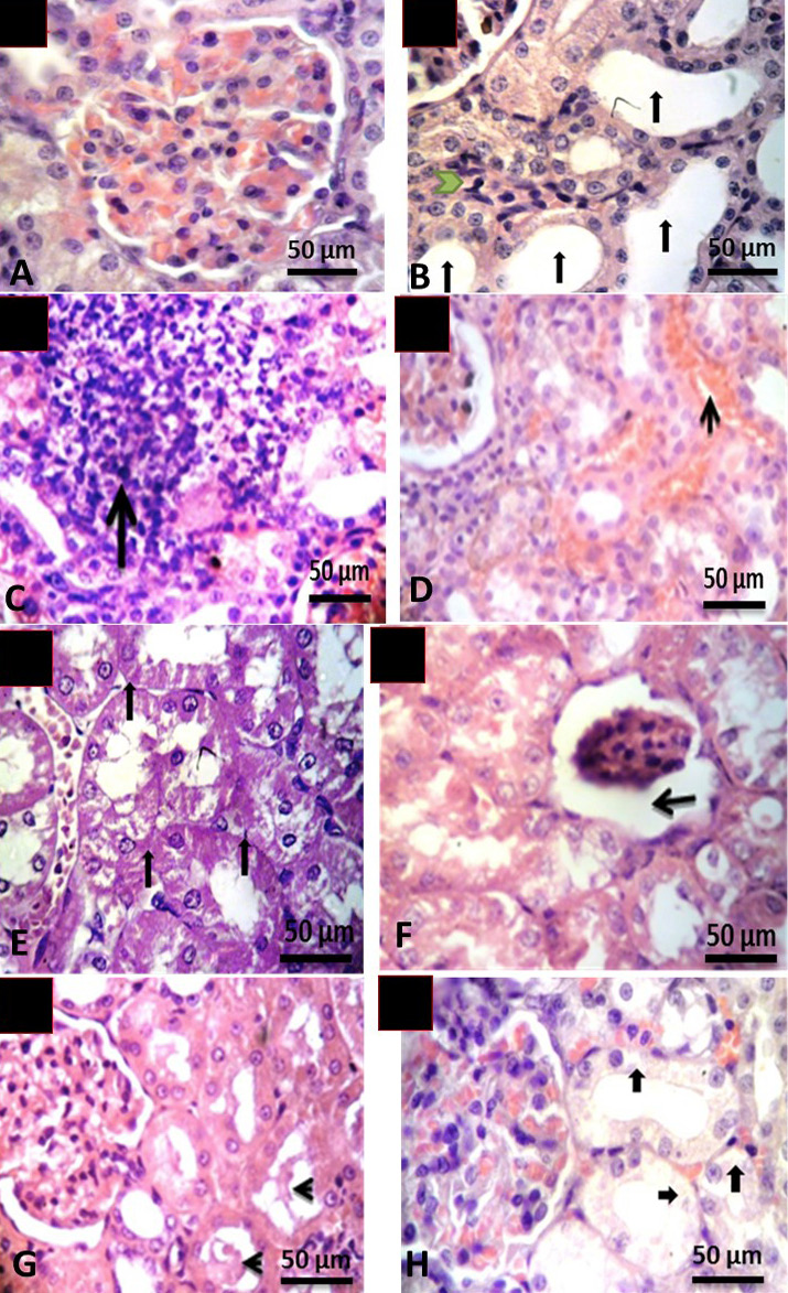
Histological structure of kidney of rat A, control group showing normal renal tissues; B-F, irradiated groups: B shows dilated renal tubules (↑) and leukocyte infiltration (→); C shows replacement of the tubules with leukocytes infiltration (↑); D shows congested intertubular blood vessels (↑); E showes necrosis of tubules with loss of its details (↑); F shows atrophy of glomeruli and increasing the capsular space (←) with loss brush border of renal tubules; G shows hyaline cast inside renal tubules (←); H shows hydropic degeneration in some distal convoluted tubules (↑). Magnification= 400X, Stain, H & E.
References
- Abdelrahman, R.S., 2017. Protective effect of apocynin against gentamicin-induced nephrotoxicity in rats. Hum. exp. Toxicol., 37: 27-37. https://doi.org/10.1177/0960327116689716[CrossRef]
- Abozaid, O.A.R., Moawed, F.S.M., Farrag, M.A. and Abdel Aziz, A.A.A., 2017. 4-(4-Hydroxy-3-methoxyphenyl)-2-butanone modulates redox signal in gamma-irradiation-induced nephrotoxicity in rats. Free Radic. Res., 51: 943-953. https://doi.org/10.1080/10715762.2017.1395025[CrossRef]
- Adaramoye, O.A., Okiti, O.O. and Farombi, E.O., 2011. Dried fruit extract from Xylopia aethiopica (Annonaceae) protects Wistar albino rats from adverse effects of whole body radiation. Exp. Toxicol. Pathol., 63: 635-643. https://doi.org/10.1016/j.etp.2010.05.005[CrossRef]
- Adhikari, M. and Arora, R., 2015. Nano-silymarin provides protection against γ-radiation-induced oxidative stress in cultured human embryonic kidney cells. Mutat. Res. Genet. Toxicol. Environ. Mutagen, 792: 1-11. https://doi.org/10.1016/j.mrgentox.2015.08.006[CrossRef]
- Aggarwal, B.B. and Shishodia, S., 2006. Molecular targets of dietary agents for prevention and therapy of cancer. Biochem. Pharmacol., 71: 1397–1421. https://doi.org/10.1016/j.bcp.2006.02.009[CrossRef]
- Akhtar, M.F., Saleem, A., Yuchi, A. and Saleem, M., 2019. A comprehensive review on ethnomedicinal, pharmacological and phytochemical basis of anticancer medicinal plants of Pakistan. Curr. Cancer Drug Targets, 19: 120-151 https://doi.org/10.2174/1568009618666180706164536[CrossRef]
- Alibakhshi, T., Khodayar, M.J., Khorsandi, L., Rashno, M. and Zeidooni, L., 2018. Protective effects of zingerone on oxidative stress and inflammation in cisplatin-induced rat nephrotoxicity. Biomed. Pharmacother., 105: 225-232. https://doi.org/10.1016/j.biopha.2018.05.085[CrossRef]
- Amir, M., Khan, M.A., Ahmad, S., Akhtar, M., Mujeeb, M., Ahmad, A., Khan, S.A. and Al-Abbasi, F.A., 2016. Ameliorating effects of Tamarindus indica fruit extract on anti-tubercular drugs induced liver toxicity in rats. Nat. Prod. Res., 30: 715-719. https://doi.org/10.1080/14786419.2015.1039001[CrossRef]
- Bagul, M.B., Sonawane, S.K. and Arya, S.S., 2018. Bioactive characteristics and optimization of tamarind seed protein hydrolysate for antioxidant-rich food formulations. 3 Biotech., 8: 218. https://doi.org/10.1007/s13205-018-1240-0[CrossRef]
- Bhadoriya, S.S., Ganeshpurkar, A., Narwaria, J., Rai, G. and Jain, A.P., 2011. Tamarindus indica: Extent of explored potential. Pharm. Rev., 5: 73-81. https://doi.org/10.4103/0973-7847.79102[CrossRef]
- Buchholz, T. and Melzig, M.F., 2016. Medicinal plants traditionally used for treatment of obesity and diabetes mellitus - Screening for pancreatic lipase and α-amylase inhibition. Phytother. Res., 30: 260-266. https://doi.org/10.1002/ptr.5525[CrossRef]
- Ekici, K., Temelli, O., Parlakpinar, H., Samdanci, E., Polat, A., Beytur, A., Tanbek, K., Ekici, C. and Dursun, I.H., 2016. Beneficial effects of aminoguanidine on radiotherapy-induced kidney and testis injury. Andrologia, 48: 683-692. https://doi.org/10.1111/and.12500[CrossRef]
- Elkady, A.A. and Ibrahim, E.M., 2016. Protective effects of erdosteine against nephrotoxicity caused by gamma radiation in male albino rats. Human exp. Toxicol., 35: 21-28. https://doi.org/10.1177/0960327115574919[CrossRef]
- Cigala, R.M., Crea, F., De Stefano, C., Lando, G., Milea, D. and Sammartano S., 2012. Modeling the acid-base properties of glutathione in different ionic media, with particular reference to natural waters and biological fluids. Amino Acids, 43: 629-648. https://doi.org/10.1007/s00726-011-1110-0[CrossRef]
- Escalona-arranz, J.C., Garcia-Diaz, J., Perez-Rosés, R., De la Vega, J., Rodríguez-Amado, J. and Morris-Quevedo, H.J., 2014. Effect of Tamarindus indica L. leaves fluid extract on human blood cells. Nat. Prod. Res., 54: 1489-1488. https://doi.org/10.1080/14786419.2014.911296[CrossRef]
- Kiang, J.G., Smith, J.T., Hegge, S.R. and Ossetrova, N.I., 2018. Circulating cytokine/chemokine concentrations respond to ionizing radiation doses but not radiation dose rates: granulocyte-colony stimulating factor and interleukin-18. Radiat. Res., 189: 634-643. https://doi.org/10.1667/RR14966.1[CrossRef]
- Kokubo, T., Kakinuma, S., Kobayashi, T., Watanabe, F., Iritani, R., Tateno, K., Nishimura, M., Nishikawa, T., Hino, O. and Shimada, Y., 2010. Age dependence of radiation-induced renal cell carcinomas in an Eker rat model. Cancer Sci., 101: 16-23. https://doi.org/10.1111/j.1349-7006.2009.01456.x[CrossRef]
- Lawrence, R.A. and Burk, R.F., 2012. Glutathione peroxidase activity in selenium-defficient rat liver. 1976. Biochem. biophys. Res. Commun., 425: 503-509. https://doi.org/10.1016/j.bbrc.2012.08.016[CrossRef]
- Lowry, O.H., Rosebrough, N.J., Farr, A.L. and Randall, R.J., 1951. Protein measurement with the Folin phenol reagent. J. biol. Chem., 193: 265-675.
- Nakchat, O., Meksuriyen, D. and Pongsamart, S., 2014. Antioxidant and anti-lipid peroxidation activities of Tamarindus indica seed coat in human fibroblast cells. Ind. J. ex. Biol., 52: 125-132.
- Natukunda, S., Muyonga, J.H. and Mukisa, I.M., 2015. Effect of tamarind (Tamarindus indica L.) seed on antioxidant activity, phytocompounds, physicochemical characteristics, and sensory acceptability of enriched cookies and mango juice. Fd. Sci. Nutr., 4: 494-507. https://doi.org/10.1002/fsn3.311[CrossRef]
- Phetdee, K., Rakchai, R., Rattanamanee, K., Teaktong, T. and Viyoch, J., 2014. Preventive effects of tamarind seed coat extract on UVA-induced alterations in human skin fibroblasts. J. Cosm. Sci., 65: 11-24.
- Radwan, R.R. and Abdel Fattah, S.M., 2017. Mechanisms involved in the possible nephroprotective effect of rutin and low dose γ irradiation against cisplatin-induced nephropathy in rats. J. Photochem. Photobiol. B., 169: 56-62. https://doi.org/10.1016/j.jphotobiol.2017.02.022[CrossRef]
- Raimondi, L., Lodovici, M., Guglielmi, F., Banchelli, G., Ciuffi, M., Boldrini, E. and Pirisino, R., 2003. The polysaccharide from Tamarindus indica (TS-polysaccharide) protects cultured corneal-derived cells (SIRC cells) from ultraviolet rays. J. Pharm. Pharmacol., 55: 333-338. https://doi.org/10.1211/002235702630[CrossRef]
- Ramlagan, P., Rondeau, P. Planesse, C., Neergheen-Bhujun, V.S., Bourdon, E. and Bahorun, T., 2017. Comparative suppressing effects of black and green teas on the formation of advanced glycation end products (AGEs) and AGE-induced oxidative stress, Fd. Funct. 8: 4194-4209. https://doi.org/10.1039/C7FO01038A[CrossRef]
- Razali, N., Mat Junit, S., Ariffin, A., Ramli, N.S. and Abdul Azizm, A., 2015. Polyphenols from the extract and fraction of T. indica seeds protected HepG2 cells against oxidative stress. BMC Complem. Altern. Med., 15: 438. https://doi.org/10.1186/s12906-015-0963-2[CrossRef]
- Sieber, F., Muir, S.A., Cohen, E.P., North, P.E., Fish, B.L., Irving, A.A., Mäder, M. and Moulder, J.E., 2009. High-dose selenium for the mitigation of radiation injury: a pilot study in a rat model. Radiat. Res., 171: 368-373. https://doi.org/10.1667/0033-7587-171.3.368[CrossRef]
- Titov, V.Y. and Osipov, A.N., 2017. Nitrite and nitroso compounds can serve as specific catalase inhibitors. Redox Rep., 22: 91-97. https://doi.org/10.1080/13510002.2016.1168589[CrossRef]
- Snecdecor, G.W. and Cochran, W.G., 1989. Statistical methods, 8th Edition, Iowa State University Press, USA, pp. 503.
- Sudjaroen, Y., Haubner, R., Würtele, G., Hull, W.E., Erben, G., Spiegelhalder, B., Changbumrung, S., Bartsch, H. and Owen, R.W., 2005. Isolation and structure elucidation of phenolic antioxidants from Tamarind (Tamarindus indica L.) seeds and pericarp. Fd. Chem. Toxicol., 43: 1673-1682. https://doi.org/10.1016/j.fct.2005.05.013[CrossRef]
- Sundaram, M.S., Hemshekhar, M., Santhosh, M.S., Paul, M., Sunitha, K., Thushara, R.M., Naveen Kumar, S.K., Naveen, S., Devaraja, S., Rangappa, K.S., Kemparaju, K. and Girish, K.S., 2015. Tamarind seed (Tamarindus indica) extract ameliorates adjuvant-induced arthritis via regulating the mediators of cartilage/bone degeneration, inflammation and oxidative stress. Sci. Rep., 5: 11117. https://doi.org/10.1038/srep11117[CrossRef]
- Suvarna, K.S., Layton, C. and Bancroft, J.D., 2013. Bancroft’s theory and practice of histological techniques, 7th Edition, Churchill Livingstone, USA, pp. 766.
- Yadav, N., Sharma, S., Sharma, KP., Pandey, A., Pareek, P. and Sharma, S., 2016. Protective role of diet supplements Spirulina and Tamarind fruit pulp on kidney in sodium fluoride exposed Swiss albino mice: Histological and biochemical indices. Ind. J. exp. Biol., 54: 44-55.
- Mohamed, E.T., Elkady, A.A. and El Kiki, S.M., 2016. Protective effect of symplocos racemosa roxb against gamma radiation induced cardiotoxicity in male albino rats. Pakistan J. Zool., 48: 1831-1837.
Services
Article usage statistics
Loading…