
Hemotropic mycoplasmas (hemoplasmas) are obligate Gram-negative bacteria that target red blood cells, and infect a wide range of hosts including cats, dogs, domestic ruminants, pigs, rodents and humans. The present study was designed for the molecular detection of Mycoplasma haemofelis and Candidatus Mycoplasma haemominutum in feline blood samples collected from various pet clinics in Pakistan, by Polymerase Chain Reaction (PCR), using 16S rDNA as the target sequence. Clinical and epidemiological data was collected in all animals included in the study. M. haemofelis and C. Mycoplasma haemominutum DNA was detected by PCR respectively in 6.8% (10/148) and in 18.2% (27/148) of cat blood samples. Of these, two animals were co-infected with both agents. Sequencing and phylogenetic analysis was performed in M. haemofelis infected samples. Analysis of risk factors revealed that risk of M. haemofelis and of C. Mycoplasma haemominutum infection was significantly higher during summer months than during the winter season (P ≤ 0.01 in both agents). Cats older than one year of age were significantly more predisposed to C. Mycoplasma haemominutum infection than younger cats (P ≤ 0.001), and fever manifestation was significantly associated with M. haemofelis infection (P ≤ 0.001) in enrolled subjects. In conclusion, this study represents the first report of molecular detection of M. haemofelis and C. Mycoplasma haemominutum in cats from Pakistan. Therefore, this agents should be considered in cats from Pakistan presented with compatible clinical signs, mainly those presented during summer season, and in adult cats and cats presented with fever.
Short Communication · Volume 52, Issue 3, 21 March 2020 · Open Access
Prevalence and Risk Factor Analysis of Haemoplasmas Infection in Cats from Lahore
· · · · · · · ·
Affiliations & Notes ⌄
- 1Institute of Pure and Applied Biology, Zoology Division, Bahauddin Zakariya University, Multan 60800, Pakistan 2Department of Veterinary Medicine, Vasco da Gama Universitary School, Av. José R. Sousa Fernandes, 3020-210, Lordemão, Coimbra, Portugal 3Baixo Vouga Veterinary Hospital, EN1, 355, 3750-742 Segadães-Águeda, Portugal 4Animal and Veterinary Research Center (CECAV), Trás-os-Montes and Alto Douro University, Quinta de Prados, 5001-801, Vila Real, Portugal 5Global Health and Tropical Medici
Article Info ⌄
- Journal:
- Pakistan Journal of Zoology
- Published:
- 21 March 2020

From the article
- Information on gender was available for 143 cats, of which 77 (53.8%) were females and 66 (46.2%) were males.
- Ages (data available for 146 cats) ranged from 0.2 to 6 years old (mean 2.2 ± 1.2 years).
- M. haemofelis DNA was detected in 10 out of 148 (6.8%) and of Candidatus Mycoplasma haemominutum detected in 27 out of 148 (18.2%) cat blood samples.
- Considering the 148 cats in which both agents were tested, 35 (25.0%) animals were infected with at least one hemoplasma species, and two cats (1.4%) were co-infected with both agents.
Selected from the article's own text.
Abstract
Materials and methods
Blood samples were collected from 148 apparently healthy or diseased client-owned cats, presented to the Veterinary Clinic of the University of Veterinary and Animal Sciences (UVAS) in Lahore, Pakistan. None of the enrolled cat was diagnosed with feline mycoplasmosis. Blood samples (approximately 2 ml) were collected from jugular vein into eppendorf tubes containing 0.5M EDTA solution as anticoagulant. Clinical and epidemiological data was collected in all animals included in the study. DNA extraction and PCR were performed in Institute of Pure and Applied Biology, Bahauddin Zakariya University Multan. Sequencing and phylogenetic analysis were performed in blood samples positive for M. haemofelis at the Institute of Hygiene and Tropical Medicine of the NOVA, University of Lisbon. All the experimental protocols and animal handling procedures were approved by the ethical review board of Institute of Pure and Applied Biology, Bahauddin Zakariya University Multan (IPAB/Eth/2016-MH 163).
DNA extraction from the collected blood sample was carried by using an inorganic method as described by Qamar et al. (2017). Detection of M. haemofelis and C. Mycoplasma haemominutum DNA was assessed by PCR as described by Braga et al. (2012). In all amplification series, positive (containing genomic DNA of M. haemofelis and of C. Mycoplasma haemominutum) and negative (without DNA) controls, were included. PCR amplifications were carried out in a DNA thermal cycler (MultiGENE OptiMAX, Labnet Inc.).
To confirm the PCR results of M. haemofelis infection, three representative PCR products were randomly selected for DNA sequencing. PCR products were purified from agarose gel slices with NZYGelpure® (Nzytech, Portugal), and subsequently sequenced (LIGHTrun™ Sequencing Service, GATC-biotech, Germany) with the same primers used for DNA amplification. The nucleotide sequences were compared with those available in the GenBank database, NCBI web server, by using BLAST algorithm (MegaBLAST option). Multiple alignments of nucleotide sequences were performed using the iterative G-INS-i method as implemented in MAFFT v7 . Resulting alignments was treated via Gblocks , (using the least stringent options available) through Seaview v4.6.1 software . The obtained DNA sequences were deposited at GenBank.
Phylogenetic tree was constructed using a maximum likelihood (ML) approach using the best fitting evolutionary model (GTR+I), on the basis of the AIC selection criterion, as suggested by jModelTest v2.0 . Phylogenetic reconstructions based on the ML optimization criterion were carried out using Mega v6.0 and the stability of the obtained tree topology assessed by bootstrapping with 1000 re-samplings of the original sequence data. The final tree were edited for display using FigTree v1.4.3.
All the data is represented as Mean ± SEM. Significance level was set at P < 0.05. Results were statistically analyzed by statistical package Minitab (version 17). Previously described risk factors for M. haemofelis and C. Mycoplasma haemominutum infection, i.e. gender, age, breed, body temperature, mucous membranes color, presence of haemolysis and/or haemoglobinuria, hydration status, deworming and vaccination habits, history of ectoparasites infestation and sampling seasons were analysed by the Fischer̕ s exact test.
Results and discussion
One hundred and forty eight animals were enrolled in the study (Table I). Information on gender was available for 143 cats, of which 77 (53.8%) were females and 66 (46.2%) were males. Ages (data available for 146 cats) ranged from 0.2 to 6 years old (mean 2.2 ± 1.2 years). Most cats were domestic short-haired cats (95.6%).
M. haemofelis DNA was detected in 10 out of 148 (6.8%) and of Candidatus Mycoplasma haemominutum detected in 27 out of 148 (18.2%) cat blood samples. Considering the 148 cats in which both agents were tested, 35 (25.0%) animals were infected with at least one hemoplasma species, and two cats (1.4%) were co-infected with both agents.
Hemotropic mycoplasmas present a worldwide distribution, and several studies have been conducted around the world regarding the prevalence of this bacterium in cats ; ; . Prevalence of infection presents geographical variation, probably due to differences in climatic conditions, vector prevalence and sampling season. The prevalence of M. haemofelis by PCR ranged from 1.49% in cats from Belem to 35.3% in cats from Brazil . C. Mycoplasma haemominutum is the most frequent feline hemoplasma in most studies, with reported prevalence ranging from 15.2% in Brazil (Alexandre- de- Santis et al., 2014) to 46.7% in cats from Japan . Similarly, in the present study, C. Mycoplasma haemominutum presented a higher prevalence of infection in cats from Pakistan than M. haemofelis (18.2% and 6.8%, respectively). Prevalence of infection detected in the present study was similar to prevalence reported in cats from Iran although prevalence of infection in present study was slightly higher for M. haemofelis than C. Mycoplasma haemominutum.
Partial DNA sequence of the 16S rDNA gene of M. haemofelis was obtained and submitted to the EMBL/GenBank database under the Accession number KY709688. A BLAST analysis revealed nucleotide sequence identities of 99% (100% query cover; 3-108 E-value) with
Table I. Analysis of risk factors for M. haemofelis and Candidatus Mycoplasma haemominutum feline infection.
| Mycoplasma haemofelis | Candidatus Mycoplasma haemominutum | ||||||
| n | Infected (%) | P-value | n | Infected (%) | P-value | ||
| Gender | Male Female | 60 75 | 06 (10%) 04 (5%) | 0.3 | 74 63 | 17 (23%) 10 (16%) | 0.3 |
| Age | >1 year <1 year | 96 41 | 07 (7%) 03 (7%) | 1.0 | 44 96 | 17 (39%) 10 (10%) | 0.0001 *** |
| Breed | Pure breed DSH | 8 103 | 00 (0%) 10 (10%) | 1.0 | 07 114 | 01 (14%) 26 (23%) | 1.0 |
| Body temperature | Normal Fever | 120 28 | 04 (3%) 06 (21%) | 0.003 ** | 120 28 | 19 (16%) 08 (29%) | 0.17 |
| Mucous membrane | Normal Pale | 136 12 | 08 (7%) 02 (17%) | 0.2 | 136 12 | 27 (20%) 00 (00%) | 0.1 |
| Hydration status | Normal Dehydrated | 146 02 | 10 (7%) 00 (0%) | 1.0 | 146 02 | 27 (18%) 00 (0%) | 1.0 |
| Haemolysis | Present Absent | 01 147 | 00 (0%) 10 (7%) | 1.0 | 01 147 | 00 (0%) 27 (19%) | 1.0 |
| Vomiting | Present Absent | 18 130 | 01 (6%) 09 (7%) | 1.0 | 18 130 | 02 (11%) 25 (19%) | 0.5 |
| Vaccinated | Yes No | 65 83 | 03(5%) 07 (8%) | 0.5 | 66 82 | 11(17%) 16(20%) | 0.6 |
| Dewormed | Yes No | 59 89 | 03 (5%) 07 (8%) | 0.7 | 60 88 | 10 (17%) 17 (19%) | 0.8 |
| History of ectoparasites Infestation | Present Absent | 19 129 | 02 (11%) 08 (6%) | 0.6 | 19 129 | 02 (11%) 25 (19%) | 0.5 |
| Season | Winter Summer | 57 89 | 00 (0%) 10 (11%) | 0.007 ** | 57 90 | 04 (7%) 23 (25%) | 0.004 ** |
DSH, Domestic short-hair cats; P > 0.05, Non significant; P < 0.01, significant; P < 0.001, highly significant.
homologous sequences of M. haemofelis isolates registered in GenBank. The phylogenetic analysis of the obtained DNA sequence placed it in one stable monophyletic cluster (supported by maximum bootstrap value) along with related 16S rRNA sequences of M. haemofelis and M. haemocanis downloaded from public databases (Fig. 1).
M. haemofelis and C. Mycoplasma haemominutum infections were significantly more frequent in summer months than during the winter season (P ≤ 0.01 for both agents). Our results are in agreement with those reported in cats from Brazil and Italy and are probably related with the higher number of vector fleas during summer. Manifestation of fever was significantly associated with M. haemofelis infection (P ≤ 0.001) during present investigation. Our results are in agreement with those of Kewish et al. (2004) who had reported that most cats having fever were harbouring the microorganism. Cats older than one year of age were significantly more predisposed to C. Mycoplasma haemominutum infection than younger cats (P ≤ 0.001). No other significant associations were detected between presences of M. haemofelis or C. Mycoplasma haemominutum and the risk factors analysed (Table I). This result is in agreement with those of Raimundo et al. (2016) who had reported that adult cats had more chances to be infected by C. Mycoplasma haemominutum than young cats.
In the present study, cat blood samples were collected from domestic short haired cats and pure-breed cats included siamese and persian cats. Hemoplasmas infection were detected in domestic short hair, persian and siamese cats. The prevalence of M. haemofelis (17%) in blood of stray cats was considerably higher (P = 1) than that found in other cat breeds. Our results are in agreement with Kamrani et al. (2008) who had reported that the prevalence of M. haemofelis (47%) in blood from stray cats (n = 45) was considerably higher than in pet cats. In addition, and although not statistically significant, males were more likely to be infected with both agents, corroborating previous data obtained by Sykes et al. (2008) who had reported that male cats were at higher risk for hemoplasmas infection than females.
In conclusion, this is first molecular report of M. haemofelis and C. Mycoplasma haemominutum infection in cats from Pakistan. Prevalence of hemoplasmas infection detected in cats from Pakistan was considerable. Therefore, this agents should be considered in cats from Pakistan presented with compatible clinical signs, mainly those presented during summer season, and in adult cats and cats presented with fever.
Acknowledgements
Authors are grateful to pet owners and veterinarians who helped in blood sample and epidemiological data collection. Positive control DNA of M. haemofelis in this study kindly provided by Prof. Dr. Ricardo G. Maggi (Intracellular Pathogens Research Laboratory, North Carolina State University, USA).
This work of CM and AP was partially supported by Funds to GHTM (UID/Multi/04413/2013). AP and CM have the support of the Portuguese Ministry of Education and Science (via Fundação para a Ciência e a Tecnologia), through PhD grant SFRH/BD/116516/2016 and an Investigator Starting Grant IF/01302/2015, respectively. The work of C. Maia was done under the frame of EurNegVec COST Action TD1303.
Statement of conflict of interest
Authors declare no conflict of interest.
Figures
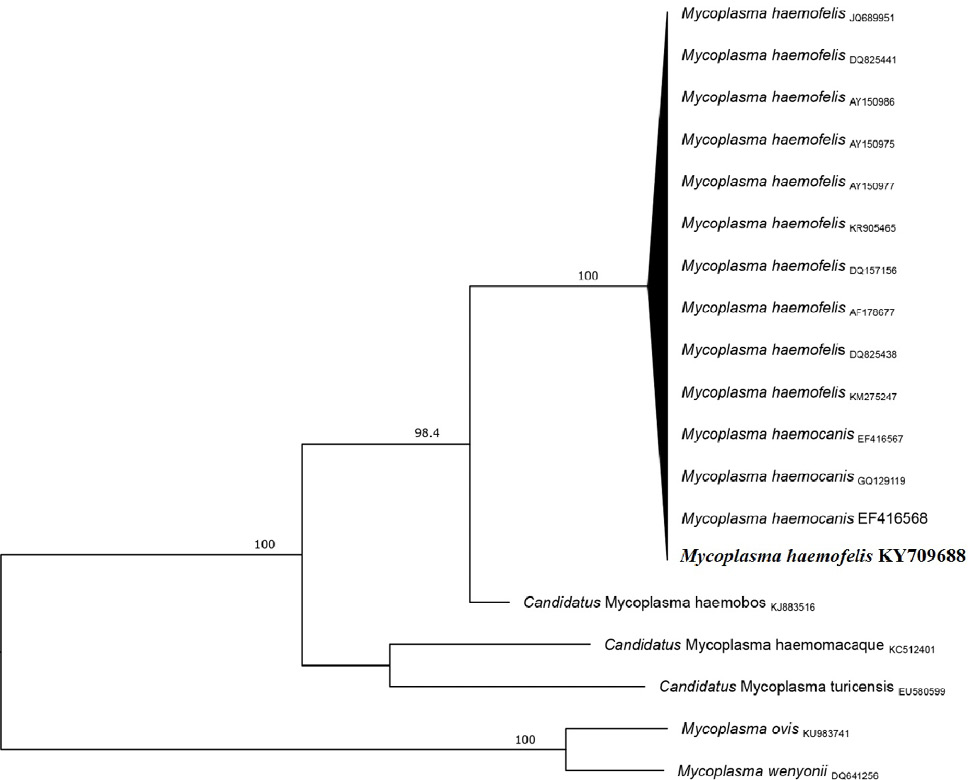
Maximum likelihood phylogenetic tree (midpoint rooted) of M. haemofelis partial 16s rDNA gene sequence amplified from cat blood sample. The percentages of significant (≥ 75) bootstrap values of 1000 resamplings of the original data are indicated at specific branch-nodes. The size bar indicates 0.02 substitutions per site.
References
- Aktas, M. and Ozubek, S., 2018. Vet. Microbiol., 221: 94-97. https://doi.org/10.1016/j.vetmic.2018.06.004[CrossRef]
- Alexandre- de- Santis, A.C.G., Herrera, H.M., Marques- de- Sousa, K.C., Gonçalves, L.Lc., Denardi, N.C.B., Domingos, L.H., Campos, J.B.V., Machado, R.Z. and Andre, M.R., 2014. Braz. J. Vet. Parasitol., 23: 231-236. https://doi.org/10.1590/S1984-29612014039[CrossRef]
- Aragão-de-Sousa, S.K.S., Sampaio-Junior, F.D., Sousa, L., Gonçalves, E.C. and Scofield, A., 2013. Braz. J. Vet. Res., 33: 1116-1120. https://doi.org/10.1590/S0100-736X2013000900011[CrossRef]
- Braga, M.S.C.O., André, M.R., Freschi, C.R., Teixeira, M.C.A. and Machado, R.Z., 2012. Braz. J. Microbiol., 43: 569-575. https://doi.org/10.1590/S1517-83822012000200018[CrossRef]
- Castresana, J., 2000. Mol. Biol. Evol., 17: 540-552. https://doi.org/10.1093/oxfordjournals.molbev.a026334[CrossRef]
- Darriba, D., Taboada, G.L., Doallo, R. and Posada, D., 2012. Nat. Meth., 9: 72-74. https://doi.org/10.1038/nmeth.2109[CrossRef]
- Firmino, F.P., Aquino, L.C., Marcola, T.G., Bittencourt, M.V., McManus, C.M. and Paludo, A.Z., 2016. Braz. J. Vet. Res., 36: 731-736. https://doi.org/10.1590/S0100-736X2016000800009[CrossRef]
- Fujihara, M., Watanabe, M., Yamada, T. and Harasawa, R., 2007. J. Vet. Med. Sci., 69: 1061-1063. https://doi.org/10.1292/jvms.69.1061[CrossRef]
- Ghazisaeedi, F., Atyabi, N., Salehi, T.Z., Gentilini, F., Tamai A.I., Akbaraein, H. and Tasker, S., 2014. J. Veter. Clin. Pathol. 43: 381-386. https://doi.org/10.1111/vcp.12166[CrossRef]
- Gouy, M., Guindon, S. and Gascuel, O., 2010. Mol. Biol. Evol., 27: 221-224. https://doi.org/10.1093/molbev/msp259[CrossRef]
- Hammer, A.S. and Wellman, M., 1999. Am. J. Anim. Hosp. Assoc., 35: 471-473. http://tree.bio.ed.ac.uk/software/figtree/
- Kamrani, A., Parreira, V.R., Greenwood, J. and Prescot, J.F., 2008. Canad. J. Vet. Res., 72: 411-419.
- Katoh, K. and Toh, H., 2008. Br. Bioinform., 9: 286–298. https://doi.org/10.1093/bib/bbn013[CrossRef]
- Kewish, K.E., Appleyard, G.D., Myers, S.J., Kidney, B.A. and Jackson, M.L., 2004. Canad. J. Vet. Res., 45: 749-752.
- Otranto, D. and Dantas-Torres, F., 2010. J. Paras.Vect., 3: 2-8. https://doi.org/10.1186/1756-3305-3-2[CrossRef]
- Qamar, M., Malik, M.I., Latif, M., Ane, Q.U., Aktas, M., Shaikh, R.S. and Iqbal, F., 2017. J. Vect.-Bor. Zoon. Dis., 17: 179-184.
- Raimundo, J.M., Guimaraes, A., Rodrigues, R.B., Botelho, C.F.M., Peixoto, M.P., Pires, M.S., 2016. Braz. J. Vet. Parsit., 25: 441-449. https://doi.org/10.1590/s1984-29612016086[CrossRef]
- Sykes, J.E., Terry, J.C., Lindsay, L.L. and Owens, S.D., 2008. J. Am. Vet. med. Assoc., 232: 372-379. https://doi.org/10.2460/javma.232.3.372[CrossRef]
- Taimura, K., Stecher, G., Peterson, D., Filipski, A. and Kumar, S., 2013. J. mol. Biol. Evol., 30: 2725–2729. https://doi.org/10.1093/molbev/mst197[CrossRef]
- Willi, B., Filoni, C. and Catao-Diaz, J.L., 2005. J. clin. Microbiol., 45: 1159–1166.
Services
Article usage statistics
Loading…