Research Article · 26 June 2026 · Open Access
Prevalence and Associated Risk Factors of Lumpy Skin Disease in the Cattle of Narsingdi in Bangladesh
· · ·
Affiliations & Notes ⌄
- 1Faculty of Animal Science and Veterinary Medicine, Patuakhali Science and Technology University, Barishal-8210, Bangladesh
- 2Department of Microbiology and Public Health, Faculty of Animal Science and Veterinary Medicine, Patuakhali Science and Technology University, Barishal-8210, Bangladesh
- 3Department of Pathology and Parasitology, Jashore University of Science and Technology, Jhenaidah Campus, Bangladesh. *Correspondence | Md. Yeasin Arafat, Department of Microbiology and Public Health, Faculty of Animal Science and Veterinary Medicine, Patuakhali Science and Technology University, Bangladesh
- 4Email: arafatyeasin@pstu.ac.bd
Article Info ⌄
- Journal:
- South Asian Journal of Life Sciences
- Published:
- 26 June 2026

Abstract
INTRODUCTION
Livestock is a crucial component of the Bangladesh economy, and its overall population is about 45.36 million, 25.01 million of which are cattle (DLS, 2023-24). The industry is the source of 1.8% of the national GDP, 13% of foreign exchange earnings and full-time employment to approximately 20% of the rural population (BBS, 2023-24). Although this is crucial, local milk and meat production is inadequate to address the needs of the states (only a percentage of the national needs are satisfied) and the availability of milk and meat in the country is 33.95 ml/day and 12.61 g/day, respectively, which is many times less than the recommended intake of 250 ml/day and 120 g/day, respectively (DLS, 2021-22). The livestock sector is almost one-third of the global agricultural production and nutritional security and livelihood of close to one billion people .
Different infectious diseases affect cattle in Bangladesh, and Lumpy Skin Disease (LSD) has become an important challenge especially in young calves (0-6 months). LSD is a virus transmitted by the Lumpy Skin Disease Virus (LSDV) which belongs to the genus Capri poxvirus in close relation to sheep pox and goat pox viruses. The clinical manifestation is high fever (104-1060F), widespread nodules on the skin, and depression , and females are said to be more vulnerable compared to men (Geri et al., 2010). Its mortality rate is variable as it is between 1-3% in most cases whilst it may go up to 40% in an extreme outbreak . LSD still spreads in the Middle East and has lately become a severe menace to the Asian and the European regions, imposing serious dilemma to the health of the cattle, as well as to the livestock farming system.
Objectives of the study
- To know the prevalence of LSD in cattle according to age, sex, breed, body region etc.
- To identify the risk factors associated with LSD prevalence in the cattle population
MATERIALS AND METHODS
Study area and duration
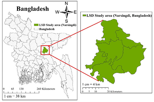
The study was carried out in Narsingdi from August to September 2022, which served as the designated study location (Figure 1).
Investigation and data recording
This research utilized the patient data present in Upazila Livestock Office and Veterinary Hospital’s register book together with on-site investigation conducted during research timelines. Each affected animal received documentation for age, sex and breed information. Physical examination included observation and manual assessment to evaluate sick cattle.
Epidemiological investigation
LSD occurs both endemically and occasionally in epidemic outbreaks. It spreads through cattle fairs, arthropod vectors, the movement of infected animals, contaminated syringes reused on affected cattle, and, most significantly, through the transportation of cattle from India to Bangladesh. Tentative diagnosis It is based on clinical history, animal examination and clinical findings.
Clinical signs for case identification
The following clinical signs were carefully observed to identify LSD cases:
- High fever (104–106°F)
- Cutaneous nodules covering the body
- Enlargement of superficial lymph nodes
- Nasal discharge
- Excessive salivation
- Weakness and loss of appetite
Rupture of cutaneous nodules
RESULTS AND DISSCUSSION
Overall Prevalence of LSD
A total of 116 disease cases of cattle were recorded at Narsindi district during August to September, 2022. Among them 65 cases were infected with LSD (Table 1).
Table 1: Prevalence of LSD in cattle.
| Total no. of cases (N) | 116 |
| Total no. of LSD affected | 65 |
| Prevalence (%) | 65*100/116= 56.03% |
This study revealed that the overall prevalence of LSD in Narsindi was 56.03%, which is comparable to the 63.33% reported in Monirampur and 52.38% in Avoynagor Upazila of Jashore district , and slightly higher than the 41.06% reported in Dinajpur Sadar . However, other studies have reported varying prevalence rates of LSD, including 4.13% in Barisal , 21% in Barishal , 13.65% in Sylhet Sadar Upazila , and 8.7% in Uganda .
LSD in different months
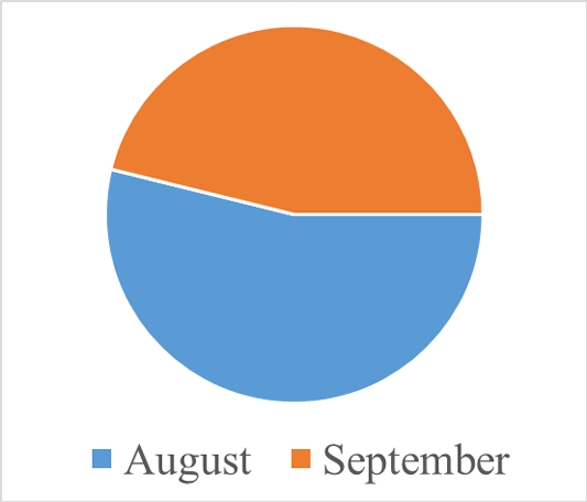
Most animals affected with LSD were found in this region. The results show that the majority of cases occurred in August (53.85%) and September (46.15%), which were higher than the prevalence reported by Kabir et al. (2022) in August (18.46%) and September (20.63%) (Table 2 and Figure 2).
Table 2: Outbreak of LSD in different months.
| Months | No. of LSD affected | Percentage (%) |
| August | 35 | 53.85% |
| September | 30 | 46.15% |
| Total | 65 | 100 % |
LSD in different age group
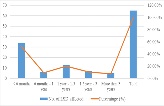
Calves (<6 Months) were the most susceptible to LSD infections with a rate of 52.31%, whereas the infection rate among the cattle above the age of 3 years was the lowest with 7.63%. Our findings correspond to Azam et al. (2024), who observed an infection rate of LSD of 41.2% and 35.2% in calves and old cattle (>3 years), respectively. Sarkar et al. (2020) have shown that older cattle (11.2%) was less susceptible than calves (68%). The sensitivity of the calves, as compared to the lower rates of infection in older cattle, can be explained by the fact that the practice of adequate care is insufficient and the immune system of young animals is weaker (Table 3 and Figure 3).
Table 3: LSD in different age group.
| Age group | No. of LSD affected | Percentage (%) |
| < 6 months | 34 | 52.31% |
| 6 months - 1 year | 06 | 9.23% |
| 1 year - 1.5 years | 13 | 20% |
| 1.5 year - 3 years | 07 | 10.77% |
| More than 3 years | 05 | 7.63% |
| Total | 65 | 100% |
Table 4: LSD in different sex group
| Sex | Number | Percentage (%) |
| Male | 38 | 58% |
| Female | 27 | 42% |
| Total | 65 | 100% |
LSD in different sex group
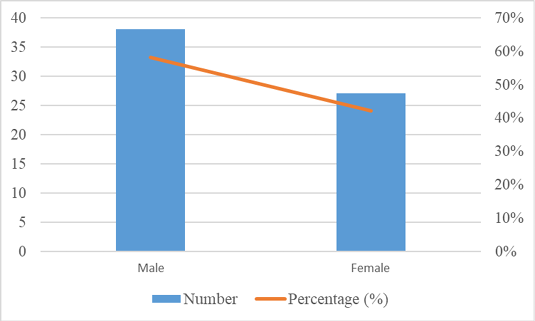
In the study, LSD infections were higher in male cattle (58%) than in female cattle (42%). These results are similar to the findings of Sarkar et al. (2020), where the occurrence was higher in males (44%) than in females (38.5%), but differ from Shake and Akimul (2025), who reported a higher prevalence in females (63.16%) than in males (36.84%) (Table 4 and Figure 4).
LSD in different breeds
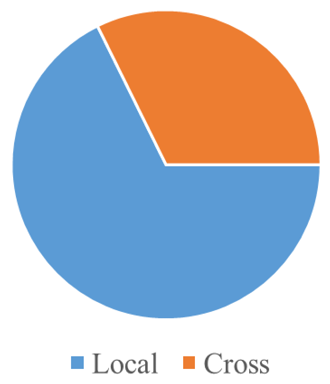
The local cattle were more susceptible to LSD infection with an infection rate of 67.69%, compared to the crossbred cattle which had the rate of 32.31%. Conversely, Shake and Akimul (2025) indicated that the infection rate was higher in the crossbred cattle (70.53%) than in the indigenous cattle (29.47%). On the same note, Sarkar et al. (2020) reported 50% infection in local cattle and 26.9% infection in the crossbreed cattle. In general, poor management practices and weak immune systems were the likely predisposing factors to all the cattle breeds (Table 5 and Figure 5).
Table 5: LSD in different breeds.
| Breed | No. of affected animal | Percentage (%) |
| Local | 44 | 67.69% |
| Cross | 21 | 32.31% |
| Total | 65 | 100% |
LSD in relation to different body region
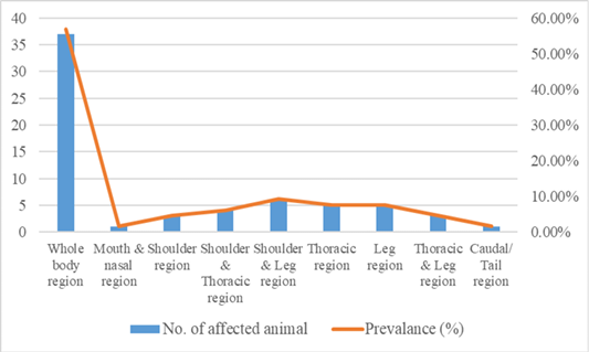
Most of the affected LSD cattle (56.92%) had lesions in the entire body. The prevalence of localized infections was smaller, with the highest prevalence being reported in the shoulder and the leg area (9.23% each), then the thoracic area (7.69%), and the leg area (7.69%). Combination of regions, e.g. shoulder and thoracic or thoracic and leg were less common. The least affected areas were the mouth, nose and the tail (1.53% and 1.53%, respectively) (Figure 6 and Table 6). These findings suggest that LSD is mainly known to cause widespread lesions, which is an expression of its systemic property and spreads via the lymphatic and circulatory system.
Table 6: LSD in relation to different body region
| Affected region of body | No. of affected animal | Prevalance (%) |
| Whole body region | 37 | 56.92 % |
| Mouth and nasal region | 01 | 1.53% |
| Shoulder region | 03 | 4.61% |
| Shoulder and Thoracic region | 04 | 6.15% |
| Shoulder and Leg region | 06 | 9.23% |
| Thoracic region | 05 | 7.69% |
| Leg region | 05 | 7.69% |
| Thoracic and Leg region | 03 | 4.61% |
| Caudal/ Tail region | 01 | 1.53% |
| Total | 65 | 100% |
CONLUSION
The prevalence of LSD was high in the investigated cattle population, and the most susceptible were calves and the local breeds. Males were more affected than females and lesions were mainly seen all over the entire body. The research has also identified the factors of age, breed, and management practices in the prevalence of the disease. Preventive interventions such as vaccination and better husbandry must be applied in order to mitigate the effects of LSD among cattle herds.
Limitations of this study
- The study had a small sample size, limiting generalizability.
- Data were collected from a single region, restricting geographic representation.
- The study covered a short period, missing potential seasonal variations.
- Diagnosis relied mainly on clinical observation, without laboratory confirmation.
- There was no facility for bacteriological and virological test in laboratory
ACKNOWLEDGEMENTS
The authors grateful to the livestock farmers of Narsingdi and the staff of the Livstock Office for their kind cooperation and support throughout this study.
NOVELTY STATEMENT
This paper provides updated epidemiological evidence on LSD in the Narsingdi region of Bangladesh, both in terms of age, breed, and sex related susceptibility patterns and in terms of specific lesion patterns in body regions. The results provide new knowledge on the local disease prevalence and risk factors, which can be useful in preventing and controlling local diseases through specific prevention and management approaches in cattle populations.
Author’s Contribution
Data collection: JI; Data analysis: JI, MKA, MYA; Writing the original draft: JI, MKA, SIB; Review and editing: MKA, MYA. All authors read and approved the final manuscript.
Generative AI and AI assisted technology statement
No generative AI or AI-assisted technology was used in the collection of data, its analysis, or even the preparation of the manuscript. All the analyses, interpretations, and writings have been done by the authors only.
Conflict of interest
The authors have declared no conflict of interest.
Figures

LSD Study area (Narsingdi, Bangladesh).

Outbreak of LSD in different Months

LSD at different ages

LSD in different sexes.

LSD in relation to breed.

References
- Abera Z, Degefu H, Gari G, Ayana Z (2015). Review on epidemiology and economic importance of lumpy skin disease. Int. J. Basic Appl. Virol., 4: 8-21.
- Abera Z, Degefu H, Gari G, Kidane M (2015). Sero-prevalence of lumpy skin disease in selected districts of West Wollega zone, Ethiopia. BMC Vet. Res., 11: 135. https://doi.org/10.1186/s12917-015-0432-7[CrossRef]
- Alemayehu G, Zewde G, Admassu B (2013). Risk assessments of lumpy skin diseases in Borena bull market chain and its implication for livelihoods and international trade. Trop. Anim. Health Prod., 45(5): 1153–1159. https://doi.org/10.1007/s11250-012-0340-9[CrossRef]
- Anil TSV, Durga ASK (2021). Antibiotic versus no antibiotic approach in the management of lumpy skin disease (LSD) in cattle. J. Entomol. Zool. Stud., 9(1): 1612-1614. https://doi.org/10.22271/j.ento.2021.v9.i1w.8366[CrossRef]
- Ansari WK, Arafat MY, Akimul MK, Rahman MS, Islam MJ, Hasan M, Mridha MI, Islam MA, Kayesh MEH (2024). Prevalence of lumpy skin disease and associated risk factors in the cattle of Barishal district in Bangladesh. Vet. Sci. Res. Rev., 10(2): 72-81. https://doi.org/10.17582/journal.vsrr/2024/10.2.72.81[Full text][CrossRef]
- Azam MG, Uddin MZ, Islam MM, Ali MH, Rahman M, Salauddin M (2024). Epidemiology and risk factors of lumpy skin disease outbreak in cattle in the North-Westarea of Bangladesh. Ger. J. Vet. Res., 4(1): 83-94. https://doi.org/10.51585/gjvr.2024.1.0078[CrossRef]
- Bangladesh Bureau of Statistics (BBS) (2023-24). Yearbook of Agricultural Statistics 2024.
- Biswas D, Saha SS, Biswas S, Sayeed MA (2020). Outbreak of lumpy skin disease of cattle in south-west part of Bangladesh and its clinical management. Vet. Sci. Res. Rev., 6(2): 100-108. https://doi.org/10.17582/journal.vsrr/2020.6.100.108[Full text][CrossRef]
- Department of Livestock Services (DLS). Livestock Economy at a Glance 2023-2024. Veterinary Sciences: Research and Review sat district veterinary hospital in Sylhet district of Bangladesh. Int. J. Res. Innov. Appl. Sci., 6(10): 111–11.
- Gari G, Waret-Szkuta A, Grosbois V, Jacquiet P, Roger F (2010). Risk factors associated with observed clinical lumpy skin disease in Ethiopia. Epidemiol. Infect., 138(11): 1657–1666. https://doi.org/10.1017/S0950268810000506[CrossRef]
- Hakimul MH, Roy RK, Yeasmin F, Fakhruzzaman M, Yeasmin T, Sazib MRI, Uddin MN, Sarker S (2021). Prevalence and management practices of lumpy skin disease (LSD) in cattle at Natore District of Bangladesh. Eur. J. Agric. Food Sci., 3(6). https://doi.org/10.24018/ejfood.2021.3.6.420[CrossRef]
- Kabir MH, Sarder MJU, Islam MH, Ali MS, Ahmed ABMT, Rahman MM, Sarker CK, Rahman MM (2022). Prevalence of lumpy skin disease (LSD) at Bagatipara Upazila, Natore, Bangladesh. Bangladesh Livest. J., 1(1): 19–22.
- Khalil MI, Sarker MFR, Hasib FMY, Chowdhury S (2021). Outbreak investigation of lumpy skin disease in dairy farms at Barishal, Bangladesh. Turk. J. Agric. Food Sci. Technol., 9(1): 205–209. https://doi.org/10.24925/turjaf.v9i1.205-209.3827[CrossRef]
- Kiplagat SK, Kitala PM, Onono JO, Beard PM, Lyons NA (2020). Risk factors for outbreaks of lumpy skin disease and the economic impact in cattle farms of Nakuru County, Kenya. Front. Vet. Sci., 7: 259. https://doi.org/10.3389/fvets.2020.00259[CrossRef]
- MacLachlan NJ, Dubovi EJ (2011). Fenner’s veterinary virology, 4th ed.
- Mulatu E, Feyisa A (2018). Review: Lumpy skin disease. J. Vet. Sci. Technol., 9: 535. https://doi.org/10.4172/2157-7579.1000535[CrossRef]
- Ochwo S, Vander-Waal K, Munsey A, Nkamwesiga J, Ndekezi C, Auma E, Mwiine FN (2019). Seroprevalence and risk factors for lumpy skin disease virus seropositivity in cattle in Uganda. BMC Vet. Res., 15(1): 236. https://doi.org/10.1186/s12917-019-1983-9[CrossRef]
- Pory FS, Lasker RM, Islam MN, Siddiqui MSI (2021). Prevalence of lumpy skin disease June 2025 | Volume 11 | Issue 1 | Page 103
- Sarkar S, Meher MM, Parvez MMM, Akther M (2020). Occurrences of lumpy skin disease in cattle in Dinajpur Sadar of Bangladesh. Res. Agric. Livest. Fish., 7(3): 445-455. https://doi.org/10.3329/ralf.v7i3.51364.[CrossRef]
- Şevik M, Doğan M (2017). Epidemiological and Molecular Studies on Lumpy Skin Disease Outbreaks in Turkey during 2014-2015. Transb. Emerg. Dis., 64(4): 1268–1279. https://doi.org/10.1111/tbed.12501[CrossRef]
- Shake MH, Akimul MK (2025). Epidemiology and clinical management of lumpy skin disease in the cattle of Dupchanchia Upazila, Bogura, Bangladesh. Vet. Sci. Res. Rev., 11(1): 97-103. https://doi.org/10.17582/journal.vsrr/2025/11.1.97.103[Full text][CrossRef]
- Tageldin MH, Wallace DB, Gerdes GH, Putterill JF, Greyling RR, Phosiwa MN, Al-Busaidy RM, Al-Ismaaily SI (2014). Lumpy skin disease of cattle: An emerging problem in the Sultanate of Oman. Trop. Anim. Health Prod., 46(1): 241–246. https://doi.org/10.1007/s11250-013-0483-3[CrossRef]
- Thornton PK (2010). Livestock production: Recent trends, future prospects. Philos. Trans. R. Soc. Lond. Ser. B, Biol. Sci., 365(1554): 2853–2867. https://doi.org/10.1098/rstb.2010.0134[CrossRef]
- Tuppurainen, E.S.M. and Oura, C.A.L., 2012. Lumpy skin disease: An emerging threat to Europe, the Middle East and Asia. Transboundary and Emerging Diseases, 59(1), pp.40–48.
- Zeynalova S, Asadov K, Guliyev F, Vatani M, Aliyev V (2016). Epizootology and molecular diagnosis of lumpy skin disease among livestock in Azerbaijan. Front. Microbiol., 7: 1022. https://doi.org/10.3389/fmicb.2016.01022[CrossRef]
Services
Article usage statistics
Loading…