Specail Issue: Artificial Intelligence in Veterinary Medicine: Current Status and Future Prospects · Volume 13, Issue 1, 10 December 2025 · Open Access
Preserving Male Fertility during Doxorubicin Chemotherapy: Integrating Mechanistic Insights with Antioxidant and Nanotechnological Therapeutic Strategies
· ·
Affiliations & Notes ⌄
1Department of Biochemistry and Molecular Biology, Faculty of Veterinary Medicine, Zagazig University, Zagazig 44511, Egypt
2Department of Pharmacy, Al-Farahidi University, Baghdad 10021, Iraq. *Correspondence | Mahran Mohamed Abd El-Emam, Department of Biochemistry and Molecular Biology, Faculty of Veterinary Medicine, Zagazig University, Zagazig 44511, Egypt
Collectively, these findings underscore the potential of antioxidant- and nanotechnology-based interventions in mitigating DOX-induced testicular damage.
To maintain male reproductive health without compromising DOX’s anticancer effectiveness, future research should concentrate on translational approaches that integrate these treatment modalities.
The influence of doxorubicin-induced damage and the effectiveness of antioxidant or nanotechnological treatments may be influenced by species-specific variations in testicular microarchitecture, redox balance, and drug metabolism.
Also, Future investigations should focus on confirming that antioxidant and nanomedicine-based strategies do not compromise DOX’s antitumor efficacy.
Selected from the article's own text.
Abstract
Doxorubicin (DOX) is a powerful anthracycline chemotherapeutic drug that is frequently used to treat hematological and solid cancers. In veterinary medicine, doxorubicin is the most widely used antineoplastic agent in dogs and cats. It may be useful in the treatment of a variety of lymphomas, carcinomas, leukemias, and sarcomas in both the dog and cat. Nevertheless, dose-limiting toxicities, particularly gonadotoxicity that results in testicular dysfunction and infertility, frequently restrict its therapeutic usefulness. This review provides a comprehensive analysis of the molecular mechanisms underlying DOX-mediated testicular toxicity, focusing on oxidative and endoplasmic reticulum (ER) stress pathways, blood–testis barrier disruption, and impaired steroidogenesis. The preventive potential of novel nanotherapeutic approaches and natural antioxidants is given particular attention. These drugs show promise in preserving testicular architecture, reducing inflammatory and apoptotic signaling, and restoring oxidative–antioxidative balance. Together, antioxidant and nanomedicine-based strategies may provide novel therapeutic pathways to reduce DOX-induced male reproductive damage and enhance fertility after chemotherapy. The novelty of this review lies in its unique integration of mechanistic insights with translational therapeutic perspectives, bridging antioxidant pharmacology and nanotechnology to propose innovative strategies for preserving male reproductive function during chemotherapy in both human and veterinary medicine. However, there are still questions about whether these treatments can specifically shield testicular tissue without reducing DOX’s anticancer effectiveness or running into the delivery issues that come with nanomedicine. To move these tactics closer to clinical use, it will be crucial to answer these issues by thorough pharmacokinetic, tumour efficacy, and translational research.
Chemotherapy remains a cornerstone in the management of various malignancies . Among chemotherapeutic agents, doxorubicin (DOX) is one of the most potent and widely used anthracycline antibiotics, approved by the U.S. Food and Drug Administration (FDA) for the treatment of several solid tumors including breast and lung cancers as well as hematological malignancies such as acute leukemia and lymphomas .
Despite its clinical efficacy, chemotherapy is often gonadotoxic, posing a significant risk to fertility and overall quality of life in cancer survivors . Increasing evidence suggests that DOX directly induces testicular toxicity, which manifests as disruptions in lipid biosynthesis, impaired steroidogenesis, DNA damage, and mutagenesis within the testicular tissue .
One of the principal mechanisms underlying the cytotoxic effects of anthracyclines, including DOX, is the excessive generation of reactive oxygen species (ROS). These ROS overwhelm the antioxidant defense system, resulting in oxidative stress, cellular injury, and apoptosis . Due to their high mitotic activity, spermatogonia are particularly vulnerable to DOX-induced damage, similar to what has been observed following etoposide exposure . Consequently, gonadal damage caused by chemotherapy may be transient, permanent, or a combination of both.
DOX inhibits topoisomerase II activity, leading to DNA double-strand breaks and the subsequent generation of ROS, which contribute to testicular necrosis and degeneration. Several studies have demonstrated that DOX administration alters male reproductive parameters, including serum sex hormone levels, sperm morphology, motility, and count, along with significant histopathological changes in the testes ; ; .
The mechanisms of DOX-induced testicular toxicity are multifactorial, involving excessive ROS production, lipid peroxidation, inflammation mediated by proinflammatory cytokines, and apoptosis. Mammalian spermatozoa are especially susceptible to oxidative damage due to their high content of polyunsaturated fatty acids . Furthermore, DOX treatment has been associated with a reduction in body and testicular weight, accompanied by histopathological findings such as azoospermia, intertubular edema, seminiferous tubule atrophy, hyperemia, necrosis, and degeneration of spermatogonia .
Given the rising concern over chemotherapy-induced infertility, understanding the molecular mechanisms underlying DOX-mediated testicular injury is of paramount importance. This review aims to provide a comprehensive overview of the pathophysiological pathways involved in DOX-induced testicular toxicity, with particular emphasis on oxidative stress, inflammation, apoptosis, and dysregulated blood-testis barrier (BTB) and autophagy. Furthermore, the review highlights recent advancements in therapeutic interventions including natural antioxidants, nanocarriers, and pharmacological agents that may mitigate DOX-induced testicular toxicity and preserve male reproductive health in cancer patients. Thus, molecular understandings of oxidative stress, apoptosis, autophagy, and disruption of the blood-testis barrier are critically integrated with new treatment approaches intended to block these processes in this review. By connecting molecular knowledge to translational implementation, the review seeks to pinpoint important molecular targets and therapeutic prospects, especially via antioxidant regulation and delivery systems based on nanocarriers, that could direct the creation of clinically successful methods to stop DOX-induced testicular damage and maintain male fertility.
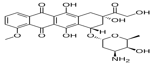
DOX consists of both aglycone and sugar moieties. The aglycone portion contains a tetracyclic ring system with adjacent quinone–hydroquinone groups, a methoxy substituent, and a short side chain bearing a carbonyl group. The sugar moiety, known as daunosamine, is a 3-amino-2,3,6-trideoxy-L-fucosyl residue covalently linked to the aglycone via a glycosidic bond.
Pharmacodynamics of DOX
DOX is an anthracycline antibiotic naturally derived from Streptomyces species and widely recognized as one of the most potent chemotherapeutic agents used in cancer treatment (Naushen et al., 2014). Owing to its broad spectrum of activity, DOX is employed either alone or in combination with other drugs for the management of both solid tumors and hematological malignancies, including cancers of the breast, bile ducts, prostate, uterus, ovary, esophagus, stomach, and liver, as well as childhood solid tumors, osteosarcomas, soft tissue sarcomas, Kaposi’s sarcoma, acute myeloid and lymphoblastic leukemia, and Wilms’ tumor .
The antineoplastic activity of DOX primarily stems from its ability to inhibit the synthesis of DNA, RNA, and proteins, thereby inducing apoptosis in rapidly dividing cells . This inhibition occurs through intercalation into the DNA double helix and/or covalent binding to DNA-associated proteins involved in transcription and replication. By intercalating between DNA base pairs, DOX disrupts the normal helical structure, impeding the progression of DNA and RNA polymerases and ultimately triggering programmed cell death.
Furthermore, DOX has been classified as a topoisomerase II poison. The topoisomerase II enzyme transiently cleaves and rejoins DNA strands to alleviate torsional stress during replication and transcription. DOX stabilizes the intermediate “cleavable complex” formed between topoisomerase II and DNA, thereby preventing the re-ligation of DNA strands and resulting in irreversible double-strand breaks that contribute to cytotoxicity .
In addition to its interaction with DNA and topoisomerase II, DOX undergoes redox cycling within cells. Several cellular oxidoreductases such as nitric oxide synthase, xanthine oxidase, NADPH-cytochrome P450 reductase (CPR), and NADH dehydrogenase can catalyze the one-electron reduction of DOX to generate semiquinone radicals (Pawlowska et al., 2003). These unstable intermediates react non-enzymatically with molecular oxygen (O₂), regenerating DOX and producing superoxide anions (O₂⁻) and hydrogen peroxide (H₂O₂) . The accumulation of these reactive oxygen species (ROS) contributes significantly to oxidative stress, lipid peroxidation, and damage to cellular macromolecules, further amplifying DOX-induced cytotoxicity.
Pharmacokinetics of DOX
Absorption
DOX is considered a substrate for both cytochrome P450 metabolic enzymes and the P-glycoprotein (P-gp) efflux transporter, which together significantly influence its pharmacokinetic behavior . Due to extensive first-pass metabolism and active efflux by intestinal P-gp, DOX exhibits poor oral absorption and consequently limited oral bioavailability. Therefore, it is administered intravenously, allowing direct systemic delivery and improved therapeutic control. However, this route also contributes to the rapid distribution and dose-dependent toxicity characteristic of anthracycline therapy.
Distribution of DOX
Following intravenous administration, DOX exhibits triphasic plasma clearance, characterized by an initial rapid distribution phase followed by slower redistribution and elimination phases . The distribution half-life of approximately 3–5 minutes reflects the rapid cellular uptake of the drug, whereas the terminal elimination half-life ranges from 24 to 36 hours, indicating prolonged tissue retention and delayed clearance. DOX accumulates extensively in tissues, with the liver serving as the principal site of metabolism and storage. Remarkably, DOX concentrations in the bone marrow and white blood cells are reported to be 200–500 times higher than those in plasma, underscoring its preferential sequestration in proliferative and metabolically active tissues .
Metabolism
Approximately 50% of administered DOX is eliminated from the body in its unchanged (parent) form, reflecting its limited metabolic conversion . The biotransformation of DOX is mediated primarily by hepatic enzymes, including members of the aldo-keto reductase (AKR) superfamily and cytochrome P450 (CYP) isoenzymes. These enzymes catalyze the reduction of the carbonyl group on DOX to form doxorubicinol, a secondary alcohol metabolite that retains pharmacological activity but is associated with enhanced cardiotoxic potential.
Elimination
DOX is rapidly cleared from the plasma and exhibits extensive tissue distribution following administration. Urinary excretion of the parent compound and its metabolites is relatively limited, accounting for less than 10% of the administered dose, whereas biliary excretion represents the major elimination pathway . In patients with hepatic dysfunction, dose adjustment is essential, as plasma concentrations of DOX and its metabolites increase markedly, while the systemic clearance rate decreases significantly under conditions of severe liver impairment.
Uses of Doxorubicin in small animals and experimental animals
Numerous studies conducted in the last few years have progressively detailed the effectiveness of doxorubicin in treating a range of clinical malignancies in cats (William et al., 2021; ; . In treating canine lymphoma, doxorubicin is thought to be the most effective single medication . Doxorubicin is regarded as the most effective medication for treating canine lymphoma and is used to treat a wide range of cancers ; ; . According to reports, dogs with lymphoma that get doxorubicin alone had a response rate of 59% to 81% .
In experimental research, animal models are crucial for comprehending different facets of disease pathophysiology and for developing novel treatments . Doxorubicin is a powerful chemotherapy medication belonging to the anthracycline class that can be used to treat a variety of cancers, such as breast cancer, sarcoma, and lymphoma.in experimental animal rats .
Mechanistic pathways in DOX–induced testicular damage
A hierarchical model of doxorubicin (DOX)-induced testicular injury in which the primary event is the rapid generation of reactive oxygen species (ROS) and mitochondrial dysfunction driven by DOX redox cycling. These early alterations initiate secondary events, including oxidative damage to lipids, proteins, and DNA, along with calcium dysregulation and activation of endoplasmic reticulum (ER) stress pathways (PERK/ATF4/CHOP). The resulting cellular stress triggers tertiary events, such as activation of inflammatory signaling (NF-κB, NLRP3), disruption of autophagic flux, and induction of the intrinsic apoptotic cascade through BAX activation, BCL-2 downregulation, and caspase cleavage. Collectively, this sequence underscores mitochondrial ROS generation as the central initiating event that propagates downstream ER stress, inflammation, and apoptosis.
Oxidative stress
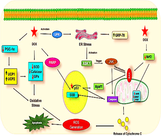
The anti-tumor activity of doxorubicin (DOX) is accompanied by significant off-target toxicity, particularly in the testes, where it promotes oxidative stress and suppresses antioxidant defense systems . DOX generates reactive oxygen species (ROS) through two principal mechanisms. In the first step, DOX undergoes one-electron reduction by several NADPH-dependent reductases, forming a semiquinone radical. In the presence of oxygen, this intermediate participates in redox cycling, producing superoxide anions (O₂⁻) and other ROS. The second mechanism involves a non-enzymatic, iron-dependent process in which DOX forms a ferrous–DOX radical complex. This complex reduces molecular oxygen to generate hydrogen peroxide (H₂O₂) and other reactive oxygen intermediates, amplifying oxidative damage (Figure 2).
The oxidative stress induced by DOX disrupts multiple cellular signaling pathways. In the testes, DOX interferes with the mTOR signaling cascade, leading to reduced ATP synthesis and impaired spermatogenesis and sperm motility. Clinical and experimental data show increased levels of oxidative and apoptotic markers, including p53 and Bax/Bcl-2, following DOX exposure (Alafif et al., 2022; ; . Dysregulation of steroidogenic enzymes is also evident: Increased StAR (steroidogenic acute regulatory) protein expression and decreased aromatase (P450arom) levels contribute to reduced estrogen biosynthesis and lower progesterone and pregnenolone levels, collectively impairing steroid hormone balance.
Experimental findings further support these biochemical alterations. Intraperitoneal administration of DOX (7.5 mg/kg) to sexually mature male Wistar rats led to elevated levels of reactive oxygen and nitrogen species (ROS/RNS), H₂O₂, and lipid peroxidation (LPO), along with altered activities of testicular marker enzymes such as glucose-6-phosphate dehydrogenase (G6PD), lactate dehydrogenase (LDH), acid phosphatase (ACP), and alkaline phosphatase (ALP) . These biochemical disturbances were accompanied by marked reductions in sperm motility, daily sperm production, and sperm count, as well as an increase in sperm morphological abnormalities.
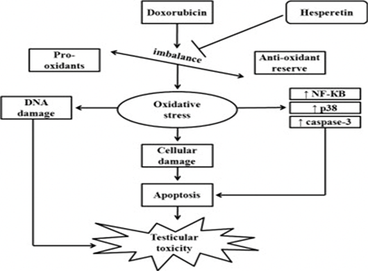
At the molecular level, DOX-induced oxidative stress triggers apoptosis via NF-κB activation, accompanied by upregulation of p38, p53, and caspase-3, and suppression of the anti-apoptotic protein Bcl-2 ; . Sperm motility progressively declines after the seventh day of DOX treatment and continues to deteriorate through day 28, likely due to poly (ADP-ribose) polymerase-1 (PARP-1) activation, which increases ATP consumption and induces energy depletion .
DOX also exerts direct cytotoxic effects on testicular cell populations. In vitro studies demonstrate dose-dependent G₂/M cell cycle arrest in spermatogonia, Leydig (TM3), and Sertoli (TM4) cells . Exposure of immature Sertoli cells (Ser-W3) to DOX results in oxidative stress and a reduction in intracellular glutathione (GSH) levels; however, this depletion does not significantly affect Ser-W3 cell survival . DOX-induced DNA damage is further evidenced by elevated expression of p53 and Ku70, and activation of PARP-1, which mediates caspase-independent cell death ; .
Recent evidence suggests that DOX alters mTOR complex signaling, decreasing mTORC2 expression while upregulating mTORC1, thereby disturbing the balance between these complexes. Such dysregulation results in abnormal spermatogonial maturation and impaired testicular function . The decline in serum sex hormone levels, particularly testosterone, correlates strongly with reduced sperm count and impaired spermatogenesis, as testosterone is a principal regulator of germ cell development . In addition, DOX-induced loss of epididymal adipose tissue may further contribute to suppressed spermatogenesis .
A growing body of evidence also highlights the interplay between oxidative stress and endoplasmic reticulum (ER) stress in DOX toxicity . DOX induces ER stress, marked by elevated expression of glucose-regulated protein 78 (GRP78) and activation of the unfolded protein response (UPR), which, when sustained, culminates in cellular apoptosis (Son et al., 2016). Collectively, these molecular and biochemical disturbances underline the multifactorial nature of DOX-induced testicular toxicity involving oxidative damage, apoptosis, metabolic disruption, and ER stress–mediated cell death.
Inflammation
DOX therapy has been reported to upregulate several inflammatory mediators, including inducible nitric oxide synthase (iNOS), interleukin-1β (IL-1β), matrix metalloproteinase-9 (MMP-9), and tumor necrosis factor-α (TNF-α) in treated rats . Moreover, DOX-induced reactive oxygen species (ROS) enhance the expression of the pro-inflammatory transcription factor nuclear factor kappa B (NF-κB) . The resulting oxidative stress promotes TNF-α release, which activates multiple signaling cascades, including the NF-κB inflammatory pathway.
DOX-induced oxidative stress also contributes to pyroptosis, an inflammatory form of programmed cell death characterized by DNA damage, release of inflammatory cytokines, and rapid plasma membrane rupture . This process is mediated by gasdermin family proteins, which are cleaved by caspase-1 and caspase-4/5/11 to form pore-generating N-terminal fragments . Central to this process is the NLRP3 inflammasome, a multiprotein complex comprising the NOD-like receptor family pyrin domain-containing 3 (NLRP3), the adaptor protein ASC (apoptosis-associated speck-like protein containing a caspase recruitment domain), and procaspase-1 . Upon activation, the NLRP3 inflammasome recruits and activates caspase-1, which then cleaves pro-IL-1β and pro-IL-18 into their mature forms, leading to pyroptosis and inflammatory amplification. ROS play a critical role in inflammasome activation, as blocking ROS generation with chemical scavengers has been shown to attenuate NLRP3 activation .
Moreover, reactive oxygen species (ROS) have been shown to play a pivotal role in the activation of the NLR pyrin domain-containing 3 (NLRP3) inflammasome, as inhibition of ROS production by chemical scavengers effectively prevents NLRP3 activation. When exposed to various cellular stressors or activators, multimolecular complexes known as inflammasomes are assembled within the cytoplasm . The activation of the NLRP3 inflammasome causes procaspase-1 to cleave and create active caspase-1, which results in the release of IL-1β and IL-18 (Kayagaki et al., 2011) and lead to a rapid inflammatory form of cell death called “pyroptosis” . Among these, the NLRP3 inflammasome is the most extensively characterized and consists of three core components: the sensor molecule NLRP3, the adaptor protein ASC (apoptosis-associated speck-like protein containing a caspase recruitment domain), and the effector enzyme procaspase-1. Upon activation, procaspase-1 is cleaved to form active caspase-1, which subsequently processes pro-inflammatory cytokines such as pro-IL-1β and pro-IL-18, initiating inflammatory cell death and immune responses .
Disruption of blood-testis barrier (BTB)
The BTB is a specialized physical barrier that separates the seminiferous tubules from the systemic circulation. It is composed of the capillary endothelium, the basement membrane of the seminiferous tubules, and the tight junctions (TJs) between Sertoli cells. This highly organized structure regulates the microenvironment necessary for spermatogenesis by restricting the passage of molecules from the bloodstream into the seminiferous epithelium. Consequently, it protects developing germ cells from harmful blood-borne substances, antigens, and antibodies that could otherwise trigger autoimmune reactions and lead to infertility .
DOX has been shown to impair BTB integrity by interfering with key signaling pathways and junctional proteins. According to Gurel et al. (2019), DOX inhibits the expression of the mammalian target of rapamycin (mTOR), a critical regulator of BTB dynamics and spermatogenesis. Within the testes, mTORC1 plays a vital role in spermatogonial mitosis and regulates BTB restructuring by inducing matrix metalloproteinase-9 (MMP-9) production in Sertoli cells. MMP-9, a collagenase enzyme, degrades adhesion complex proteins, thereby facilitating BTB remodeling . In contrast, mTORC2 supports BTB stability by upregulating connexin 43 (Cx43), a gap junction protein essential for intercellular communication between Sertoli cells and germ cells . DOX exposure first induces excessive ROS generation and mitochondrial injury, leading to reduced cellular ATP levels and activation of AMPK, which in turn negatively regulates mTOR signaling. This suppression of mTOR contributes to impaired BTB integrity through the downregulation of connexin-43 (Cx43) and increased MMP-9 activity, promoting tight junction disassembly.
DOX treatment disrupts this delicate balance by enhancing MMP-9 expression while downregulating Cx43, resulting in impaired gap junctions and BTB dysfunction . Furthermore, aberrant expression and mislocalization of claudin-11, a key tight junction protein, have been associated with defective spermatogenesis . Collectively, these findings indicate that DOX-induced alterations in mTOR signaling and junctional proteins compromise BTB integrity, contributing to testicular dysfunction and male infertility.
Autophagy
Autophagy, a vital cellular degradation and recycling process, has been increasingly implicated in DOX-induced organ toxicity . Several studies have demonstrated that DOX can activate autophagy-related genes, contributing to its cytotoxic effects . In testicular tissue, excessive generation of reactive oxygen species (ROS) following DOX administration has been shown to induce autophagy, linking oxidative stress to autophagic dysregulation . However, when autophagy becomes excessive or aberrant, it may contribute to impaired steroidogenesis and decreased testosterone production .
Autophagy and apoptosis are closely interconnected cellular processes, and DOX has been shown to influence both pathways through several overlapping mechanisms . Dysregulated autophagy has been associated with DOX-induced multi-organ toxicity, suggesting that aberrant activation of autophagic signaling may exacerbate tissue damage . In the testes, DOX-induced disturbances in the mTOR/Beclin-1 axis appear to play a key role in mediating reproductive toxicity. Experimental studies have demonstrated that DOX exposure increases Beclin-1 activity while inhibiting the mammalian target of rapamycin (mTOR), a central regulator of cell growth, metabolism, and redox homeostasis ; Hasan et al., 2021; .
Inhibition of mTOR signaling disrupts cellular metabolism and autophagy balance, leading to oxidative stress, lipid peroxidation, protein oxidation, and mitochondrial dysfunction in testicular cells. These alterations collectively result in sperm DNA fragmentation, apoptosis, and structural damage to the blood–testis barrier, ultimately impairing spermatogenesis and fertility . Moreover, studies have linked defective autophagy genes to mitochondrial abnormalities and increased apoptotic signaling in germ cells .
Autophagy can cause cell death either alone if apoptosis is unsuccessful or in combination with it . The spermatogenic or endocrine processes may be impacted by the autophagic and apoptotic pathways in response to thechemo therapy stimuli. Since EDCs disrupt the body’s endocrine system, special attention has been given to them ; .
Collectively, these findings indicate that DOX-induced testicular injury involves a complex interplay between oxidative stress, autophagy, apoptosis, and inflammation. Modulating the mTOR/Beclin-1 signaling pathway may therefore represent a promising therapeutic strategy to mitigate DOX-induced reproductive toxicity (Lv et al., 2021).
Apoptosis
Apoptosis, or programmed cell death, is a key mechanism underlying DOX-induced germ cell loss, primarily driven by DNA damage and oxidative stress. The spermatogonia, due to their high mitotic activity, are particularly vulnerable to free radical-induced DNA alterations compared with spermatocytes . Among the key molecular regulators of apoptosis, p53 plays a pivotal role in tumor suppression, DNA repair, and cell cycle control. Expression of p53 mRNA and protein in primary spermatocytes during spermatogenesis suggests its involvement in meiotic regulation .
DOX activates apoptosis through several interconnected signaling pathways, including oxidative stress, tumor necrosis factor-α (TNF-α), p53, and the PI3K/Akt axis . In particular, p53-mediated apoptosis involves activation of the Apaf-1/caspase-9 intrinsic pathway, where mitochondrial release of cytochrome c triggers caspase-9 activation, subsequently initiating downstream caspase cascades that execute cell death.
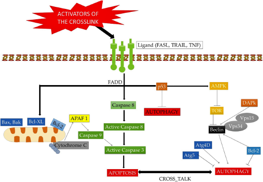
Autophagy and apoptosis are often interrelated processes, both influencing spermatogenic and endocrine functions under pathological or toxic conditions (Figure 3). Notably, environmental endocrine-disrupting chemicals (EDCs) widespread in the environmenthave been shown to impair testicular function and hormonal balance, further aggravating reproductive toxicity ; ; .
The Bcl-2 protein family serves as a central regulator of apoptosis, balancing pro-apoptotic (e.g., Bax) and anti-apoptotic (e.g., Bcl-2) signaling ; Bunz et al., 1998). Activation of caspase-3 marks the final execution phase of apoptosis . Consistent with this mechanism, DOX has been shown to upregulate pro-apoptotic genes such as p53, Bax, and caspase-3, while downregulating anti-apoptotic Bcl-2 expression, leading to testicular apoptosis and impaired spermatogenesis (Yeh et al., 2008; ; .
Treatment strategies for DOX-induced testicular damage
Despite numerous investigations, the therapeutic management of doxorubicin (DOX)-induced testicular damage remains largely ineffective. Exogenous follicle-stimulating hormone (FSH) administration has been reported to enhance spermatogenesis following low-dose DOX exposure . Additionally, co-treatment with selenium and thiocyanoacetamide demonstrated a protective effect against DOX-induced testicular toxicity in murine models . Furthermore, several plant-derived bioactive compounds and small molecules have shown promise in alleviating DOX-induced testicular injury by attenuating oxidative stress, inflammation, and apoptosis, thereby preserving spermatogenic function and hormonal balance. In recent years, nanotechnology-based approaches have also gained significant attention as innovative strategies to mitigate DOX-induced toxicity. Nanocarrier systems such as liposomes, polymeric nanoparticles, and metal-based nanostructures offer improved drug delivery, targeted release, and reduced off target accumulation of DOX, thereby minimizing its deleterious effects on testicular tissue.
Quercetin
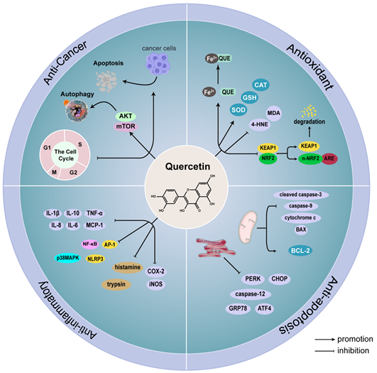
According to Cazarolli et al. (2008), flavonoids are plant-derived polyphenolic compounds that exhibit a wide range of biological activities, including anti-inflammatory, antiallergic, antiviral, antibacterial, antitumoral, and antioxidant effects. Among them, quercetin (QUE; 3,3′,4′,5,7-pentahydroxyflavone) is one of the most abundant and potent flavonoid antioxidants (Figure 4) . Quercetin supplementation has been shown to protect against DOX-induced testicular toxicity by reducing apoptotic cell counts and ameliorating histopathological alterations . In various studies worldwide, flavonoids have been extensively investigated as potential therapeutic agents for managing male reproductive dysfunction (Farombi et al., 2021). Notably, In rats, quercetin was administered via oral gavage for 10 days, with a single dose of DOX given intraperitoneally on day 6. to restore testosterone levels and testicular function by inhibiting the enzyme responsible for converting testosterone into testosterone glucuronide, thereby preserving androgenic activity .
Hesperetin (HST), a naturally occurring citrus flavonoid, exhibits a wide spectrum of pharmacological activities, including anti-inflammatory, anti-carcinogenic, antihypertensive, anti-atherogenic, and antioxidant effects ; . HST enhances the cellular antioxidant defense system primarily through its potent free radical–scavenging activity, thereby mitigating oxidative damage and preserving redox homeostasis . Notably, Trivedi et al. (2011) reported that rat Rats received DOX (4 mg/kg) weekly for five weeks. Concurrently, they were treated with hesperetin (25, 50, or 100 mg/kg) for five days per week over the same period.significantly attenuated doxorubicin (DOX)-induced oxidative stress in rat testes, suggesting its potential protective role against DOX-mediated gonadotoxicity (Figure 5).
Propolis
Propolis, also known as bee glue or bee gum, is a resinous substance collected and deposited by various bee species. Due to its rich composition of flavonoids, esters, and phenolic compounds, propolis exhibits potent antioxidant and therapeutic properties ; . Administration of propolis in combination with DOX, with a weekly injection of 4 mg/kg DOX and daily oral dose of 30 mg/kg propolis using a stomach tube, significantly improved the seminiferous tubule architecture and restored germ cell populations, with histological features closely resembling those of the control group. These findings indicate that the antioxidant capacity of propolis plays a vital role in mitigating DOX-induced testicular toxicity. In addition to its antioxidant action, propolis possesses anti-inflammatory, antibacterial, antifungal, anticancer, anti-ulcer, tonic, and immunomodulatory properties, supporting its potential therapeutic application against a wide spectrum of pathological conditions ; ; .
Silymarin
Silymarin, commonly known as milk thistle, is a medicinal plant extract derived from the seeds of Silybum marianum (L.) Gaertn. . Silymarin exerts its antioxidant effects by directly scavenging reactive oxygen species (ROS) and enhancing the activity of endogenous antioxidant enzymes. Several studies have demonstrated the protective role of silymarin against DOX-induced testicular toxicity. Co-administration of silymarin has been reported to significantly improve sperm count, daily sperm production, spermatid head count, spermatogenesis quality, and serum testosterone levels, while reducing malondialdehyde (MDA) formation, a marker of lipid peroxidation (Janbaz et al., 2012; .
Moreover, Shafiei-Roudbari et al. (2017) found that administration of silymarin (50 mg/kg/day for four weeks) concurrently with DOX markedly reduced sperm abnormalities, DNA fragmentation, and nitric oxide levels induced by DOX (cumulative dose 15 mg/kg). This treatment also significantly increased total antioxidant capacity, sperm motility, and viability, while downregulating p53 mRNA expression, indicating attenuation of DOX-induced apoptosis. Collectively, these findings highlight silymarin’s potent antioxidant and anti-apoptotic properties, supporting its use as a promising adjuvant therapy to protect against DOX-induced reproductive toxicity.
Among the natural antioxidants reviewed, quercetin, hesperetin, propolis, and silymarin exhibit the most consistent and compelling evidence for mitigating doxorubicin (DOX)-induced testicular toxicity in preclinical studies. Many positive animal studies use high oral or parenteral doses that produce tissue exposures not achievable with standard human oral dosing; first-pass metabolism and rapid phase II conjugation (esp. for quercetin) mean plasma/tissue exposure in humans will be substantially lower than in rodents given the same dose. Thus, efficacy in animals may reflect supra-physiological exposures or active metabolites not replicated clinically. Nanotechnological delivery systems offer a solution by enhancing solubility, protecting against rapid metabolism, and enabling targeted or controlled release. For instance, quercetin-loaded nanoparticles exhibit superior protection against DOX-induced toxicity compared to free quercetin by enhancing intracellular uptake, prolonging circulation time, and reducing oxidative and apoptotic markers ; . Similarly, silymarin-loaded nanoliposomes showed greater restoration of spermatogenic cell architecture and antioxidant enzyme activity than unformulated silymarin . Curcumin and resveratrol nanocarriers have likewise demonstrated improved mitigation of ROS and inflammatory responses in gonadal and hepatic models ; . The conceptual framework of this review, therefore, proposes co-delivery of antioxidants via targeted delivery of antioxidants to the testes for example, using ligand-directed (peptide/aptamer/antibody) or BTB-transcytosis focused nanoparticles combined with stimulus-responsive release (e.g., ultrasound-triggered shells) is intended to concentrate protective drug exposure in the testis while minimizing systemic and tumor exposure.
Zinc oxide nanoparticles
Zinc is recognized as a protective antioxidant due to its ability to restore malondialdehyde (MDA) levels toward normal physiological values ; . Raajshreer and Durairaj (2017) demonstrated that synthesized zinc oxide nanoparticles (ZnO NPs) exhibit notable antioxidant activity, functioning through both free radical scavenging and reducing mechanisms. Similarly, El-Maddawy and Naby (2019) reported that DOX induced oxidative damage and toxicity in testicular and sperm tissues, while administration of ZnO NPs (3 mg/kg body weight) concurrently with DOX significantly attenuated this toxicity, confirming their antioxidant potential.
Furthermore, Zhao et al. (2014) found that an optimal concentration of nano-ZnO enhances Cu-Zn superoxide dismutase (SOD) activity, thereby reducing reactive oxygen species (ROS) generation.
Different oxidant/antioxidant responses were observed in testicular homogenates treated with ZnO NPs of varying particle sizes. Treatment with small-sized ZnO NPs markedly increased SOD activity relative to controls, reflecting an adaptive antioxidant response to elevated oxidative stress. Conversely, large-sized ZnO NPs significantly reduced MDA levels, indicating protection against lipid peroxidation and potential stabilization of cell membrane integrity.
Previous studies have reported inconsistent findings regarding the effects of ZnO NPs on testicular oxidative stress. Some investigations revealed that ZnO NPs decrease oxidative stress and enhance antioxidant defense systems ; .
Selenium nanoparticles
Selenium (Se) has been shown to play a crucial protective role against DOX-induced gonadotoxicity. According to Boussada et al. (2017), selenium administration reduces oxidative stress and associated apoptosis in male rats, thereby mitigating DOX-induced testicular injury. These findings emphasize the importance of Se in male reproductive function and its potential to counteract chemotherapy-induced reproductive toxicity.
Previous studies demonstrated that selenium nanoparticles (SeNPs) exert superior biological activity compared to inorganic Se. Abd-Allah and Hashem (2015) reported that SeNPs enhanced sperm concentration, motility, and viability, while also elevating testicular antioxidant enzyme activity. Additionally, SeNPs have shown protective efficacy against testicular injury caused by environmental pollutants and the anticancer drug cisplatin .
In DOX-treated rats, SeNP administration significantly decreased testicular germ cell apoptosis, evidenced by the downregulation of p53, Bax, and caspase-3 proteins (p < 0.05) and upregulation of the anti-apoptotic Bcl-2 protein (p < 0.05). This anti-apoptotic effect aligns with findings by Zhang et al. (2019), who observed that SeNPs attenuated NiSO₄-induced testicular damage by modulating the expression of pro- and anti-apoptotic genes.
Moreover, co-administration of DOX and SeNPs markedly activated the Nrf2/HO-1 signaling pathway, consistent with previous findings by Yuan et al. (2022), who demonstrated that SeNP treatment upregulated Nrf2/HO-1, thereby inhibiting neuronal apoptosis and conferring neuroprotection against oxidative injury. Collectively, these results suggest that rats receiving SeNPs at dose of 0.5 mg/kg/day of SeNPs by oral gavage for four weeks with 3 mg/kg/week IP of DOX protect against DOX-induced testicular toxicity through antioxidant and anti-apoptotic mechanisms, particularly via the Nrf2/HO-1 signaling pathway .
Silver nanoparticles loaded with oleuropein
The previous findings demonstrated that DOX administration significantly increased the expression of NF-κB p65, IL-1β, and TNF-α, indicating a strong pro-inflammatory response. In contrast, treatment with olive leaf extract (OLE) or OLE combined with silver nanoparticles (OLE+AgNPs) markedly reduced the expression of these inflammatory mediators, thereby attenuating DOX-induced inflammation. Additionally, DOX exposure significantly elevated caspase-3 expression and reduced Bcl-2 levels in testicular tissue, consistent with enhanced apoptotic activity. Conversely, rats given Doxorubicin+Silver Nanoparticle: 2.5 mg/kg i.p. every 2 days + Silver Nanoparticle 100 mg/ kg via gavage for 11 days showed effective inhibition of apoptosis by upregulating Bcl-2 and downregulating caspase-3 expression. These findings support previous evidence that OLE and AgNPs exert potent anti-inflammatory and anti-apoptotic effects across various tissues . While nanoparticles have been reported to modulate oxidative stress markers in testicular tissue, their mechanisms of action appear to be primarily systemic rather than through active testis-specific targeting.
CONCLUSION AND RECOMMENDATIONS
Despite its strong antitumor effects, DOX severely damages the testicles by causing oxidative stress, inflammation, mitochondrial malfunction, and apoptosis. This ultimately hinders the generation of testosterone and spermatogenesis. Natural antioxidants such as quercetin, hesperetin, propolis, silymarin, and selenium nanoparticles have demonstrated protective effects by modulating oxidative, inflammatory, and apoptotic pathways. Recent developments in nanotechnology have reduced systemic toxicity while improving these drugs’ tailored delivery and effectiveness. Collectively, these findings underscore the potential of antioxidant- and nanotechnology-based interventions in mitigating DOX-induced testicular damage. To maintain male reproductive health without compromising DOX’s anticancer effectiveness, future research should concentrate on translational approaches that integrate these treatment modalities. While animal models have shown promise, it is crucial to recognize their inherent limitations in their ability to accurately represent the intricacy of human reproductive physiology. The influence of doxorubicin-induced damage and the effectiveness of antioxidant or nanotechnological treatments may be influenced by species-specific variations in testicular microarchitecture, redox balance, and drug metabolism. Future research should therefore use sophisticated human-relevant systems, such as testicular organoids, ex vivo tissue cultures, and microfluidic models, and perform pharmacokinetic and safety assessments in higher-order species in order to close the translational gap prior to clinical translation. Also, Future investigations should focus on confirming that antioxidant and nanomedicine-based strategies do not compromise DOX’s antitumor efficacy. This requires integrated experimental designs that simultaneously evaluate tumor response, reproductive outcomes, and systemic pharmacokinetics. Comparative in vivo studies using tumor-bearing models, along with biodistribution and cytotoxicity analyses, will be essential to ensure that testicular protection is achieved without diminishing the chemotherapeutic potency of DOX.
ACKNOWLEDGEMENT
The present study was supported by Zagazig University, College of Veterinary Medicine, for which the researchers are grateful.
NOVELTY STATEMENT
This work uniquely bridges antioxidant pharmacology with nanomedicine, highlighting translational strategies to preserve male reproductive function during chemotherapy in both human and veterinary medicine..
AUTHOR’s CONTRIBUTION
Mahran Mohamed Abd El-Emam: Conceptualization, supervision, writing review and editing. Aya Elhady Atia: writing original draft preparation. Hussein I Elbelbesy: supervision. All authors have read and agreed to the published version of the manuscript.
Generative AI and AI-assisted technology statement
No generative artificial intelligence (AI) or AI-assisted tools were used
Conflicts of interest
The authors have declared no conflicts of interest.
Overall events leading to male reproductive toxicity by Doxorubicin adapted from Mohan et al. 2021, with permission from Elsevier.
Figure 3:.
Crosstalk between autophagy and apoptosis (Rotimi and Singh, 2022). The figure illustrates shared regulatory molecules such as Bcl-2 and p53, highlighting how cellular stress can shift the balance toward either survival (autophagy) or cell death (apoptosis).
Figure 4:.
The primary biological functions and mechanisms of quercetin (QUE).
Figure 5:.
Proposed pathway illustrating the protective mechanism of hesperetin against DOX-induced testicular toxicity, adapted from Trivedi et al., 2011, with permission from Elsevier.
References
Abd El-Gany, A.M., Rasha, M.N., Bakry, N., Shimaa, B. 2016; Amelioration of cisplatin-induced testicular injury by different garlic preparations in experimental rats. World J. Pharm. Res., 5(9): 160–181.
Abd-Allah, S., Hashem, K.S. 2015; Selenium nanoparticles increase testicular antioxidant activity and spermatogenesis in male rats as compared to ordinary selenium. International Journal of Advanced Research, 3: 792–802.
Abdel Ghfar, S.S., Ali, M.E., Momenah, M.A., Al-Saeed, F.A., Al-Doaiss, A.A., Mostafa, Y.S., Ahmed, A.E., Abdelrahman, M. 2022; Effect of Allium sativum and Nigella sativa on alleviating aluminum toxicity state in albino rats. Frontiers in Veterinary Science, 9: 1042640. https://doi.org/10.3389/fvets.2022.1042640[CrossRef]
Abdel-Megeed, R.M., Abdel-Hamid, A.H.Z., Kadry, M.O. 2024; Titanium dioxide nanostructure-loaded Adriamycin surmounts resistance in breast cancer therapy: ABCA/P53/C-myc crosstalk. Future Science OA, 10(1): FSO979. https://doi.org/10.2144/fsoa-2023-0107[CrossRef]
Abidli, N., Ghaly, I.S., Hassanane, M.M., Ahmed, E.S., Khalil, W.K.B. 2015; Myrtus species prevents reproductive toxicity induced by doxorubicin in male mice. Asian Journal of Pharmaceutical and Clinical Research, 8(3): 169–175.
Ahmed, R.F., Moussa, R.A., Eldemerdash, R.S., Zakaria, M.M., Abdel-Gaber, S.A. 2019; Ameliorative effects of silymarin on HCl-induced acute lung injury in rats: role of the Nrf2/HO-1 pathway. Iranian Journal of Basic Medical Sciences, 22(12): 1483–1492.
Ahmed, Z.A., Abtar, A.N., Othman, H.H., Aziz, T.A. 2019; Effects of quercetin, sitagliptin alone or in combination in testicular toxicity induced by doxorubicin in rats. Drug Design, Development and Therapy, 13: 3321–3329. https://doi.org/10.2147/DDDT.S222127[CrossRef]
Akinjo, O.O., Gant, T.W., Marczylo, E.L. 2018; Perturbation of microRNA signalling by doxorubicin in spermatogonial, Leydig and Sertoli cell lines in vitro. Toxicol. Res. (Camb)., 7: 639–648. https://doi.org/10.1039/C7TX00314E[CrossRef]
Aksu, E.H., et al. 2019; Palliative effect of curcumin on doxorubicin-induced testicular damage in male rats. J. Biochem. Mol. Toxicol., 33(10): e22384. https://doi.org/10.1002/jbt.22384[CrossRef]
Alafifi, S., Wahdan, S., Elsherbiny, D., Azab, S.S. 2022; Doxorubicin-induced testicular toxicity: possible underlying mechanisms and promising pharmacological treatments in experimental models. Arch. Pharm. Sci. Ain Shams Univ., 6: 196–207. https://doi.org/10.21608/aps.2022.155127.1098[CrossRef]
Ali, M.E., Abd-Elkariem, M.F.A., Fahmy, S., Hussein, H.A., Abdelrahman, M., Ahmed, A.E. 2023; Dietary supplementation with different nano and organic selenium doses improves ovarian activity, fertility rate, and progesterone level in Ossimi ewes. Italian Journal of Animal Science, 22(1): 651–659. https://doi.org/10.1080/1828051X.2023.2231946[CrossRef]
Alrushaid, S., Sayre, C.L., Yáñez, J.A., Forrest, M.L., Senadheera, S.N., Burczynski, F.J., Löbenberg, R., Davies, N.M. 2017; Pharmacokinetic and toxicodynamic characterization of a novel doxorubicin derivative. Pharmaceutics, 9: 35. https://doi.org/10.3390/pharmaceutics9030035[CrossRef]
Alyane, M., Kebsa, L.B.W., Boussenane, H., Rouibah, H., Lahouel, M. 2008; Cardioprotective effects and mechanism of action of polyphenols extracted from propolis against doxorubicin toxicity. Pakistan Journal of Pharmaceutical Sciences, 21(3): 201–209.
Babalola, A.A., Adelowo, A.R., Da-Silva, O.F., Ikeji, C.N., Owoeye, O., Rocha, J.B.T., Adedara, I.A., Farombi, E.O. 2023; Attenuation of doxorubicin-induced hypothalamic-pituitary-testicular axis dysfunction by diphenyl diselenide involves suppression of hormonal deficits, oxido-inflammatory stress, and caspase-3 activity in rats. J. Trace Elem. Med. Biol., 79: 127254. https://doi.org/10.1016/j.jtemb.2023.127254[CrossRef]
Bains, O.S., Grigliatti, T.A., Reid, R.E., Riggs, K.W. 2010; Naturally occurring variants of human aldo-keto reductases with reduced in vitro metabolism of daunorubicin and doxorubicin. J. Pharmacol. Exp. Ther., 335: 533–545. https://doi.org/10.1124/jpet.110.173179[CrossRef]
Baokbah, T.A. 2023; Protective effects of selenium nanoparticles against doxorubicin-induced testicular apoptosis in rats. Medical Science, 27: e188ms2975. https://doi.org/10.54905/disssi/v27i134/e188ms2975[CrossRef]
Barber LG, Sorenmo KU, Cronin KL, Shofer FS. 2000, Combined doxorubicin and cyclophosphamide chemotherapy for nonresectable feline fibrosarcoma. J. Am. Anim. Hosp. Assoc., 36: 416–421. https://doi.org/10.5326/15473317-36-5-416[CrossRef]
Begleiter, A., Leith, M. 1990; Activity of quinine alkylating agents in quinine-resistant cells. Cancer Res., 50: 2872–2876.
Bharti, S., Misro, M.M., Rai, U. 2014; Quercetin supplementation restores testicular function and augments germ cell survival in the estrogenized rats. Molecular and Cellular Endocrinology, 383(1–2): 10–20. https://doi.org/10.1016/j.mce.2013.11.021[CrossRef]
Boussada, M., Ali, R.B., Said, A.B., Bokri, K., Akacha, A.B., Dziri, C., El-May, M.V. 2017; Selenium and a newly synthesized thiocyanoacetamide reduce doxorubicin gonadotoxicity in male rats. Biomedicine & Pharmacotherapy, 89: 1005–1017. https://doi.org/10.1016/j.biopha.2017.03.002[CrossRef]
Boussada, M., Dias, T.R., Crisóstomo, L., Akacha, A.B., Ali, R.B., Michèle, V. et al. 2019, A new thiocyanoacetamide (2-cyano-2-p-nitrophenyl-N-benzylthioamide) reduces doxorubicin-induced in vitro toxicity in Sertoli cells by decreasing apoptosis and autophagy. Theriogenology. 140, 188–200. https://doi.org/10.1016/j.theriogenology.2019.08.030[CrossRef]
Breslow, N., Ou, S., Beckwith, M., Haase, G., Kalapurakal, J., Ritchey, M., Shamberger, R., Thomas, P., D’Angio, G., Green, D. 2004; Doxorubicin for favorable histology, stage II–III Wilms tumor: results from the National Wilms Tumor Studies. Cancer, 101: 1072–1080. https://doi.org/10.1002/cncr.20433[CrossRef]
Bunz F, Dutriaux A, Lengauer C, Waldman T, Zhou S, Brown JP, Sedivy JM, Kinzler KW, Vogelstein B. 1998; Requirement for p53 and p21 to sustain G2 arrest after DNA damage. Science 282(5393):1497-501. https://doi.org/10.1126/science.282.5393.1497[CrossRef]
Burawat, J., Uabandit, N., Arun, S., Nualkaew, S., Iamsaard, S. 2018; Effects of Leucaena leucocephala (Lamk.) shoot tips plus young leaf extract containing mimosine on the reproductive system of male rats. International Journal of Morphology, 36(3): 956–963. https://doi.org/10.4067/S0717-95022018000301062[CrossRef]
Cardinale, D.; Colombo, A.; Bacchiani, G.; Tedeschi, I.; Meroni, C.A.; Veglia, F.; Civelli, M.; Lamantia, G.; Colombo, N.; Curigliano, G.; et al. 2015, Early detection of anthracycline cardiotoxicity and improvement with heart failure therapy. Circulation 131, 1981–1988. https://doi.org/10.1161/CIRCULATIONAHA.114.013777[CrossRef]
Carvalho, C., Santos, R., Cardoso, S., Correia, S., Oliveira, P., Santos, M., Moreira, P. 2009; Doxorubicin: the good, the bad and the ugly effect. Curr. Med. Chem., 16: 3267–3285. https://doi.org/10.2174/092986709788803312[CrossRef]
Castaldo, S., Capasso, F. 2002; Propolis, an old remedy used in modern medicine. Fitoterapia, 73(Suppl. 1): S1–S6. https://doi.org/10.1016/S0367-326X(02)00185-5[CrossRef]
Cazarolli, L.H., Zanatta, L., Alberton, E.H. 2008; Flavonoids: prospective drug candidates. Mini Reviews in Medicinal Chemistry, 8(13): 1429–1440. https://doi.org/10.2174/138955708786369564[CrossRef]
Christidi, E., Brunham, L.R. 2021; Regulated cell death pathways in doxorubicin-induced cardiotoxicity. Cell Death and Disease, 12(4): 339. https://doi.org/10.1038/s41419-021-03614-x[CrossRef]
Chun R, Garrett LD, Vail DM. 2000; Evaluation of a high-dose chemotherapy protocol with no maintenance therapy for dogs with lymphoma. J Vet Intern Med 14: 120–124 .https://doi.org/10.1111/j.1939-1676.2000.tb02224.x[CrossRef]
Cui, Z., Zhao, X., Amevor, F.K., Du, X., Wang, Y., Li, D., Shu, G., Tian, Y., Zhao, X. 2022; Therapeutic application of quercetin in aging-related diseases: SIRT1 as a potential mechanism. Frontiers in Immunology, 13: 943321. https://doi.org/10.3389/fimmu.2022.943321[CrossRef]
Dani, V., Dhawan, D.K. 2005; Radioprotective role of zinc following single-dose radioiodine exposure to red blood cells of rats. Indian Journal of Medical Research, 122(4): 338–342.
Den Hond, E., Tournaye, H., De Sutter, P., Ombelet, W., Baeyens, W., Covaci, A., D’Hooghe, T. 2015; Human exposure to endocrine disrupting chemicals and fertility: a case–control study in male subfertility patients. Environment International, 84: 154–160. https://doi.org/10.1016/j.envint.2015.07.017[CrossRef]
Dowd, A.F.J., Johnson, B.S., Mariotti, A. 2016; Pharmacology and Therapeutics for Dentistry. 7th ed., Elsevier, pp. 530–562.
Dutta, D., Dutta, D., Xu, J., Dirain, M.L.S., 2014; Leeuwenburgh, C. Calorie restriction combined with resveratrol induces autophagy and protects 26-month-old rat hearts from doxorubicin-induced toxicity. Free Radical Biology and Medicine, 74: 252–262. https://doi.org/10.1016/j.freeradbiomed.2014.06.011[CrossRef]
Dutta, S., Sengupta, P., Slama, P., Roychoudhury, S. 2021; Oxidative stress, testicular inflammatory pathways, and male reproduction. International Journal of Molecular Sciences, 22(18): 10043. https://doi.org/10.3390/ijms221810043[CrossRef]
El-Maddawy, Z.K., Abd El Naby, W.S.H. 2019; Protective effects of zinc oxide nanoparticles against doxorubicin-induced testicular toxicity and DNA damage in male rats. Toxicology Research, 8(5): 654–662. https://doi.org/10.1039/c9tx00052f[CrossRef]
Erbaş, Elif, et al. 2024, Silver nanoparticles loaded with oleuropein reduce doxorubicin-induced testicular damage by regulating endoplasmic reticulum stress, and apoptosis. Biological Trace Element Research, 202.10: 4687-4698. https://doi.org/10.1007/s12011-024-04058-y[CrossRef]
Farombi, E.O., Adedara, I.A., Akinrinde, S.A., Ojo, O.O., Eboh, A.S. 2012; Protective effects of kolaviron and quercetin on cadmium-induced testicular damage and endocrine pathology in rats. Andrologia, 44: 273–284. https://doi.org/10.1111/j.1439-0272.2012.01279.x[CrossRef]
Galati, E.M., Trovato, A., Kirjavainen, S., Forestieri, A.M., Rossitto, A., Monforte, M.T. 1996; Biological effects of hesperidin, a citrus flavonoid: antihypertensive and diuretic activity in rats. Farmaco, 51(3): 219–221. https://doi.org/10.1016/S0944-7113(96)80046-8[CrossRef]
Garg, A., Garg, S., Zaneveld, L.J., Singla, A.K. 2001; Chemistry and pharmacology of the citrus bioflavonoid hesperidin. Phytotherapy Research, 15(8): 655–669. https://doi.org/10.1002/ptr.1074[CrossRef]
Garrett, L.D., Thamm, D.H., Chun, R., et al. 2002; Evaluation of a 6-month chemotherapy protocol with no maintenance therapy for dogs with lymphoma. J. Vet. Intern. Med., 16:704–709. https://doi.org/10.1111/j.1939-1676.2002.tb02411.x[CrossRef]
Goma, A.A., Tohamy, H.G., El-Kazaz, S.E., et al. 2020; Comparative study between zinc oxide nanoparticles and bulk zinc oxide on reproductive performance, antioxidant levels, gene expression, and histopathology of testes in male rats. Antioxidants, 10(1): 41. https://doi.org/10.3390/antiox10010041[CrossRef]
Gungor-Ordueri, N.E., Kuscu, N., Tasatargil, A., Burgucu, D., Karacan, M., Celik Ozenci, C. 2019; Doxorubicin-induced testicular damage is related to PARP-1 signaling molecules in mice. Pharmacol. Rep., 71(4): 591–602. https://doi.org/10.1016/j.pharep.2019.02.018[CrossRef]
Gurel, C., Kuscu, G.C., Buhur, A., Dagdeviren, M., Oltulu, F., Karabay Yavasoglu, N.U. et al. 2019, Fluvastatin attenuates doxorubicin-induced testicular toxicity in rats by reducing oxidative stress and regulating the blood–testis barrier via mTOR signaling pathway. Hum. Exp. Toxicol., 38, 1329–1343. https://doi.org/10.1177/0960327119862006[CrossRef]
Gurel, C., Kuscu, G.C., Buhur, A., Dagdeviren, M., Oltulu, F., Karabay Yavasoglu, N.U., Ozyurt, H., Gurel, E., Ozkan, Y. 2019; Fluvastatin attenuates doxorubicin-induced testicular toxicity in rats by reducing oxidative stress and regulating the blood–testis barrier via mTOR signaling pathway. Human and Experimental Toxicology, 38(12): 1329–1343. https://doi.org/10.1177/0960327119862006[CrossRef]
Hafez, H.M., Hassanein, H. 2022; Montelukast ameliorates doxorubicin-induced cardiotoxicity via modulation of P-glycoprotein and inhibition of ROS-mediated TNF/NF-κB pathways. Drug Chem. Toxicol., 45: 548–559. https://doi.org/10.1080/01480545.2020.1730885[CrossRef]
Hagiuda, J., Ishikawa, H., Kaneko, S., Okazaki, M., Oya, M., Nakagawa, K. 2015; Follicle-stimulating hormone enhances recovery from low-dose doxorubicin-induced spermatogenic disorders in mice. Journal of Assisted Reproduction and Genetics, 32(8): 1301–1310. https://doi.org/10.1007/s10815-015-0472-8[CrossRef]
Harada H, Grant S. 2003; Apoptosis regulators. Rev Clin Exp Hematol 7(2):117-138
Hasan, H., Bhushan, S., Fijak, M., Meinhardt, A. 2022; Mechanism of inflammatory associated impairment of sperm function, spermatogenesis and steroidogenesis. Frontiers in Endocrinology, 13: 897029. https://doi.org/10.3389/fendo.2022.897029[CrossRef]
Hosoya K, Kisseberth WC, Lord LK, et al. 2007; Comparison of COAP and UW-19 protocols for dogs with multicentric lymphoma. J. Vet. Intern. Med., 21:1355–1363. https://doi.org/10.1111/j.1939-1676.2007.tb01959.x[CrossRef]
Hosseini, S.M., Jafari, S.M., Ghorbani, M., Mirzaei, H. 2022; Silymarin-loaded nanoliposomes: Preparation, characterization, and evaluation of their potential for controlled release and improved bioavailability. Journal of Drug Delivery Science and Technology, 70: 103259. https://doi.org/10.1016/j.jddst.2022.103259[CrossRef]
Hussein, M.A., Ali, H.A., Saadeldin, I.M., et al. 2016; Quercetin alleviates zinc oxide nanoreprotoxicity in male albino rats. Journal of Biochemical and Molecular Toxicology, 30(10): 489–496. https://doi.org/10.1002/jbt.21812[CrossRef]
Jagetia, G.C., Venkatesh, P. 2015. The indigenous plant bael (Aegle marmelos (L.) Correa) extract protects against doxorubicin-induced cardiotoxicity in mice. Biochemistry and Physiology, 4(163): 2–9. https://doi.org/10.4172/2168-9652.1000163[CrossRef]
Jalali, A.S., Hasanzadeh, S. 2013; Crataegus monogyna fruit aqueous extract as a protective agent against doxorubicin-induced reproductive toxicity in male rats. Avicenna J. Phytomed., 3: 159–166.
Janbaz-Acyabar, H., Malekinejad, H., Sadrrkhanlou, R., Razi, M. 2012; Silymarin prevents and protects from doxorubicin-induced oxidative stress in testis and improves sperm quality in rats. International Journal of Fertility and Sterility, 6(Suppl. 1): 61.
Kabel, A.M. 2018; Zinc/alogliptin combination attenuates testicular toxicity induced by doxorubicin in rats: role of oxidative stress, apoptosis and TGF-β1/NF-κB signaling. Biomedicine & Pharmacotherapy, 97: 439–449. https://doi.org/10.1016/j.biopha.2017.10.144[CrossRef]
Kang, J., Lee, Y., No, K., Jung, E., Sung, J., Kim, Y., Nam, S. 2002; Ginseng intestinal metabolite-I (GIM-I) reduces doxorubicin toxicity in mouse testis. Reprod. Toxicol., 16(3): 291–298. https://doi.org/10.1016/S0890-6238(02)00021-7[CrossRef]
Kayagaki, N., Warming, S., Lamkanfi, M., Van Walle, L., Louie, S., Dong, J., Newton, K., Qu, Y., Liu, J., Heldens, S., Zhang, J. Non-canonical inflammasome activation targets caspase-11. Nature, 2011; 479(7371): 117–121.
Khan, J., Deb, P.K., Priya, S., Medina, K.D., Devi, R., Walode, S.G., Rudrapal, M., Tekade, R.K. 2021; Doxorubicin-induced toxicity and its amelioration by nanostructured drug delivery systems: A review. Life Sciences, 279: 119685.
Kim, Y.C., Guan, K.L. 2015; mTOR: a pharmacologic target for autophagy regulation. Journal of Clinical Investigation, 125(1): 25–32. https://doi.org/10.1172/JCI73939[CrossRef]
Knez, J. 2013; Endocrine-disrupting chemicals and male reproductive health. Reproductive Biomedicine Online, 26(5): 440–448. https://doi.org/10.1016/j.rbmo.2013.02.005[CrossRef]
Kranti, V., Mahesh, V., Srinivas, P., Ganesh, Y., Godwin, A., Lahkar, M. 2013; Evaluation of the protective effect of silymarin on doxorubicin-induced chronic testicular toxicity in rats. International Journal of Pharmaceutical and Biological Sciences, 4(1): 473–484.
Kumar, S.S., Manna, K., Das, A. 2018; Tender coconut water attenuates heat stress-induced testicular damage through modulation of the NF-κB and Nrf2 pathways. Food & Function, 9(10): 5463–5479. https://doi.org/10.1039/C8FO01207E[CrossRef]
Lee, K.M., et al. 2012. Melatonin attenuates doxorubicin-induced testicular toxicity in rats. Andrologia, 44: 796–803. https://doi.org/10.1111/j.1439-0272.2011.01269.x[CrossRef]
Lee, S.J., Schover, L.R., Partridge, A.H., Patrizio, P., Wallace, W.H., Hagerty, K., Beck, L.N., Brennan, L.V., Oktay, K. 2006; American Society of Clinical Oncology recommendations on fertility preservation in cancer patients. J. Clin. Oncol., 24: 2917–2931. https://doi.org/10.1200/JCO.2006.06.5888[CrossRef]
Li, N., Cheng, C.Y. 2016; Mammalian target of rapamycin complex (mTOR) pathway modulates blood-testis barrier (BTB) function through F-actin organization and gap junction. Histology and Histopathology, 31(8): 901–910.
Li, N., Wang, C., Zhang, P., You, Y., Lin, Y., Wang, Y. 2020; Quercetin-loaded nanocarriers for improved anticancer therapy: A comprehensive review. International Journal of Nanomedicine, 15: 7491–7505. https://doi.org/10.2147/IJN.S239548[CrossRef]
Li, X., Guo, S., Zhu, C., Zhu, Q., Luo, F., Liu, W. 2018; Resveratrol-loaded solid lipid nanoparticles with enhanced solubility and controlled release: Preparation and in vitro and in vivo evaluation. Drug Delivery, 25(1): 112–124.
Li, Z., Mo, F., Wang, Y., Li, W., Chen, Y., Liu, J. et al. 2022, Enhancing Gasdermin-induced tumor pyroptosis through preventing ESCRT-dependent cell membrane repair augments antitumor immune response. Nat. Commun. 13, 6321. https://doi.org/10.1038/s41467-022-34036-8[CrossRef]
Liu, J., et al. 2008; ERKs/p53 signal transduction pathway is involved in doxorubicin-induced apoptosis in H9c2 cells and cardiomyocytes. Am. J. Physiol. Heart Circ. Physiol., 295(5): H1956–H1965. https://doi.org/10.1152/ajpheart.00407.2008[CrossRef]
Lockshin, R. A., & Zakeri, Z. 2004. Apoptosis, autophagy, and more. The International Journal of Biochemistry & Cell Biology, 36(12), 2405–2419. https://doi.org/10.1016/j.biocel.2004.04.011[CrossRef]
Luo, J.-L., Kamata, H., Karin, M. 2005; IKK/NF-κB signaling balancing life and death: a new approach to cancer therapy. J. Clin. Invest., 115: 2625–2632. https://doi.org/10.1172/JCI26322[CrossRef]
Lu, Y., Xu, S., Chen, H., He, M., Deng, Y., Cao, Z., Pi, H., Chen, C., Li, M., Ma, Q., Gao, P. CdSe/ZnS quantum dots induce hepatocyte pyroptosis and liver inflammation via NLRP3 inflammasome activation. Biomaterials, 2016; 90: 27–39.
Lv, C., Wang, X., Guo, Y., Yuan, S. 2020; Role of selective autophagy in spermatogenesis and male fertility. Cells, 9(11): 2523. https://doi.org/10.3390/cells9112523[CrossRef]
Ma, Y., Yang, L., Ma, J., Lu, L., Wang, X., Ren, J., Fan, Y., Wu, J., Li, W., Zheng, Q. 2017; Rutin attenuates doxorubicin-induced cardiotoxicity via regulating autophagy and apoptosis. Biochimica et Biophysica Acta (BBA) - Molecular Basis of Disease, 1863(8): 1904–1911. https://doi.org/10.1016/j.bbadis.2016.12.021[CrossRef]
Mackova. K., Da Cunha, M.G., Krofta, L., Albersen, M., Deprest, J. 2019; The importance of developing relevant animal models to assess existing and new materials. Curr Opin Urol. 29(4): 400-406. https://doi.org/10.1097/MOU.0000000000000625[CrossRef]
Malekirad, A.A., Oryan, S., Fani, A., Babapor, V., Hashemi, M., Baeeri, M. Bayrami, Z., Abdollahi, M. 2010, Study on clinical and biochemical toxicity biomarkers in a zinc-lead mine workers. Toxicology and Industrial Health, 26(6), 331–337. https://doi.org/10.1177/0748233710365697[CrossRef]
Mantawy, E.M., El-Bakly, W.M., Esmat, A., Badr, A.M., El-Demerdash, E. 2014; Chrysine alleviates acute doxorubicin cardiotoxicity in rats via suppression of oxidative stress, inflammation and apoptosis. Eur. J. Pharmacol., 728: 107–118. https://doi.org/10.1016/j.ejphar.2014.01.065[CrossRef]
Markowska, A., Antoszczak, M., Markowska, J., Huczyński, A. 2024; Gynotoxic effects of chemotherapy and potential protective mechanisms. Cancers, 16: 2288. https://doi.org/10.3390/cancers16122288[CrossRef]
Martinon, F., Burns, K., Tschopp, J. 2002, The inflammasome: A molecular platform triggering activation of inflammatory caspases and processing of pro–IL-β. Mol. Cell 10, 417–426. https://doi.org/10.1016/S1097-2765(02)00599-3[CrossRef]
Minotti, G., Menna, P., Salvatorelli, E., Cairo, G., Gianni, L. 2004; Anthracyclines: molecular advances and pharmacologic development in antitumor activity and cardiotoxicity. Pharmacol. Rev., 56: 185–229. https://doi.org/10.1124/pr.56.2.6[CrossRef]
Mohan, U.P., Tirupathi Pichiah, P.B., Iqbal, S.T.A., Arunachalam, S. 2021; Mechanisms of doxorubicin-mediated reproductive toxicity. A review. Reprod. Toxicol., 102: 80–89. https://doi.org/10.1016/j.reprotox.2021.04.003[CrossRef]
Mok, K.W., Mruk, D.D. Cheng, C.Y. 2014, rpS6 regulates blood–testis barrier dynamics through Akt-mediated effects on MMP-9. J. Cell Sci., 127(22):4870–4882. https://doi.org/10.1242/jcs.152231[CrossRef]
Molehin, O.R. 2020; Alleviation of doxorubicin-induced nephrotoxicity by Clerodendrum volubile leaf extract in Wistar rats: A preliminary study. Journal of Herbmed Pharmacology, 9(2): 138–144. https://doi.org/10.34172/jhp.2020.18[CrossRef]
Moreira, B.P., Oliveira, P.F., Alves, M.G. 2019; Molecular mechanisms controlled by mTOR in male reproductive system. International Journal of Molecular Sciences, 20(7): 1633. https://doi.org/10.3390/ijms20071633[CrossRef]
Murakami, A., Ashida, H., Terao, J. 2008; Multitargeted cancer prevention by quercetin. Cancer Letters, 269: 315–325. https://doi.org/10.1016/j.canlet.2008.03.046[CrossRef]
Mutsaers, A.J., Glickman, N.W., DeNicola, D.B., et al. 2002; Evaluation of treatment with doxorubicin and piroxicam or doxorubicin alone for multicentric lymphoma in dogs. J. Am. Vet. Med. Assoc., 220: 1813–1817. https://doi.org/10.2460/javma.2002.220.1813[CrossRef]
Nausheen, Q.N., Ali, S.A., Subur, K. Cardioprotective and antioxidant activity of onion’ (Allium cepa) leaves extract in doxorubicin-induced cardiotoxicity in rats. Annals of Experimental Biology, 2014; 2: 37–42.
Nowrouzi, F., Azadbakht, M., Kalehoei, E., Modarresi, M. 2019; Protective effect of Rosa canina extract against doxorubicin-induced testicular toxicity in mice. Brazilian Archives of Biology and Technology, 62: e19180656. https://doi.org/10.1590/1678-4324-2019180017[CrossRef]
Pasupuleti, V.R., Sammugam, L., Ramesh, N., Gan, S.H. 2017; Honey, propolis, and royal jelly: a comprehensive review of their biological actions and health benefits. Oxidative Medicine and Cellular Longevity, 2017: 1259510. https://doi.org/10.1155/2017/1259510[CrossRef]
Pawłowska, J., Tarasiuk, J., Wolf, C., Paine, M., Borowski, E. 2003; Differential ability of cytostatics from the anthraquinone group to generate free radicals in three enzymatic systems: NADH dehydrogenase, NADPH cytochrome P450 reductase, and xanthine oxidase. Oncol. Res., 13: 245–252. https://doi.org/10.3727/096504003108748294[CrossRef]
Pichiah, P.B.T., Sankarganesh, A., Kalaiselvi, S., Indirani, K., Kamalakkannan, S., SankarGanesh, D. et al. 2012, Adriamycin-induced spermatogenesis defect is due to the reduction in epididymal adipose tissue mass: a possible hypothesis. Med. Hypotheses, 78, 218–220. https://doi.org/10.1016/j.mehy.2011.10.027[CrossRef]
Pirker, R. 2020; Chemotherapy remains a cornerstone in the treatment of non-small cell lung cancer. Curr. Opin. Oncol., 32: 63–67. https://doi.org/10.1097/CCO.0000000000000592[CrossRef]
Prahalathan, C., Selvakumar, E., Varalakshmi, P. 2006; Lipoic acid modulates adriamycin-induced testicular toxicity. Reprod. Toxicol., 21(1): 54–59. https://doi.org/10.1016/j.reprotox.2005.07.002[CrossRef]
Raajshreer, K., Durairaj, B. 2017, Evaluation of The Antityrosinase and Antioxidant potential of Zinc Oxide Nanoparticles synthesized from the brown seaweed Turbinaria conoides. Int. J. Appl. Pharm., 9(5), 116–120, https://doi.org/10.22159/ijap.2017v9i5.20847[CrossRef]
Raajshreer, K., Durairaj, B. 2017; Evaluation of the antityrosinase and antioxidant potential of zinc oxide nanoparticles synthesized from the brown seaweed– Turbinaria conoides. International Journal of Applied Pharmaceutics, 9(5): 116–120. https://doi.org/10.22159/ijap.2017v9i5.20847[CrossRef]
Raimondo, S., Gentile, T., Gentile, M., Morelli, A., Donnarumma, F., Cuomo, F., Montano, L. 2019; P53 protein evaluation on spermatozoa DNA in fertile and infertile males. Journal of Human Reproductive Sciences, 12(2): 114–121. https://doi.org/10.4103/jhrs.JHRS_170_18[CrossRef]
Renu, K., Gopalakrishnan, A.V. 2019; Deciphering the molecular mechanism during doxorubicin-mediated oxidative stress, apoptosis through Nrf2 and PGC-1α in a rat testicular milieu. Reprod. Biol., 19(1): 22–37. https://doi.org/10.1016/j.repbio.2019.02.004[CrossRef]
Renu, K., Pureti, L.P., Vellingiri, B., Gopalakrishnan, A.V. 2022; Toxic effects and molecular mechanism of doxorubicin on different organs – an update. Toxin Rev., 41: 650–674. https://doi.org/10.1080/15569543.2021.1912099[CrossRef]
Renu, K., Valsala Gopalakrishnan, A. 2019; Deciphering the molecular mechanism during doxorubicin-mediated oxidative stress, apoptosis through Nrf2 and PGC-1α in a rat testicular milieu. Reproductive Biology, 19(1): 22–37. https://doi.org/10.1016/j.repbio.2019.02.004[CrossRef]
Rezvanfar, M.A., Rezvanfar, M.A., Shahverdi, A.R., Ahmadi, A., Baeeri, M., Mohammadirad, A., Abdollahi, M. 2013; Protection of cisplatin-induced spermatotoxicity, DNA damage and chromatin abnormality by selenium nanoparticles. Toxicology and Applied Pharmacology, 266(3): 356–365. https://doi.org/10.1016/j.taap.2012.11.025[CrossRef]
Rizk, S.M., Zaki, H.F., Mina, M.A. 2014; Propolis attenuates doxorubicin-induced testicular toxicity in rats. Food and Chemical Toxicology, 67: 176–186. https://doi.org/10.1016/j.fct.2014.02.031[CrossRef]
Rotimi, D.E., Singh, S.K. 2022; Interaction between apoptosis and autophagy in testicular function. Andrologia, 54(11): e14602. https://doi.org/10.1111/and.14602[CrossRef]
Sabeti, P., Pourmasumi, S., Rahiminia, T., Akyash, F., Talebi, A.R. 2016; Etiologies of sperm oxidative stress. International Journal of Reproductive Biomedicine, 14(4): 231–240. https://doi.org/10.29252/ijrm.14.4.231[CrossRef]
Safaei-Pourzamani, M., Oryan, S.H., Yaghmaei, P., Jalili, C., Ghanbari, A. 2022; Royal jelly alleviates side effects of doxorubicin on the male reproductive system: A mouse model simulating human chemotherapy cycles. Research Journal of Pharmacognosy, 9(1): 77–87.
Salvesen, G.S. 2002; Caspases and apoptosis. Essays in Biochemistry, 38: 9–19. https://doi.org/10.1042/bse0380009[CrossRef]
Sarker, S.D., Nahar, L., Miron, A., Guo, M. 2020. Anticancer Natural Products. 1st ed. Elsevier Inc.,
Schroder, K., Tschopp, J. 2010, The inflammasomes. Cell, 140, 821–832. https://doi.org/10.1016/j.cell.2010.01.040[CrossRef]
Shabir, I., Pandey, V.K., Shams, R., Dar, A.H., Dash, K.K., Khan, S.A., Bashir, I., Jeevarathinam, G., Rusu, A.V., Esatbeyoglu, T., et al. 2022; Promising bioactive properties of quercetin for potential food applications and health benefits: A review. Frontiers in Nutrition, 9: 999752. https://doi.org/10.3389/fnut.2022.999752[CrossRef]
Shafiei-Roudbari, S.K., Malekinejad, H., Janbaz-Acyabar, H., Razi, M. 2017; Crosstalk between E2F1 and p53 transcription factors in doxorubicin-induced DNA damage: Evidence for preventive/protective effects of silymarin. Journal of Pharmacy and Pharmacology, 69(9): 1116–1124. https://doi.org/10.1111/jphp.12745[CrossRef]
Shagirtha, K., Pari, L. 2011; Hesperetin, a citrus flavonone, protects against cadmium-induced oxidative testicular dysfunction in rats. Ecotoxicology and Environmental Safety, 74(7): 2105–2111. https://doi.org/10.1016/j.ecoenv.2011.06.002[CrossRef]
Sharma, R.A., Gescher, A.J., Steward, W.P. 2020; Curcumin: The story so far. Frontiers in Pharmacology, 11: 748.
Sikora, T., Morawska, K., Lisowski, W., Rytel, P., Dylong, A. 2022; Application of optical methods for determination of concentration of doxorubicin in blood and plasma. Pharmaceuticals, 15(2): 112. https://doi.org/10.3390/ph15020112[CrossRef]
Simon, K.L., Cook, M.R., Husbands, B.D., Long, M.E., Hokamp, J.A., Helms, T.H. 2022, Use of doxorubicin chemotherapy following debulking surgery in the treatment of a cat withmesenteric hemangiosarcoma. JFMS Open Rep., 8: 20551169221121900. https://doi.org/10.1177/20551169221121900[CrossRef]
Singh, S., Patel, S.K., Singh, S.K. Endocrine disrupting chemicals and male reproductive health. In: Haldar, C., et al. 2016. Updates on Integrative Physiology and Comparative Physiology, Volume II. Banaras Hindu University Press, pp. 18–47.
Sokolova, R., Ramesova, S., Degana, I., Hromadova, M., Gal, M., Zabka, J. 2012; The oxidation of natural flavonoid quercetin. Chemical Communications, 48: 3433–3435. https://doi.org/10.1039/c2cc18018a[CrossRef]
Soni, K.K., Kim, H.K., Choi, B.R., Karna, K.K., You, J.H., Cha, J.S., et al. 2016; Dose-dependent effects of cisplatin on the severity of testicular injury in Sprague Dawley rats: Reactive oxygen species and endoplasmic reticulum stress. Drug Des. Devel. Ther., 10: 3951–3963. https://doi.org/10.2147/DDDT.S120014[CrossRef]
Soni, K.K., Kim, H.K., Choi, B.R., Karna, K.K., You, J.H., Cha, J.S. et al. 2016. Dose-dependent effects of cisplatin on the severity of testicular injury in Sprague Dawley rats: Reactive oxygen species and endoplasmic reticulum stress. Drug Des. Devel. Ther., https://doi.org/10.2147/DDDT.S120014[CrossRef]
Stammler, A.; Lüftner, B.U.; Kliesch, S.; Weidner, W.; Bergmann, M.; Middendor, R.; Konrad, L. 2016, Highly Conserved Testicular Localization of Claudin-11 in Normal and Impaired Spermatogenesis. PLoS ONE 11, e0160349. https://doi.org/10.1371/journal.pone.0160349[CrossRef]
Stanton, P.G. 2016, Regulation of the blood–testis barrier. Semin. Cell Dev. Biol. 59, 166–173. https://doi.org/10.1016/j.semcdb.2016.06.018[CrossRef]
Swift, L.P., Rephaeli, A., Nudelman, A., Phillips, D.R., Cutts, S.M. 2006; Doxorubicin DNA adducts induce a non-topoisomerase II-mediated form of cell death. Cancer Res., 66: 4863–4871. https://doi.org/10.1158/0008-5472.CAN-05-3410[CrossRef]
Tacar, O., Sriamornsak, P., Dass, C.R. 2013; Doxorubicin: an update on anticancer molecular action, toxicity, and novel drug delivery systems. J. Pharm. Pharmacol., 65: 157–170. https://doi.org/10.1111/j.2042-7158.2012.01567.x[CrossRef]
Tian, Y., Song, W., Xu, D., Chen, X., Li, X., Zhao, Y. 2020; Autophagy induced by ROS aggravates testis oxidative damage in diabetes via breaking the feedforward loop linking p62 and Nrf2. Oxidative Medicine and Cellular Longevity, 2020: 1–13. https://doi.org/10.1155/2020/7156579[CrossRef]
Tirupathi Pichiah, P.B., Sankarganesh, A., Kalaiselvi, S., Indirani, K., Kamalakkannan, S., SankarGanesh, D., et al. 2012; Adriamycin-induced spermatogenesis defect is due to reduction in epididymal adipose tissue mass: a possible hypothesis. Med. Hypotheses, 78(2): 218–220. https://doi.org/10.1016/j.mehy.2011.10.027[CrossRef]
Traila, A., Dima, D., Achimas-Cadariu, P., Micu, R. 2018; Fertility preservation in Hodgkin’s lymphoma patients that undergo targeted molecular therapies: an important step forward from the chemotherapy era. Cancer Manag. Res., 10: 1517–1526. https://doi.org/10.2147/CMAR.S154819[CrossRef]
Tremblay, A.R., Delbes, G. 2018; In vitro study of doxorubicin-induced oxidative stress in spermatogonia and immature Sertoli cells. Toxicol. Appl. Pharmacol., 348: 32–42. https://doi.org/10.1016/j.taap.2018.04.014[CrossRef]
Trivedi, P.P., Tripathi, D.N., Jena, G.B. 2011; Hesperetin protects against doxorubicin-induced testicular toxicity in rats: role of NF-κB, p38 and caspase-3. Food and Chemical Toxicology, 49(4): 838–847. https://doi.org/10.1016/j.fct.2010.12.005[CrossRef]
Ujah, G.A., Nna, V.U., Suleiman, J.B., Eleazu, C., Nwokocha, C., Rebene, J.A., Imowo, M.U., Obi, E.O., Amachree, C., Udechukwu, E.C., Mohamed, M. 2021; Tert-butylhydroquinone attenuates doxorubicin-induced dysregulation of testicular cytoprotective and steroidogenic genes and improves spermatogenesis in rats. Sci. Rep., 11: 1–13. https://doi.org/10.1038/s41598-021-85026-7[CrossRef]
Valerius, K.D., Ogilvie, G.K., Mallinckrodt, C.H., Getzy, D.M. 1997; Doxorubicin alone or in combination with asparaginase, followed by cyclophosphamide, vincristine, and prednisone for treatment of multicentric lymphoma in dogs: 121 cases (1987–1995). J. Am. Vet. Med. Assoc., 210:512–516. https://doi.org/10.2460/javma.1997.210.04.512[CrossRef]
Varela-López, A., Battino, M., Navarro-Hortal, M.D., Giampieri, F., Forbes-Hernández, T.Y., Romero-Márquez, J.M., Collado, R., Quiles, J.L. 2019; An update on the mechanisms related to cell death and toxicity of doxorubicin and the protective role of nutrients. Food Chem. Toxicol., 134: 110834. https://doi.org/10.1016/j.fct.2019.110834[CrossRef]
Vernet, P., Aitken, R.J., Drevet, J.R. 2004; Antioxidant strategies in the epididymis. Mol. Cell. Endocrinol., 216(1–2): 31–39. https://doi.org/10.1016/j.mce.2003.10.069[CrossRef]
Vertika, S., Singh, K.K., Rajender, S. 2020; Mitochondria, spermatogenesis, and male infertility–an update. Mitochondrion, 54: 26–40. https://doi.org/10.1016/j.mito.2020.06.003[CrossRef]
Williams, A.G., Hohenhaus, A.E., Lamb, K.E. 2021, Incidence and treatment of feline renal lymphoma: 27 cases. J. Feline Med. Surg., 23: 936–944. https://doi.org/10.1177/1098612X20984363[CrossRef]
Xia, W., Li, Y., Wu, M., Jin, Q., Wang, Q., Li, S. et al. 2021, Gasdermin E deficiency attenuates acute kidney injury by inhibiting pyroptosis and inflammation. Cell Death Dis., 12, 139. https://doi.org/10.1038/s41419-021-03431-2[CrossRef]
Xu, H., Shen, L., Chen, X., Ding, Y., He, J., Zhu, J. et al. 2016.mTOR/P70S6K promotes spermatogonia proliferation and spermatogenesis in Sprague Dawley rats. Reprod. Biomed. Online, https://doi.org/10.1016/j.rbmo.2015.11.007[CrossRef]
Xu, Z., Han, X., Ou, D., Liu, T., Li, Z., Jiang, G., Zhang, J., Jiang, H. 2020; Targeting PI3K/AKT/mTOR-mediated autophagy for tumor therapy. Applied Microbiology and Biotechnology, 104(2): 575–587. https://doi.org/10.1007/s00253-019-10257-8[CrossRef]
Yang, C.C., Chen, Y.T., Chen, C.H., Chiang, J.Y., Zhen, Y.Y., Yip, H.K. 2017; Assessment of doxorubicin-induced mouse testicular damage by the novel second-harmonic generation microscopy. Am. J. Transl. Res., 9(12): 5432–5443.
Yeh, Y.C., Liu, T.J., Wang, L.C., Lee, H.W., Ting, C.T., Lee, W.L., Hung, C.J., Wang, K.Y., Lai, H.C., Lai, H.C. 2009; A standardized extract of Ginkgo biloba suppresses doxorubicin-induced oxidative stress and p53-mediated mitochondrial apoptosis in rat testes. British Journal of Pharmacology, 156(1): 48–61. https://doi.org/10.1111/j.1476-5381.2008.00042.x[CrossRef]
Yuan, X., Fu, Z., Ji, P., Guo, L., Al-Ghamdy, A.O., Alkandiri, A., Habotta, O.A., Abdel-Moneim, A.E., Kassab, R.B. 2020; Selenium nanoparticles pre-treatment reverses behavioural, oxidative damage, neuronal loss and neurochemical alterations in pentylenetetrazole-induced epileptic seizures in mice. International Journal of Nanomedicine, 15: 6339–6353. https://doi.org/10.2147/IJN.S259134[CrossRef]
Zeeshan, H.M.A., Lee, G.H., Kim, H.R., Chae, H.J. 2016, Endoplasmic reticulum stress and associated ROS. Int. J. Mol. Sci., 17, 3: 237. https://doi.org/10.3390/ijms17030327[CrossRef]
Zhang, X., Gan, X., E, Q., Zhang, Q., Ye, Y., Cai, Y., Han, A., Tian, M., Wang, Y., Wang, C., Su, L., Liang, C. 2019; Ameliorative effects of nano-selenium against NiSO₄-induced apoptosis in rat testes. Toxicology Mechanisms and Methods, 29(7): 467–477. https://doi.org/10.1080/15376516.2019.1611979[CrossRef]
Zhang, X., Tang, Y., Lu, G., Gu, J. 2023; Pharmacological activity of flavonoid quercetin and its therapeutic potential in testicular injury. Nutrients, 15(9): 2231. https://doi.org/10.3390/nu15092231[CrossRef]
Zhao, C.Y., Tan, S.X., Xiao, X.Y., Liu, J., Wang, Q., Chen, H., Li, P., Zhang, Y., Huang, X. 2014; Effects of dietary zinc oxide nanoparticles on growth performance and antioxidative status in broilers. Biological Trace Element Research, 160(3): 361–367. https://doi.org/10.1007/s12011-014-0052-2[CrossRef]
Zhao, X., Xu, W., Wu, J., Zhang, D., Abou-Shakra, A., Di, L., Li, C., Zhang, Y. 2018; Nicotine induced autophagy of Leydig cells rather than apoptosis is the major reason for the decrease of serum testosterone. International Journal of Biochemistry and Cell Biology, 100: 30–41. https://doi.org/10.1016/j.biocel.2018.05.001[CrossRef]
Zheng, D., Liwinski, T., Elinav, E. 2020, Inflammasome activation and regulation: Toward a better understanding of complex mechanisms. Cell Discov., 6, 36. https://doi.org/10.1038/s41421-020-0167-x[CrossRef]
Zheng, Z., Pavlidis, P., Chua, S., D’Agati, V.D., Gharavi, A.G. 2006; An ancestral haplotype defines susceptibility to doxorubicin nephropathy in the laboratory mouse. J. Am. Soc. Nephrol., 17: 1796–1800. https://doi.org/10.1681/ASN.2005121373[CrossRef]