
Drugs used in chemotherapy that target rapidly dividing cells, also exert toxic effects on healthy tissues. Doxorubicin (DOX) is a commonly used chemotherapeutic drug. Oxidative stress plays an important role in DOX-induced toxicity. This study was aimed at analyzing the expression levels of genes that are induced in response to oxidative stress in heart and liver tissues following DOX administration to rats. In this study, DOX was administered to rats intraperitoneally (i. p.) at 3 mg/kg at alternate days for two weeks. Heart and liver tissues were harvested and the mRNA levels of NAD(P)H quinone dehydrogenase 1 (Nqo1), glutathione peroxidase (Gpx1) and isocitrate dehydrogenase-1 (Idh1) were analyzed by RT-PCR. We observedvariable pattern of gene expression was observed. Nqo1 and Idh1 genes were upregulated significantly in heart but not in liver tissue. Gpx1 seems to be unaffected both in heart and liver tissues. It is concluded from this study that toxicity due to doxorubicin is variable in terms of expression of certain oxidative stress induced genes and is tissue dependent. These genes therefore can be a potential target for future treatment of cardiotoxicity induced by doxorubicin.
Research Article · Volume 51, Issue 5, 14 July 2019 · Open Access
Expression Analysis of Oxidative Stress Induced Genes in Liver and Heart Tissues in Response to Doxorubicin
· · · ·
Affiliations & Notes ⌄
- 1Department of Biochemistry, and Department of Biotechnology Federal Urdu University of Arts Science and Technology, Gulshan-e-Iqbal Campus, Karachi-75300 2Dr. Panjwani Center for Molecular Medicine and Drug Research, International Center for Chemical and Biological Sciences, University of Karachi, Karachi-75270 , * Corresponding author: asmat.salim@iccs.edu
Article Info ⌄
- Journal:
- Pakistan Journal of Zoology
- Published:
- 14 July 2019

Abstract
Introduction
The cytotoxic drugs used in chemotherapy are targeted at rapidly dividing cells, but they commonly exert toxic effect on both tumor cells and healthy tissues with rapidly proliferating cells . One of the cytotoxic drugs, doxorubicin, which is classified as an “anthracycline antiobiotic,” is a quinine-containing broad spectrum antitumor drug, and is used in many cancer therapies and induced cardiomyopathies. Oxidative stress plays a significant role in doxorubicin (DOX)-induced toxicity. Nicotinamide adenine dinucleotide phosphate reductases catalyze the formation of doxorubicin semiquinone free radical, which in the presence of oxygen, generates superoxide free radicals . Oxidative stress occurs when redox homoeostasis within the cell is altered. This imbalance is due to either an overproduction of ROS or a deficiency in an antioxidant system . Additionally, ROS may induce genomic alterations which affect cellular homoeostasis and may result in the onset of various diseases . Role of oxidative stress as a modulator of transcription factors should therefore be carefully monitored.
Clinical use of doxorubicin results in multi-organ toxicity that leads to liver and kidneys damage ; . Doxorubicin (DOX) is known to increase oxidative stress in several organs, especially in heart, liver, and kidney tissues . Doxorubicin induced cardiomyopathy is also involved in the development of systolic dysfunction of the heart which is a major limiting factor for its use in clinic .
Doxorubicin side effects, such as cardiomyopathy, have been found to be related to the formation of free radicals after reacting with oxygen . Doxorubicin-induced delayed cardiotoxicity is thought to be a complex multifactorial process, in which oxidative stress plays a crucial role. With the progress of oxidative stress, cardiomyocytes’ mitochondria become insufficient, leading to heart failure . It has been reported that a number of potential heat shock binding elements (heat shock factors) situated at the angiotensin II receptor type are involved in DOX-induced cardiomyocyte cell damage (Huang et al., 2017). Similarly, DOX-dependent ROS cellular effect could be expected in hepatocytes. Doxorubicin (DOX) intoxication promotes oxidative stress and subsequent apoptosis leading to kidney damage and induces hepatorenal toxicities via the suppression of oxidative stress .
In this current study, an optimized concentration of doxorubicin was used to induce oxidative stress in rats and expression of oxidative stress genes was analyzed in liver and heart tissues. Extensive pharmacological studies of doxorubicin have been reported however; to the best of our knowledge no study has revealed the molecular analysis of doxorubicin toxicity in terms of expression of certain oxidative stress induced genes. This study will be helpful in understanding the mechanism of action of doxorubicin toxicity in liver and heart tissues.
Materials and Methods
Animals
Male Spraque-Dawley rats (SD rats) weighing 180-200g were used in this study. All animal procedures were carried out in accordance with the international guidelines for the care and use of laboratory animals. The study is approved by the local institutional committee. Animals were allowed to acclimatize for a period of 3-4 days prior to the start of the experiment. The animals were provided with sterile water and food with 12-h light:12-h dark cycle.
Experimental groups
The animals were divided into two groups: (i) Untreated control: Rats were administered normal saline (i.p.); (ii) Treated: Rats were given doxorubicin intraperitoneally (i.p.). Doxorubicin was dissolved in saline and injected 3 mg/kg after every 2 days for two weeks.
Tissue harvesting
Rats were sacrificed and heart and liver tissues were dissected at the end of the experimental period and stored at -80oC for analysis of gene expression.
Gene expression analysis
Total RNA was isolated from liver and heart tissues by using the SV total RNA isolation system kit (Promega, USA) according to the manufacturer’s instruction. Total RNA was reverse-transcribed using Reverse Transcription Kit (Promega, USA) according to the manufacturer’s instructions with 2 µg total RNA. Amplification of cDNA was performed using GoTaq(R) PCR kit (Promega, USA) with gene specific primers corresponding to Gpx1, Nqo1, and Idh1. Rat GAPDH primer was used as an internal standard. The primer sequences and their expected product sizes and calculated annealing temperatures are listed in Table I. cDNA was denatured for 1 min at 94°C, followed by 30 cycles of amplification: 1 min denaturation at 94°C, 1 min annealing at 59-63°C, and 10 min elongation at 72°C in thermal cycler. The PCR products were identified on 1 % agarose gel electrophoresis and visualized through scanning with an ultraviolet gel documentation system. Comparison of the gene expression was done by normalizing expression bands with that of glyceraldehyde-3-phosphate dehydrogenase (GAPDH) that was used as the internal control.
Statistical analysis
All data are expressed as the means ± standard error of the mean. Data were subjected to Students’s t-test to determine significant differences in gene expression level; the level of significance was defined as p< 0.05.
Results and Discussion
Analysis of gene expression in heart and liver tissues
Analysis of oxidative stress genes, Gpx1, Nqo1, and Idh1 in liver and heart tissues was performed in response to doxorubicin. Several mechanisms of doxorubicin-induced cardiotoxicity have been studied broadly but remain arguable. However, its medical use is limited due to a serious dose-dependent cardiotoxicity that leads to irreversible degenerative cardiomyopathy and heart failure ; .
Table I.- Genes and primer sequences with annealing temperatures and expected product sizes.
| Genes | Accession No. | Primer sequence (5’-3’) | Annealing temp. (°C) | Product size (bp) |
| Gpx1 (L) | NM_030826 | ATAGAAGCCCTGCTGTCCAA | 56 | 216 |
| Gpx1 (R) | GAAACCGCCTTTCTTTAGGC | |||
| Idh1 (L) | NM_031510 | GCTTCATCTGGGCCTGTAAG | 58 | 246 |
| Idh1 (R) | GCTTTGCTCTGTGGGCTAAC | |||
| Nqo1 (L) | NM_017000 | GCCCGGATATTGTAGCTGA | 56 | 202 |
| Nqo1 (R) | GTGGTGATGGAAAGCAAGGT | |||
| GAPDH (F) | BC09593 | GGAAAGCTGTGGCGTGATGG | 60 | 414 |
| GAPDH (R) | GTAGGCCATGAGGTCCACCA |
We found differential gene expression which depends on the tissue type. It was observed that doxorubicin caused toxicity by means of oxidative stress and in response genes to this, certain genes are upregulated; however, the pattern of regulation varies or in other words the level of stress varies with the tissue type. We studied the pattern of expression of NAD(P)H quinone dehydrogenase 1(Nqo1), Glutathione peroxidase (Gpx1) and Isocitrate dehydrogenase-1 (Idh1) genes in liver and heart tissues. Following sections are the description of comparative gene analysis, their role in oxidative stress and possible relationship with doxorubicin cytotoxicity.
NAD(P)H quinone dehydrogenase 1 (Nqo1)
Nqo1 gene is an endogenous antioxidant enzyme. It catalyzes reactions having cytoprotective effect against redox cycling and oxidative stress . The capability to protect cells from oxidative challenge and the ability to reduce quinones via an obligate two electron mechanism, which precludes generation of reactive oxygen radicals, demonstrates that Nqo1 is a chemoprotective enzyme ; . In our study, we observed that Nqo1 gene expression was significantly increased in the heart tissue (Fig. 1), while it was only slightly increased in the liver (Fig. 2) as compared to untreated control. The upregulation of Nqo1 gene expression may reflect an endogenous defense response against reactive oxygen species-mediated cellular toxicity. Cardiotoxicity induced by doxorubicin can be prevented with the upregulation of Nqo1 gene expression. Nqo1 therefore, can be a potential target for future treatment of cardiotoxicity induced by doxorubicin.
Glutathione peroxidase (Gpx1)
Glutathione peroxidase (Gpx) is a class of antioxidant enzymes using glutathione as a reducing agent and is expressed in all kidney cells . It protects cells from oxidative damage by catalyzing the reduction of both organic and hydrogen peroxides to water and removes peroxides and peroxynitrite that can cause renal damage. In our study, the Gpx1 gene expression was decreased in heart (Fig. 1) and liver tissues (Fig. 2) significantly in doxorubicin treated rats than the control. It seems that GPx is not involved in the oxidative induced cytotoxicity by doxorubicin.
Isocitrate dehydrogenase-1 (Idh1)
It has also been reported that isocitrate dehydrogenases are highly expressed in heart, kidney, and brown fat but only a low level in other tissues, including liver . In our study, we observed that Idh1 expression was increased in the heart (Fig. 1), but slightly reduced in liver (Fig. 2) as compared to untreated control. Significant role of Idh1 gene in antioxidant defense function in the liver has been reported with an increase in the NADP(+)/NADPH ratio and in limiting liver inflammation . Isocitrate dehydrogenases show highest activity and expression in the heart, where it is confined to cardiomyocytes ; . Idh1 can therefore be targeted to reduce cardiotoxicity induced by doxorubicin.
We can conclude from this study that toxicity due to doxorubicin is variable in terms of expression of certain oxidative stress induced genes and is tissue dependent. Cardiotoxicity but not liver toxicity was observed in terms of upregulation of oxidative stress genes Nqo1 and Idh1 in heart tissues. Genes upregulated by doxorubicin as a homeostasis mechanism can be used to overcome toxicity and stress caused by this chemotherapeutic agent so that targeted therapy can be achieved. Nqo1 and Idh1 therefore can be a potential target for future treatment of cardiotoxicity induced by doxorubicin.
Acknowledgment
This work is supported by the Higher Education Commission (HEC), Pakistan.
Statement of conflict of interest
The authors declare no conflict of interest.
Figures
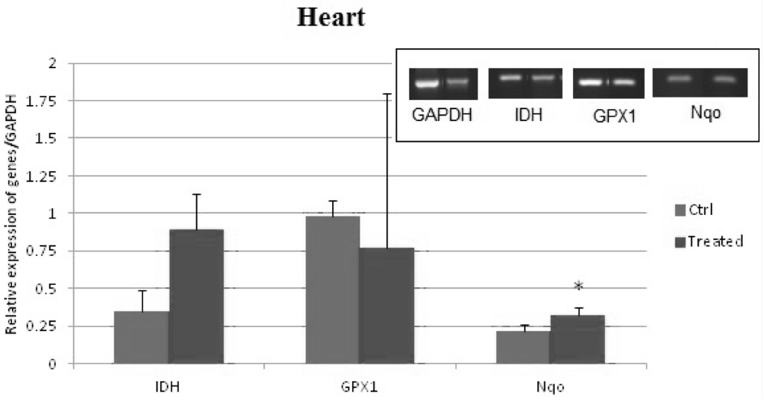
Bar diagram and representing gels showing RT-PCR gene expression analysis of the oxidative stress genes, NAD(P)H quinone dehydrogenase 1 (Nqo1), Glutathione peroxidase (Gpx1) and Isocitrate dehydrogenase-1 (Idh1) in the heart tissue after doxorubicin treatment in SD rats. Data are expressed as the means ± standard error of the mean as analyzed by Student’s t-test; the level of significance is p< 0.05.
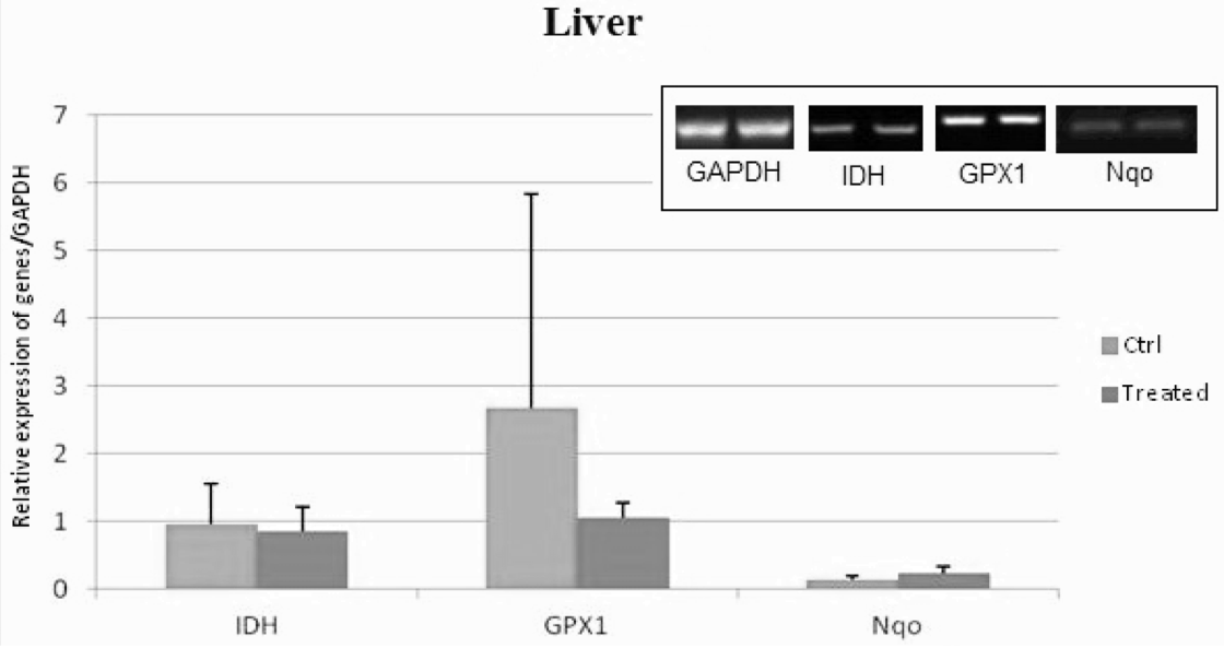
Bar diagram and representing gels showing RT-PCR gene expression analysis of the oxidative stress genes, NAD(P)H quinone dehydrogenase 1 (Nqo1), Glutathione peroxidase (Gpx1) and Isocitrate dehydrogenase-1 (Idh1) in the liver tissue after doxorubicin treatment in SD rats. Data are expressed as the means ± standard error of the mean as analyzed by Student’s t-test; the level of significance is p< 0.05.
References
- Carvalho, F.S., Burgeiro, A., Garcia, R., Moreno, A.J., Carvalho, R.A. and Oliveira, P.J., 2014. Doxorubicin-induced cardiotoxicity: From bioenergetic failure and cell death to cardiomyopathy. Med. Res. Rev., 34: 106-135. https://doi.org/10.1002/med.21280[CrossRef]
- Chmielewska, M., Symonowicz, K., Pula, B., Owczarek, T., Podhorska-Okolow, M., Ugorski, M. and Dziegiel, P., 2015. Expression of metallothioneins I and II in kidney of doxorubicin-treated rats. Exp. Toxicol. Pathol., 67: 297-303. https://doi.org/10.1016/j.etp.2015.01.006[CrossRef]
- Goyal, S.N., Mahajan, U.B., Chandrayan, G., Kumawat, V.S., Kamble, S., Patil, P., Agrawal, Y. O., Patil, C.R. and Ojha, S., 2016. Protective effect of oleanolic acid on oxidative injury and cellular abnormalities in doxorubicin induced cardiac toxicity in rats. Am. J. Transl. Res., 8: 60-69.
- Guo, H., Liu, Y., Wang, L., Zhang, G., Su, S., Zhang, R., Zhang, J., Li, A., Shang, C., Bi, B. and Li, Z., 2016. Alleviation of doxorubicin-induced hepatorenal toxicities with sesamin via the suppression of oxidative stress. Hum. exp. Toxicol., 35: 1183-1193. https://doi.org/10.1177/0960327115626581[CrossRef]
- Haraguchi, C.M., Mabuchi, T. and Yokota, S., 2003. Localization of a mitochondrial type of NADP-dependent isocitrate dehydrogenase in kidney and heart of rat: An immunocytochemical and biochemical study. J. Histochem. Cytochem., 51: 215-226. https://doi.org/10.1177/002215540305100210[CrossRef]
- Huang, C.Y., Chen, J.Y., Kuo, C.H., Pai, P.Y., Ho, T.J., Chen, T.S., Tsai, F.J., Padma, V.V., Kuo, W.W. and Huang, C.Y., 2018. Mitochondrial ROS-induced ERK1/2 Activation and HSF2-mediated AT1 R upregulation are required for Doxorubicin-induced cardiotoxicity. J. Cell. Physiol., 233: 463-475. https://doi.org/10.1002/jcp.25905[CrossRef]
- Ibrahim, M.A., El-Sheikh, A.A., Khalaf, H.M. and Abdelrahman, A.M., 2014. Protective effect of peroxisome proliferator activator receptor (PPAR)-alpha and -gamma ligands against methotrexate-induced nephrotoxicity. Immunopharmacol. Immunotoxicol., 36: 130-137. https://doi.org/10.3109/08923973.2014.884135[CrossRef]
- Ichikawa, Y., Ghanefar, M., Bayeva, M., Wu, R., Khechaduri, A., Naga-Prasad, S.V., Mutharasan, R.K. and Naik, T.J., 2014. Ardehali H. Cardiotoxicity of doxorubicin is mediated through mitochondrial iron accumulation. J. clin. Invest., 124: 617-630. https://doi.org/10.1172/JCI72931[CrossRef]
- Itsumi, M., Inoue, S., Elia, A.J., Murakami, K., Sasaki, M., Lind, E.F., Brenner, D., Harris, I.S., Chio, I.I., Afzal, S., Cairns, R.A., Cescon, D.W., Elford, A.R., Ye, J., Lang, P.A., Li, W.Y., Wakeham, A., Duncan, G.S., Haight, J., You-Ten, A., Snow, B., Yamamoto, K., Ohashi, P.S. and Mak, T.W., 2015. Idh1 protects murine hepatocytes from endotoxin-induced oxidative stress by regulating the intracellular NADP(+)/NADPH ratio. Cell Death Differ., 22: 1837-1845. https://doi.org/10.1038/cdd.2015.38[CrossRef]
- Jo, S.H., Son, M.K., Koh, H.J., Lee, S.M., Song, I.H., Kim, Y.O., Lee, Y.S., Jeong, K.S., Kim, W.B., Park, J.W., Song, B.J. and Huh, T.L., 2001. Control of mitochondrial redox balance and cellular defense against oxidative damage by mitochondrial NADP+-dependent Isocitrate Dehydrogenase. J. biol. Chem., 276: 16168-16176. https://doi.org/10.1074/jbc.M010120200[CrossRef]
- Kumral, A., Giriş, M., Soluk-Tekkeşin, M., Olgaç, V., Doğru-Abbasoğlu, S., Türkoğlu, Ü. and Uysal, M., 2015. Effect of olive leaf extract treatment on doxorubicin-induced cardiac, hepatic and renal toxicity in rats. Pathophysiology, 22: 117-123. https://doi.org/10.1016/j.pathophys.2015.04.002[CrossRef]
- Lakomkin, V.L., Abramov, A.A., Gramovich, V.V., Vyborov, O.N., Lukoshkova, E.V., Ermishkin, V.V. and Kapelko, V.I., 2017. The time course of formation of systolic dysfunction of the heart in doxorubicin cardiomyopath. Kardiologiia, 1: 59-64.
- Minotti, G., Menna, P., Salvatorelli, E., Cairo, G. and Gianni, L., 2004. Anthracyclines: Molecular advances and pharmacologic developments in antitumor activity and cardiotoxicity. Pharmacol. Rev., 56: 185-229. https://doi.org/10.1124/pr.56.2.6[CrossRef]
- Muse, K.E., Oberley, T.D., Sempf, J.M. and Oberley, L.W., 1994. Immunolocalization of antioxidant enzymes in adult hamster kidney. Histochem. J., 26: 734-753. https://doi.org/10.1007/BF00158205[CrossRef]
- Noreen, A., Bukhari, D.A. and Rehman A., 2018. Reactive oxygen species: Synthesis and their relationship with cancer-a review. Pakistan J. Zool., 50: 1951-1963.
- Ray, P.D., Huang, B.W. and Tsuji, Y., 2012. Reactive oxygen species (ROS) homeostasis and redox regulation in cellular signaling. Cell. Signall., 24: 981-990. https://doi.org/10.1016/j.cellsig.2012.01.008[CrossRef]
- Rehman, M.U., Tahir, M., Khan, A.Q., Khan, R., Oday-O-Hamiza, Lateef, A., Hassan, S.K., Rashid, S., Ali, N., Zeeshan, M. and Sultana, S., 2014. D-limonene suppresses doxorubicin-induced oxidative stress and inflammation via repression of COX-2, iNOS, and NFkappaB in kidneys of Wistar rats. Exp. Biol. Med., 239: 465-476. https://doi.org/10.1177/1535370213520112[CrossRef]
- Ross, D. and Siegel, D., 2004. NAD(P)H:quinone oxidoreductase 1 (NQO1, DT-diaphorase), functions and pharmacogenetics. Methods Enzymol., 382: 115-144. https://doi.org/10.1016/S0076-6879(04)82008-1[CrossRef]
- Sharma, M., Mehndiratta, M., Gupta, S., Kalra, O.P., Shukla, R. and Gambhir, J.K., 2016. Genetic association of NAD(P)H Quinone Oxidoreductase (NQO1*2) polymorphism with NQO1levels and risk of diabetic nephropathy. Biol. Chem., 397: 725-730. https://doi.org/10.1515/hsz-2016-0135[CrossRef]
- Tulubas, F., Gurel, A., Oran, M., Topcu, B., Caglar, V. and Uygur, E., 2015. The protective effects of omega-3 fatty acids on doxorubicin-induced hepatotoxicity and nephrotoxicity in rats. Toxicol. Ind. Hlth., 31: 638-644. https://doi.org/10.1177/0748233713483203[CrossRef]
- Van, C.K., Heyns, L., De, S.F., van Eycken, L., Gziri, M.M., van Gemert, W., Halaska, M., Vergote, I., Ottevanger, N. and Amant, F., 2010. Cancer during pregnancy: An analysis of 215 patients emphasizing the obstetrical and the neonatal outcomes. J. clin. Oncol., 28: 683-699. https://doi.org/10.1200/JCO.2009.23.2801[CrossRef]
- Wu, Y., Wang, X., Chang, S., Lu, W., Liu, M. and Pang, X., 2016. β-Lapachone induces NQO1- and oxidative stress-dependent Hsp90 cleavage and inhibits tumor growth and angiogenesis. J. Pharmacol. exp. Ther., 357: 466-475. https://doi.org/10.1124/jpet.116.232694[CrossRef]
Cited by 1 article
In journals published by ResearchersLinks.
Services
Article usage statistics
Loading…