Reputed National and International firms’ viz. Sigma Chemicals (USA), SRL (India) and Merck (India) were our sole source from where standard analytical grade of chemicals were procured for this research work. Chitosan and sodium tripolyphosphate (TTP) used in this study were purchased from Sigma, USA. Acetic acid was of HPLC grade procured from Merck. All other chemicals used in this study were of the molecular grade. We used bench top centrifuge– HermLe Z 360 K, Sorvall RC 5C plus high speed refrigerated centrifuge (Depont, USA), Micro centrifuge (Eppendorf, Germany), Magnetic stirrer– REMI, Micropipettes– Ependorff, Germany. Glass wares used were obtained from Borosil (India) and Schott Duran (Germany). Glass wares were thoroughly washed and sterilized wherever necessary following the recommended procedures.
Preparation of Blank Chitosan Nanoparticles
Chitosan nanoparticles were prepared according to the procedure developed by Zhang et al. (2004) based on the ionotropic gelation of chitosan (CS) with sodium tripolyphosphate (TPP) anions with slight modification. A total of 8.75 gm of CS was dissolved in 5mL of an aqueous solution of acetic acid (0.306 wt %) at concentrations of 0.175 wt %. Afterward, 2.0 mL of TPP solution (1mg/mL) was added drop wise to 5.0 mL of CS solution in a 20–mL glass vial under magnetic stirring at 600 rpm using an octagonal stirring bar. The mixture was then stirred for additional 10 min. The nanoparticles formed were centrifuged at 20000 rpm for 40 min. The pellet formed was resuspended in de–ionized water with constant shaking and then pipetting.
Antigen Encapsulation Efficiency
One mg of BSA was dissolved in 5mL of chitosan solution. Thereafter, the procedure for preparation of BSA loaded chitosan nanoparticles was same as described for the preparation blank nanoparticles. After preparation of BSA loaded nanoparticles, the solution was centrifuged at 24000g for 30 min at 10ºC. The concentration of antigen in the supernatant was measured using Bicinchoninic acid (BCA) method of protein estimation. Antigen encapsulation efficiency was determined by subtracting the antigen present in the supernatant from the total amount of antigen used for loading. Each batch was analyzed in triplicate.
Where A is the total amount of antigen added during preparation, B is the amount of antigen remaining in the supernatant.
Particle Size and Zeta Potential Analysis
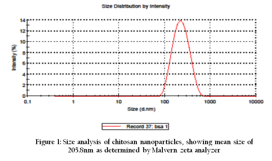
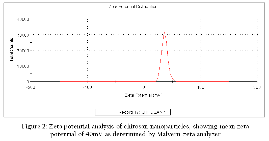
The size distribution and zeta potential of nanoparticles were determined by quasi–elastic light scattering at 25 oC, at an angle of 900, using a Zeta Sizer 3000 HS (Malvern Instruments, UK). Highly diluted colloidal dispersions of nanoparticles in pure distilled water were used for size and zeta potential measurement as per the standard procedure followed at National Institute of Immunology (NII), New Delhi. All the measurements were performed in triplicate for 60 seconds at 1000 Hz, and an electric current of 3 mA with zero field correction.
Size and Morphology
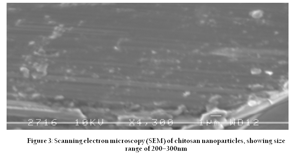
Particle size and surface morphology of nanoparticles were determined by Scanning electron microscopy (SEM). Briefly, a small pinch of the air dried nanoparticles were coated on the metallic slub by ion sputtering or gold coating and then observed under different magnifications using Jeol JSM 840 x Scanning electron microscope.
Sterility Testing
The sterility of nanoparticles was checked by inoculating in nutrient broth, nutrient agar, blood agar and Robertson’s cooked meat media and observing for growth of any micro–organisms.