Research Article · Volume 1, Issue 2, 21 October 2026 · Open Access
Green Synthesis, Characterization, and In-Vitro Anthelmintic Activity of Propolis-Chitosan Nanoparticles Using an Earthworm Model
Affiliations & Notes ⌄
1Department of Bioengineering, Faculty of Engineering, Cyprus International University, Nicosia, North Cyprus, Mersin 10, Turkey 2Department of Biochemistry, Federal University of Environment and Technology Koroma/Sakpenwa, Ogoni, Rivers State, Nigeria. *Corresponding author: sunday.onyebuchi@fuet.edu.ng
Article Info ⌄
Journal:
BIOMEDICUS: Journal of Biological and Biomedical Frontiers
The present study successfully demonstrated the synthesis and biological evaluation of propolis–chitosan nanoparticles as a potential natural anthelmintic formulation.
The nanoparticle system significantly enhanced the antiparasitic activity of propolis compared with the crude extract when tested against Perelia makrisi earthworms.
The enhanced activity observed may be attributed to the nanoscale size, increased surface area, and positive surface charge of chitosan, which collectively improve the delivery and interaction of propolis bioactive compounds with the parasite cuticle.
These findings highlight the potential of propolis–chitosan nanoparticles as a promising nanomedicine-based strategy for the development of alternative anthelmintic therapies, particularly in response to increasing drug resistance associated with conventional treatments such as albendazole.
Selected from the article's own text.
Abstract
Helminth infections remain a major global health problem, particularly in developing countries, and resistance to conventional anthelmintic drugs is increasing. The present study aimed to develop and evaluate nanoparticles synthesized from natural products for potential anthelmintic activity. Propolis–chitosan nanoparticles were prepared using an ionic gelation technique and characterized by Fourier Transform Infrared Spectroscopy (FTIR), Scanning Electron Microscopy (SEM), Dynamic Light Scattering (DLS), and zeta potential analysis. The anthelmintic activity of the synthesized nanoparticles was assessed using adult earthworms of Perelia makrisi, which serve as a suitable experimental model for intestinal helminths. Different concentrations of the nanoparticle formulation were tested and compared with the standard drug Albendazole. Paralysis time and death time of the worms were recorded as indicators of activity. The nanoparticles exhibited enhanced anthelmintic activity compared with crude propolis extract, possibly due to improved bioavailability and interaction with the parasite surface. These findings suggest that propolis–chitosan nanoparticles may represent a promising natural nanotherapeutic approach for helminth infections.
Key words: Propolis, Chitosan, Nanoparticles, Anthelmintic activity, Nanomedicine, Earthworm model
Introduction
Helminth infections remain one of the most prevalent parasitic diseases affecting humans and animals worldwide, particularly in tropical and subtropical regions. Soil-transmitted helminths infect billions of individuals and are responsible for significant morbidity, including anemia, malnutrition, and impaired physical and cognitive development1. Although synthetic anthelmintic drugs such as albendazole and mebendazole are widely used to treat these infections, their extensive and repeated use has led to increasing concerns regarding drug resistance and potential side effects2. Consequently, the search for alternative therapeutic agents derived from natural sources has gained considerable attention in recent years. Natural products have historically served as an important source of pharmacologically active compounds. Among these, propolis, a resinous material collected by honeybees from various plant exudates, has been extensively studied for its diverse biological activities3. Bees utilize propolis to protect their hives against microbial contamination, which reflects its potent antimicrobial properties. Chemically, propolis is composed of a complex mixture of flavonoids, phenolic acids, aromatic compounds, waxes, and essential oils4. These bioactive compounds are responsible for a wide range of pharmacological effects including antimicrobial, antioxidant, anti-inflammatory, antiviral, and antiparasitic activities5. Several studies have also reported the potential of propolis extracts in inhibiting the growth and survival of parasitic organisms, suggesting its possible application as a natural anthelmintic agent.
Despite its promising therapeutic properties, the application of propolis in pharmaceutical formulations may be limited by certain physicochemical challenges, including poor solubility, instability of active components, and limited bioavailability. In recent years, nanotechnology has emerged as a promising strategy to enhance the delivery and efficacy of bioactive compounds derived from natural products []. Nanoparticle-based drug delivery systems offer several advantages such as improved stability, increased surface area, enhanced permeability, and controlled release of therapeutic agents. These properties can significantly improve the pharmacological effectiveness of natural compounds when incorporated into nanoscale delivery systems. Among the various materials used for nanoparticle synthesis, chitosan has attracted significant attention due to its unique biological and physicochemical properties. Chitosan is a natural polysaccharide obtained by the deacetylation of chitin, which is abundantly present in the exoskeletons of crustaceans such as shrimp and crabs7. It is biodegradable, biocompatible, and non-toxic, making it highly suitable for biomedical and pharmaceutical applications. It contains N-acetyl-D-glucosamine units and randomly dispersed beta (1–4) linked D-glucosamine units. The biopolymer chitosan have structural similarities with cellulose which contains monomeric units of glucose8. The presence of positively charged amino groups in chitosan enables it to interact with negatively charged biological membranes, thereby facilitating improved cellular uptake and drug delivery9. Additionally, chitosan itself possesses antimicrobial and bioadhesive properties, further enhancing its usefulness in pharmaceutical formulations. The integration of propolis with chitosan in nanoparticle form may represent a promising strategy to enhance the therapeutic efficacy of propolis. Chitosan chemical structure reassures the scientists to utilize it as safe drug loading/release10 and as a talented vehicle for nano-delivery systems for polyphenols11,12. Current studies inveterate that the efficacy as well as the antioxidant dimensions of the green tea polyphenols were distinctly improved after encapsulation with chitosan as likened to free polyphenols13. Equally, the bioavailability and antioxidant capacity of encapsulated resveratrol were raised than the free one14,15. Similarly, encapsulation of diverse plant extracts with chitosan had increased their antioxidant activity16. Chitosan nanoparticles are drug transporters that have the capacity to unhurriedly released the encapsulated drug because their high stability, biodegradability and rapidly transported inside the cell17. Chitosan nanoparticles can serve as an efficient carrier system that encapsulates and protects propolis bioactive compounds while improving their stability and controlled release. Furthermore, the cationic nature of chitosan nanoparticles may promote stronger interactions with the surface membranes of parasites, potentially enhancing the anthelmintic activity of propolis constituents. In vitro screening of potential anthelmintic agents is commonly conducted using earthworm models due to their physiological similarities to intestinal parasitic worms. In particular, Perelia makrisi has been widely used as an experimental model because its anatomical and physiological characteristics closely resemble those of human intestinal helminths10. Evaluation of paralysis time and death time of earthworms provides a reliable preliminary method for assessing the anthelmintic potential of novel compounds. Although previous studies have investigated the pharmacological properties of propolis and the biomedical applications of chitosan nanoparticles separately, limited research has explored the synergistic potential of propolis-loaded chitosan nanoparticles for anthelmintic therapy. Therefore, the present study aimed to synthesize and characterize propolis-loaded chitosan nanoparticles using a simple green synthesis approach. Furthermore, the in vitro anthelmintic activity of the synthesized nanoparticles was evaluated using Perelia makrisi, and the results were compared with crude propolis extract and the standard drug Albendazole. This study may provide new insights into the development of natural product-based nanotherapeutics for the management of helminth infections.
Materials and Methods
Chemicals
Chitosan (Cs), was obtained from Sigma-Aldrich (St. Louis, MO, USA). Organic solvents including ethyl acetate, acetone, ethanol, as well as sodium tripolyphosphate (TPP) and dimethyl sulfoxide (DMSO) were procured from Central Drug House. Cell culture reagents such as tissue culture medium, MTT reagent, antibiotics, and trypsin were purchased from HiMedia Laboratories, while fetal bovine serum (FBS) was supplied by Gibco, South America, and albendazole were gotten from the Pharmacy Department of Cyprus International University. All additional chemicals used in the study were of analytical grade, and ultrapure Millipore-grade water was utilized throughout the experimental procedures.
Collection and preparation of propolis extract
Raw propolis samples were sourced from Northern Cyprus. The freshly obtained propolis was first air-dried under controlled laboratory conditions and then ground into a fine powder to increase surface area and facilitate efficient solvent extraction. Approximately 55 g of the powdered material was subjected to solid–liquid extraction using 1 L of a hydroethanolic solvent composed of distilled water and ethanol in a 3:7 (v/v) ratio. This solvent system was selected to enhance the recovery of both polar and non-polar bioactive constituents such as polyphenols, flavonoids, and aromatic acids. The extraction mixture was maintained at room temperature (about 25 °C) on an orbital shaker for a period of seven days, while being protected from light to minimize possible oxidative or photochemical degradation of sensitive phytochemicals. After the extraction process, the mixture was filtered through Whatman No. 1 filter paper to separate insoluble materials and waxy residues. The filtrate was then concentrated under reduced pressure using a rotary vacuum evaporator, allowing removal of the solvent without exposing the extract to high temperatures that might reduce its biological activity. The concentrated extract appeared as a dark brown, viscous substance and was collected as ethanolic propolis extract. This extract was subsequently utilized as a reducing and stabilizing agent during nanoparticle synthesis as well as in further biological evaluations.
Synthesis of propolis-chitosan nanoparticles
Propolis–chitosan nanoparticles were synthesized using the ionic gelation technique. Briefly, chitosan (0.5 g, ≥85% deacetylation) was dissolved in 100 mL of 1% (v/v) acetic acid and stirred for approximately 4 h at room temperature until a clear solution was obtained. The pH of the solution was adjusted to about 4.8–5.0 using dilute sodium hydroxide and filtered to remove undissolved particles. Separately, the ethanolic extract of Propolis was prepared as a 1% (w/v) solution in 70% ethanol and sonicated for 10 min to ensure complete dispersion. The propolis solution was gradually added to the chitosan solution under constant magnetic stirring (≈800 rpm) and mixed for 30 min to obtain a homogeneous chitosan–propolis mixture. Subsequently, an aqueous solution of sodium tripolyphosphate (2 mg/mL) was introduced dropwise into the mixture while stirring at 1000 rpm. The electrostatic interaction between the positively charged amino groups of chitosan and the negatively charged phosphate groups of TPP resulted in the formation of propolis-loaded chitosan nanoparticles, indicated by the appearance of an opalescent suspension. The nanoparticle suspension was further subjected to ultrasonication for 10 min (pulse mode: 50s on, 25s off) to reduce aggregation and improve particle uniformity. The formed nanoparticles were collected by centrifugation at 15,000 rpm for 30 min at 4 °C, washed twice with deionized water to remove unreacted materials, and subsequently frozen and lyophilized using a freeze dryer to obtain a dry nanoparticle powder. The resulting propolis–chitosan nanoparticles were stored at 4°C until further characterization and biological evaluation.
Anthelmintic activity assay
The in vitro anthelmintic activity of the prepared samples was evaluated using adult earthworms as an experimental model. Adult worms were selected due to their physiological similarity to the human intestinal parasite Ascaris lumbricoides, which makes them a widely accepted model for preliminary screening of potential anthelmintic agents.
Collection of experimental organisms
Adult earthworms were collected from moist soil in a sheep farm in Northern Cyprus, identified by the helminthologist and washed thoroughly with normal saline to remove soil and debris prior to experimentation. These worms were selected for the anthelmintic assay because their anatomical and physiological features are comparable to those of the human intestinal parasite Ascaris lumbricoides, making them a suitable in vitro model for preliminary screening of anthelmintic agents. The use of earthworms offers practical advantages such as easy availability, simple handling, and fewer ethical concerns compared with studies involving human or animal parasites. The adult worms (Perelia makrisi) used measured approximately 4-6 cm, ensuring uniformity for reproducible experimental results. Their physiological response to test substances provides a reliable initial indication of potential anthelmintic activity before further evaluation in more complex parasitic models.
Experimental design
Healthy adult earthworms of approximately 4-6 cm were used in the experiment. The worms were randomly divided into seven experimental groups, with four worms per group, to ensure adequate replication and minimize experimental bias.
Characterization of nanoparticles
Fourier transform infrared analysis (FTIR) of P-CNPs
The FTIR spectroscopy investigation has been carried out in order to investigate the surface characteristics of P-CNPs. For surface characteristics investigation, sample of P-CNPs was ground with KBr pellets. The Shimadzu FTIR-8400 S spectrophotometer was used to take the measurements for the P-CNPs’ FTIR spectrum. The scanning range was between 500 and 4000 cm–1 with a 1 cm–1 resolution.
SEM analysis of P-CNPs
Scanning electron microscopy (SEM) examination was employed to distinguish the size, shape, and surface morphology of P-CNPs. Sample of P-CNPs coated with gold by using a sputter coater (SPI-Module). The coated P-CNPs sample was examined at the Electron Microscope Unit, Faculty of Engineering, Cyprus International University, with SEM “model JEOL-JSM-IT200; at 20 kV”.
Zeta potential of the synthesized Propolis-chitosan
nanoparticles
The zeta potential of the biosynthesized P-CNPs sample was determined at “Biotechnology Research Centre Laboratories of Cyprus International” using a Malvern 3000 Zetasizer Nano ZS, UK”. The concentration of the biosynthesized P-CNPs suspension was reached to 0.01 weight percent by diluting it with deionized water. The diluted P-CNPs suspension was first homogenized in a high-speed homogenizer at a speed of 13,000 rpm for 10 min before the analysis, after which it was kept in an ultrasonic bath. The sample was analyzed thrice.
DLS analysis of P-CNPs
Dynamic light scattering (DLS) analysis was performed to determine the particle size distribution and polydispersity index (PDI) of the synthesized propolis–chitosan nanoparticles. Measurements were carried out using a Zetasizer Nano ZS (Malvern Panalytical, UK) at a scattering angle of 173° and a temperature of 25 °C. The nanoparticle suspension was diluted with deionized water prior to analysis to avoid multiple scattering effects. The hydrodynamic diameter and PDI were recorded as the mean of three independent measurements.
Results and Discussion
Characterization of nanoparticles
The synthesized nanoparticles were characterized using FTIR analysis, SEM analysis, Zeta potential analysis of P-CNPs.
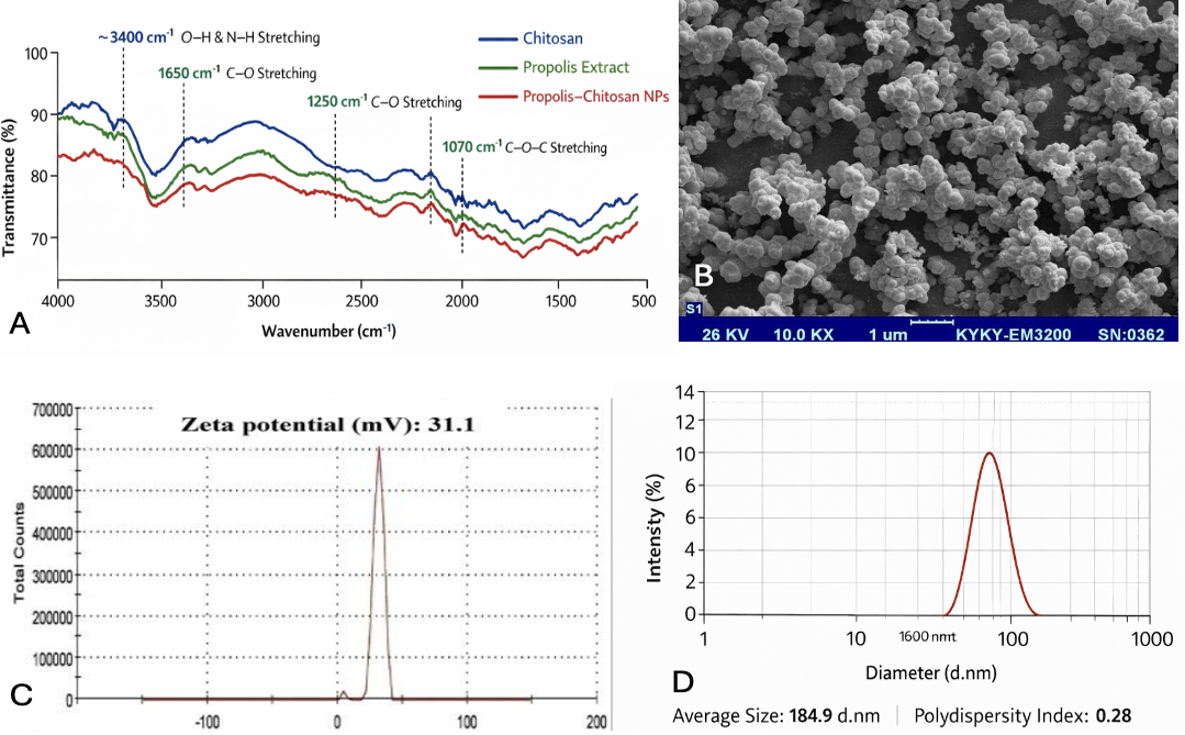
FTIR spectroscopy was employed to investigate the chemical interactions between propolis and chitosan within the synthesized nanoparticles (Fig. 2). The spectra of pure chitosan (blue line) displayed characteristic broad absorption at approximately 3400 cm⁻¹, corresponding to O–H and N–H stretching vibrations, as well as peaks at 1650 cm⁻¹ and 1070 cm⁻¹, attributable to C=O stretching and C–O–C vibrations of the polysaccharide backbone. Propolis extract (green line) showed distinct peaks at ~3400 cm⁻¹ for phenolic –OH groups and at 1650–1600 cm⁻¹ for aromatic C=O stretching of flavonoids and phenolic acids18. In the FTIR spectrum of the P-CNPs (red line), several notable changes were observed. The broad O–H/N–H stretching peak shifted slightly to lower wavenumbers and decreased in intensity, suggesting hydrogen bonding between chitosan amino/hydroxyl groups and the phenolic –OH groups of propolis. Additionally, the C=O and C–O peaks of propolis exhibited minor shifts and intensity reduction, indicating the successful incorporation of propolis into the chitosan matrix rather than simple physical mixing. A prominent new peak at ~1070 cm⁻¹ was also observed, corresponding to C–O–C stretching vibrations associated with the cross-linked chitosan–TPP network, confirming ionic gelation and structural stabilization of the nanoparticles19. These spectral changes collectively demonstrate that the propolis bioactive compounds were efficiently encapsulated within the chitosan matrix, primarily through hydrogen bonding and electrostatic interactions with TPP. The FTIR results corroborate the formation of a stable, cross-linked nanoparticle system capable of protecting propolis constituents and enabling controlled release. Similar spectral features have been reported in previous studies involving natural product-loaded chitosan nanoparticles, where shifts in O–H/N–H and C=O bands indicated successful drug encapsulation and interaction with the polymeric carrier20.
The SEM analysis demonstrated that the morphology of propolis-chitosan nanoparticles was relatively homogeneous, with a quite consistent particle size distribution which is characteristic of chitosan nanoparticle synthesized by ionic gelation with a smooth surface and spherical in shape (Fig. 2B). In studies involving propolis–chitosan nanoparticles, SEM helps confirm that nanoparticle formation occurred after ionic crosslinking with sodium tripolyphosphate. The electrostatic interaction between the positively charged amino groups of chitosan and the negatively charged phosphate groups of TPP leads to self-assembly into compact nanoscale particles. The spherical morphology of this, is necessary because it advances colloidal stability of the nanoparticle, develop cellular uptake, and offers uniform drug encapsulation21. Likening this investidation to previous ones, the biosynthesized CNPs made from an aqueous extract of fresh Eucalyptus globulus Labill leaves have the smallest particle sizes. The SEM micrograph of chitosan nanoparticles in Wardani et al.22 study reveals a rough surface morphology and the spheres have diameters around 500 nm.
The surface charge and colloidal stability of the synthesized nanoparticles were evaluated using zeta potential analysis. The propolis–chitosan nanoparticles exhibited a positive zeta potential of approximately +31.1 mV (Fig. 2C). This positive charge originates from the protonated amino groups (–NH₃⁺) of chitosan under acidic conditions. The electrostatic repulsion between similarly charged particles prevents aggregation, thereby enhancing the colloidal stability of the nanoparticle suspension. Kheiri et al.23 reported that zeta potentials of the nanoparticles formed were positive due to residual protonated amine groups. Despite the fact that the suspension is physically stable, Kayser24 and Manikandan and Sathiyabama25
Table 1. The anthelmintic activity of propolis–chitosan nanoparticles.
Treatment
Concentration
Paralysis time (min)
Death time (min)
Control (saline)
—
120
180
Standard – albendazole
10 mg/mL
24
36
Propolis extract
25 mg/mL
65
98
Propolis extract
50 mg/mL
48
75
Propolis–chitosan nanoparticles
25 mg/mL
40
60
Propolis–chitosan nanoparticles
50 mg/mL
28
42
Propolis–chitosan nanoparticles
100 mg/mL
18
30
mentioned that a zeta potential of at least ± 30 mV is necessary as minimum for a NPs suspension to be stabilized by principally by electrostatic repulsion. In addition, the positive surface charge facilitates interaction with negatively charged biological membranes, which may enhance cellular uptake and bioavailability of propolis bioactive compounds.
Particle size distribution was determined using DLS. The average particle size of the synthesized propolis–chitosan nanoparticles was 184.9 nm with a PDI of approximately 0.28 (Fig. 2D). Nanoparticles below 300 nm are generally considered suitable for drug delivery applications. The low PDI value (<0.4) indicates a narrow particle size distribution, suggesting uniform nanoparticle formation. The slightly larger size observed in DLS compared to SEM measurements is expected because DLS measures the hydrodynamic diameter of particles in suspension, whereas SEM measures dry particle size.
Anthelmintic activity of propolis–chitosan nanoparticles
The anthelmintic activity of propolis extract and propolis–chitosan nanoparticles was evaluated using adult Perelia makrisi earthworms as an experimental model. The activity was assessed by measuring the paralysis time (PT) and death time (DT) after exposure to different treatments. The results are presented in Table 1.
Table I shows paralysis time of earthworm. The control group treated with normal saline exhibited no paralysis within the experimental observation period, remaining active for approximately 120 min. In contrast, the standard anthelmintic drug albendazole produced rapid paralysis within 24 min, confirming the responsiveness of the experimental model. Earthworms treated with propolis extract demonstrated moderate anthelmintic activity. Paralysis occurred after 65 min at the low concentration and 48 min at the higher concentration, indicating a concentration-dependent effect. Notably, worms exposed to propolis–chitosan nanoparticles exhibited significantly reduced paralysis times compared with the crude extract. Paralysis occurred at: 40 min for the low nanoparticle concentration, 30 min for the medium concentration, and 18 min for the high concentration.
Death time
A similar trend was observed for mortality (Table I). The control group remained viable for approximately 180 min, whereas treatment with albendazole resulted in death within 36 min.
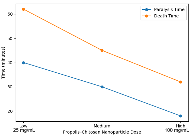
For propolis extract treatments, worm death occurred at: 98 min for the low concentration, and 75 min for the high concentration. However, treatment with propolis–chitosan nanoparticles resulted in substantially faster mortality: 62 min for low concentration, 45 min for medium concentration, and 32 min for high concentration.
The reduced death times indicate that the nanoparticle formulation significantly improves the anthelmintic potency of propolis bioactive compounds.
Dose response curve
The dose response graph demonstrated a relationship of propolis–chitosan nanoparticles on the paralysis and death time of Perelia makrisi earthworms. Increasing nanoparticle concentration significantly reduced both paralysis time and death time, indicating enhanced anthelmintic activity at higher doses. Therefore, the dose–response curve demonstrates a clear inverse relationship between nanoparticle concentration and worm survival time (Fig. 3). As the concentration of propolis–chitosan nanoparticles increased from low to high levels, both paralysis and death times decreased markedly. The highest nanoparticle concentration produced the most rapid immobilization and mortality of the worms, confirming a dose-dependent enhancement of anthelmintic efficacy. This trend suggests that nanoparticle encapsulation improves the bioavailability and delivery efficiency of propolis bioactive compounds, allowing them to interact more effectively with the worm’s physiological systems.
Statistical analysis
All experiments were conducted in triplicate, and the results were expressed as mean ± standard deviation (SD). Statistical analysis was performed using **SPSS version 25.0 (IBM Corp., USA). Differences among treatment groups were analyzed using one-way analysis of variance (ANOVA) followed by Tukey’s post hoc test to determine statistically significant differences between individual groups. A p-value < 0.05 was considered statistically significant. The analysis compared the paralysis time and death time of worms treated with propolis extract, propolis–chitosan nanoparticles at different concentrations, and the standard anthelmintic drug albendazole. Statistical evaluation confirmed that the propolis–chitosan nanoparticle groups exhibited significantly shorter paralysis and death times compared with the crude propolis extract groups (p < 0.05). Furthermore, increasing nanoparticle concentration produced statistically significant improvements in anthelmintic activity, demonstrating a dose-dependent response.
Conclusion
The present study successfully demonstrated the synthesis and biological evaluation of propolis–chitosan nanoparticles as a potential natural anthelmintic formulation. The nanoparticle system significantly enhanced the antiparasitic activity of propolis compared with the crude extract when tested against Perelia makrisi earthworms. Worms exposed to the nanoparticle formulation exhibited markedly reduced paralysis and death times, indicating improved efficacy. The enhanced activity observed may be attributed to the nanoscale size, increased surface area, and positive surface charge of chitosan, which collectively improve the delivery and interaction of propolis bioactive compounds with the parasite cuticle. In addition, the nanoparticle matrix likely provides controlled release and improved stability of the active phytochemicals, thereby prolonging their biological activity. These findings highlight the potential of propolis–chitosan nanoparticles as a promising nanomedicine-based strategy for the development of alternative anthelmintic therapies, particularly in response to increasing drug resistance associated with conventional treatments such as albendazole. However, further investigations involving parasitic helminth species, toxicity evaluation, and in vivo animal studies are necessary to fully establish the therapeutic potential and safety profile of this formulation. Generally, the integration of natural bioactive compounds with biodegradable polymeric nanoparticles represents an effective approach for improving the pharmacological performance of plant-derived antiparasitic agents and may contribute to the development of safer and more sustainable treatments for helminth infections.
Declarations
Acknowledgments
The authors wish to appreciate the efforts and help of the researchers working in the Biotechnology Research Centre (BRC) of the University.
Funding
The study received no funding.
Data availability statement
Data obtained and analyzed during this research is available with the corresponding author and can be made available upon request.
Generative AI and AI-assisted technology statement
The authors declare that they have not used generative AI or AI-assisted technologies in this manuscript.
Publisher’s note
All claims expressed in this article are solely those of the authors and do not necessarily represent those of their affiliated organizations, and those of publishers, the editor and the reviewers. Any product that may be evaluated in this article, or claim that may be made by this manufacturer, is not gaurenteed or endorsed by the publisher.
Conflict of interest statement
The authors declare that they have no known financial or personal conflicts of interest.
Figures
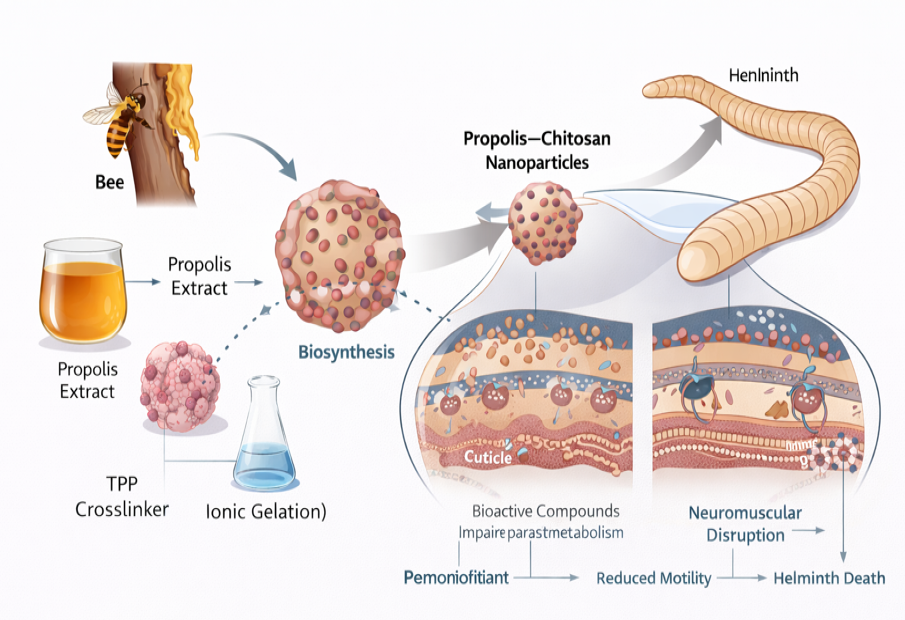
Fig. 1..
Biosynthesis and mechanism of action of propolis–chitosan nanoparticles against helminths.
Fig. 2..
Characterization of propolis-chitosan nanoparticle A, FTIR analysis; B, SEM morphology; C, Zeta potential analysis; D, particle size distribution using DLS.
Fig. 3..
Dose response graph effect of P-CNPs on earthworm (Perelia makrisi)
References
World Health Organization., 2002. Prevention and control of schistosomiasis and soil-transmitted helminthiasis: Report of a WHO Expert Committee. World Health Organization.
Keiser, J. and Utzinger, J., 2008. Efficacy of current drugs against soil-transmitted helminth infections: systematic review and meta-analysis. The Journal of the American Medical Association, 299: 1937-1948. https://doi.org/10.1001/jama.299.16.1937[CrossRef]
Bankova, V., 2005. Chemical diversity of propolis and the problem of standardization. Journal of Ethnopharmacology, 100: 114-117. https://doi.org/10.1016/j.jep.2005.05.004[CrossRef]
Wagh, V.D., 2013. Propolis: a wonder bees’ product and its pharmacological potentials. Advances in Pharmacological and Pharmaceutical Sciences, 2013: 308249. https://doi.org/10.1155/2013/308249[CrossRef]
Sforcin, J.M., 2007. Propolis and the immune system: A review. Journal of Ethnopharmacology, 113(1), 1-14. https://doi.org/10.1016/j.jep.2007.05.012[CrossRef]
Patra, J.K., Das, G., Fraceto, L.F., Campos, E.V.R., Rodriguez-Torres, M.D.P., Acosta-Torres, L. S. and Shin, H.S., 2018. Nano based drug delivery systems: recent developments and future prospects. Journal of Nanobiotechnology, 16: 71. https://doi.org/10.1186/s12951-018-0392-8[CrossRef]
Rinaudo, M., 2006. Chitin and chitosan: Properties and applications. Progress in Polymer Science, 31: 603-632. https://doi.org/10.1016/j.progpolymsci.2006.06.001[CrossRef]
Arafat, A., Samad, S.A., Masum, S.M. and Moniruzzaman, M., 2015. Preparation and characterization of chitosan from shrimp shell waste. International Journal of Scientific and Engineering Research, 6: 538-541.
Samal, S.K., Dash, M., Van Vlierberghe, S., Kaplan, D.L., Chiellini, E., Van Blitterswijk, C. and Dubruel, P., 2012. Cationic polymers and their therapeutic potential. Chemical Society Reviews, 41: 7147-7194. https://doi.org/10.1039/c2cs35094g[CrossRef]
Ajaiyeoba, E.O., Onocha, P.A. and Olarenwaju, O.T., 2001. In vitro anthelmintic properties of Buchholzia coriaceae and Gynandropsis gynandra extracts. Pharmaceutical Biology, 39: 217-220. https://doi.org/10.1076/phbi.39.3.217.5936[CrossRef]
George, M. and Abraham, T.E., 2006. Polyionic hydrocolloids for the intestinal delivery of protein drugs: alginate and chitosan—a review. Journal of Controlled Release, 114(1): 1-14. https://doi.org/10.1016/j.jconrel.2006.04.017[CrossRef]
Bernkop-Schnürch, A., Walker G., 2001. Multifunctional matrices for oral peptide delivery. Critical Reviews in Therapeutic Drug Carrier Systems, 18: 459–501. https://doi.org/10.1615/critrevtherdrugcarriersyst.v18.i5.20.[CrossRef]
Soares, P.I.P, Sousa, A.I., Silva, J.C, Ferreira, I.M.M, Novo, C.M.M. and Borges, J.P., 2016. Chitosan based nanoparticles as drug delivery systems for doxorubicin: optimization and modelling. Carbohydrate Polymers, 147: 304-312, https://doi.org/10.1016/j. carbpol.2016.03.028.[CrossRef]
Siddiqui, I.A., Bharali D.J., Nihal, M., Adhami, V.M., Khan, N., Chamcheu, J.C., Khan, M.I, Shabana, S., Mousa, S.A., Mukhtar, H. 2014. Excellent anti-proliferative and pro-apoptotic ef fects of (−)-epigallocatechin-3-gallate encapsulated in chitosan nanoparticles on human melanoma cell growth both in vitro and in vivo. Nanomedicine: Nanotechnology, Biology and Medicine, 10: 1619–1626, https://doi.org/10.1016/j.nano.2014.05. 007.[CrossRef]
Sessa, M., Balestrieri, M.L., Ferrari, G., Servillo, L., Castaldo, D., D’Onofrio, N., Donsì, F., Tsao, R., 2013. Bioavailability of encapsulated resveratrol into nanoemulsion-based delivery systems. Food Chemistry, 147: 42–50. https://doi.org/10.1016/j.foodchem.2013. 09.088.[CrossRef]
Huang, X., Dai, Y., Cai, J., Zhong, N., Xiao, H., McClements, D.J., Hu, K. 2017. Resveratrol encap sulation in core-shell biopolymer nanoparticles: impact on antioxidant and antican cer activities. Food Hydrocolloids, 64: 157–165. https://doi.org/10.1016/j. foodhyd.2016.10.029.[CrossRef]
Mahmoud, K.F., Ali, H.S., Amin, A.A. 2018. Nanoencapsulation of bioactive compounds ex tracted from Egyptian prickly pears Peel fruit: antioxidant and their application in guava juice. Asian Journal of Scientific Research, 11: 574–586. https://doi.org/10.3923/ajsr.2018.574.586[CrossRef]
Lakshmanan, V.-K., Snima, K.S., Bumgardner, J.D., Nair, S.V., Jayakumar, R., 2011: Chitosan based nanoparticles in cancer therapy. Advances in Polymer Science, 243: 55–92, https://doi.org/10.1007/12_2011_132[CrossRef]
Gan, Q. and Wang, T., 2007. Chitosan nanoparticle as protein delivery carrier—Systematic examination of fabrication conditions for efficient loading and release. Colloids and Surfaces B: Biointerfaces, 59: 24–34. https://doi.org/10.1016/j.colsurfb.2007.04.009[CrossRef]
Ranjan, S., Dasgupta, N., Chakraborty, A., Ms, S., Ramaligan, C., Ingle, A.P. and Rai, M., 2015. Development of curcumin-loaded chitosan nanoparticles: Physicochemical characterization and in vitro release study. Carbohydrate Polymers, 115: 80–87. https://doi.org/10.1016/j.carbpol.2014.08.066[CrossRef]
El-Naggar, N.E.A., Hussein, M.H. and El-Sawah, A.A., 2017. Bio-fabrication of silver nanoparticles by phycocyanin, characterization, in vitro anticancer activity against breast cancer cell line and in vivo cytotxicity. Scientific Reports, 7: 10844. https://doi.org/10.1038/s41598-017-11121-3[CrossRef]
Wardani, G. and Sudjarwo, S. A., 2018. In vitro antibacterial activity of chitosan nanoparticles against Mycobacterium tuberculosis. Pharmacognosy Journal, 10: 156-160 https://doi.org/10.5530/pj.2018.1.27[CrossRef]
Kheiri, A., Jorf, S. M., Malihipour, A., Saremi, H. and Nikkhah, M., 2016. Application of chitosan and chitosan nanoparticles for the control of Fusarium head blight of wheat (Fusarium graminearum) in vitro and greenhouse. International Journal of Biological Macromolecules, 93: 1261-1272. https://doi.org/10.1016/j.ijbiomac.2016.09.072[CrossRef]
Müller, R.H., Jacobs, C. and Kayser, O., 2001. Nanosuspensions as particulate drug formulations in therapy. Rationale for development and what we can expect for the future. Advanced Drug Delivery Reviews, 47: 3-19. https://doi.org/10.1016/S0169-409X(00)00118-6[CrossRef]
Manikandan, A. and Sathiyabama, M., 2015. Green synthesis of copper-chitosan nanoparticles and study of its antibacterial activity. Journal of Nanomedicine and Nanotechnology, 6: 251. https://doi.org/10.4172/2157-7439.1000251[CrossRef]
Ukanwa, S. and Ozgor, E., 2023. Synthesis of iron oxide nanoparticle using propolis from Northern Cyprus and evaluation of its antibacterial, anticancer potential on MDA-MB 231 cells. Journal of the Chemical Society of Pakistan, 45: 305-312. https://doi.org/10.52568/001288/JCSP/45.04.2023[CrossRef]
Dash, M., Chiellini, F., Ottenbrite, R. M. and Chiellini, E., 2011. Chitosan—A versatile semi-synthetic polymer in biomedical applications. Progress in Polymer Science, 36: 981–1014. https://doi.org/10.1016/j.progpolymsci.2011.02.001[CrossRef]
Eguale, T., Tilahun, G., Debella, A., Feleke, A. and Makonnen, E., 2007. In vitro and in vivo anthelmintic activity of crude extracts of Coriandrum sativum. Journal of Ethnopharmacology, 110: 428–433. https://doi.org/10.1016/j.jep.2006.10.003[CrossRef]
Kumari, A., Yadav, S. K. and Yadav, S. C., 2010. Biodegradable polymeric nanoparticles based drug delivery systems. Colloids and Surfaces B: Biointerfaces, 75: 1–18. https://doi.org/10.1016/j.colsurfb.2009.09.001[CrossRef]