
Crude solvent extracts of traditional medicinal plants have been used for thousands of years in different region of the globe for the treatment of various diseases. In developing countries, traditional medicines are used as source of primary health care. Keeping in view the importance of Camellia sinensis L., present investigation was aimed to evaluate the phytochemical and pharmacological potential of different morphological parts of C. sinensis L. Successive extractions of all plant parts was performed with different solvents like, petroleum ether, acetone, ethanol and water. Phytochemical analysis of all extracts showed the presence of polyphenolic compounds, flavonoids, alkaloids, glycosides and carbohydrates in all plant parts with varied strength. Phytochemical analysis showed comparatively high percentage yield of ethanolic extract of all plant parts, hence was employed for evaluating the antimicrobial potential against 11 Gram positive bacteria, 9 Gram negative bacteria, 2 yeasts, 2 dermatophytes and 7 saprophytes. The petroleum ether seed extract and methanolic leaf extract was evaluated for the comparative anti-inflammatory and analgesic potential using different parameters like licking - biting response, inflammation of hind paw and writhing effect. The results showed that different pharmacological activities were due to the presences of various phytochemicals like tannins, resin and flavonoids, observed maximally in ethanolic extract with minor quantity of alkaloids and glycosides. Fourier transform infrared spectroscopy (FTIR) of dried roots, stems, leaves and seeds provided a conclusive support to the above results. Anti-inflammatory effect was less significant (p < 0.05) in seed extract, while leaf extract displayed highly significant results both at low and high doses. Likewise, both seed and leaf showed significant analgesic effects. However, compared to seed extract which showed highly significant (p < 0.001) increase in concentration dependent manner, leaf extract displayed highly significant results even at low dose with better results at high dose compared with standard.
Research Article · Volume 55, Issue 2, 20 December 2022 · Open Access
Phytochemical and Pharmacological Potential of Camellia sinensis L.
· · · · · · · · ·
Affiliations & Notes ⌄
- 1Department of Pharmacognosy, Lahore Pharmacy College, LMDC Lahore, Pakistan 2Department of Bait-Ul-Hikmah, Hamdard University, Karachi, Pakistan 3Department of Chemistry, University of Engineering and Technology, Lahore, Pakistan 4Microbiology Lab, Department of Zoology, GC University, Lahore, Pakistan 5Department of Microbiology, University of Karachi, Karachi, Pakistan 6Department of Pharmacology, Federal Urdu University of Arts and Technology, Karachi 7Institute of Pharmacy, Lahore College fo
Article Info ⌄
- Journal:
- Pakistan Journal of Zoology
- Published:
- 20 December 2022

From the article
- The ethanolic extracts of C. sinensis L. seeds showed high percentage yield compared to other solvents.
- Phytochemical analysis of petroleum ether extracts of the parts of the plant showed positive results for primary metabolites while secondary metabolites indicated lack of lignin and steroids (Supplementary Table II).
- Our results suggested that C. sinensis L. could be a source of potential antimicrobial agent which varies with the part of the plant.
- The findings reported highlights the fact that high dose of both C. sinensis L. seed and leaf extracts are safe for consumption with analgesic and anti-inflammatory potentials compared to standard drug group.
Selected from the article's own text.
Abstract
INTRODUCTION
Improper and extensive use of antimicrobial agents has led to the emergence of unusual infections with strong adverse effects. Plants can be considered as a good option as antimicrobial agents ; . Elevated expenditure and side effects associated with synthetic medicines are attracting the researchers to search for alternative antimicrobial drugs ; .
The proper functioning of human body requires the essential components which play vital role in the growth and development. The consumption of herbal medicine has gained particular attention in medical practice due to its growing knowledge and universal tolerability ; ; . The natural constituents are relatively more compatible and healthier as compared with synthetic one ; . Secondary metabolites such as alkaloids, polyphenols tannins and flavonoids obtained from the herbal medicinal plants provide the important role in the evaluation of biological and pharmacological activities ; . There is need for the identification of the natural products obtained from the herbal medicinal plants which have significant role in treating the pain and inflammatory disorders and have non-addictive properties with lesser side effects ; . Extensive work is being done for the identification of important phytochemical, antimicrobial and antifungal components from diversities of normal flora found around the world.
Colourful fruits, vegetables, spices, wines and tears contain dietary polyphenols, which have gained the fame due to their powerful anti-inflammatory and antioxidant activity . These natural products can be utilized as therapeutic agents and replica for the compounds which are pharmacologically active and can be utilized for the synthesis of the synthetic drugs .
Pharmacognostic methods are helpful in minimizing the phytochemical variations. Phytochemical analyses are helpful in evaluating the percentage and efficacy of biomolecules present in various parts of the plant. Camellia sinensis L. (green tea) is cultivated in Shinkiari, District Mansehra, Pakistan . It is the rich source of polyphenols, flavonoids, polysaccharides and anthraquinone derivatives. Its leaves are used as beverage and the catechins are an important component . Components of this plant predominantly include polyphenols, caffeine and amino acids, which are beneficial for human health .
Therefore, present investigation was aimed to elaborate the phytoconstituents of Camellia sinensis L (green tea) and to explore their potential role towards human benefits. The variations present in the phytochemical and antimicrobial potential of different morphological parts of the green tea from the region, especially Pakistan were explored which to our knowledge have not been reported before. However, the analgesic and anti-inflammatory effects of its seed extracts have been reported occasionally.
MATERIALS AND METHODS
Preparation of plant extract
Camellia sinensis L. was collected from National Tea and High Value Crops Research Institute (NTHRI) Shinkiari, Mansehra, Pakistan. All plant parts were kept in shade for about 15 days, dried completely and ground separately to a fine powder by passing through sieve no. 120. The fine-powdered plant materials were stored in amber coloured bottles and well-preserved at ambient temperature and pressure.
All powdered plant parts (500g) like root, stem, leaf and seed were subjected to extraction at room temperature using 3 L of each solvent like petroleum ether, acetone, ethanol and water, based on their polarity the successive extractions were carried out. The extraction was done with Soxhlet apparatus at 40oC with 40 rpm and at 0.9 MPA pressure. All powdered materials were macerated for four days at room temperature and were filtered. The filtrate was dried at reduced pressure and residues were obtained which were weighed and stored in the air tight containers at room temperature.
Phytochemical analysis was performed for the determination of the presence of primary and secondary metabolites like, carbohydrates, fats, fixed oils, proteins flavonoids, glycosides, resins, alkaloids, terpenoids and steroids present in plant parts.
Screening of antimicrobial activity
Agar well diffusion method was used for the screening of the antimicrobial activity of Camellia sinensis L. against a library of microbial cultures, preserved at the Department of Microbiology, University of Karachi, Pakistan. The antimicrobial activity of the extracts was tested against 11 Gram positive bacteria (Bacillus subtilis, B. cereus, Staphylococcus aureus, S. saprophyticus, S. epidermidis, Enterococcus faecalis, Vancomycin Resistant Enterbacter (VRE), Methicillin Resistant S. aureus (MRSA), Micrococcus luteus, Corynecacterium xerosis), 9 Gram negative bacteria (Salmonella typhi, S. paratyphi A, S. paratyphi B, Shigella sp., Klebsiella pneumoniae, Proteus mirabilis, Escherichia coli, Enterobacter sp, Pseudomonas aeruginosa), two yeasts (Candida albicans and Saccharomyces cerevisae), two dermatophytes (Trichophyton mentagrophyte and Microsporum gypseum) and seven saprophytes (Aspergillus niger, Penicillium sp., Paccilomyces variola, A. flavus, Chrysporium sp., Fusarium oxysporum, Chrysporium sp., A. terreus, A. terricola).
Stock solution (20 mg/mL) of plant extracts was prepared in sterilized 40% dimethyl sulfoxide (DMSO, Sigma Aldrich, UK). DMSO was used as a negative control, while ciprofloxacin (CIP, Oxoid, UK) and Otosporin (Oxoid, UK) was used as a positive control for bacteria and fungi, respectively.
The cell suspension was prepared by inoculating 24 h old bacterial culture and yeast (0.5 McFarland standards) in 5 mL saline. For fungal cultures, spore suspension (5 x 10⁵ spores/mL) was prepared from 5 days old mould plates. Confluent lawn was made on Mueller Hinton Agar (MHA, Oxoid, UK) plates and Sabourauds Dextrose Agar (SDA, Oxoid, UK) for bacterial and fungal cultures, respectively, and permitted to dry for 5-10 mins. Wells (8 mm) were made with the help of sterile borer. Plant extracts, DMSO solution, positive or negative controls (20 µL) were added into the wells. Plates were incubated at 37 °C for 24 h for bacteria and 5 days for fungi .
Determination of minimum inhibitory concentration (MIC)
MIC was determinded via agar well diffusion assay. The extracts exhibited a significant zone of inhibition (> 15 mm. The extracts were dissolved in sterile DMSO with 20 mg/mL concentration and serially diluted to1.25 mg/mL. The above-mentioned method was repeated to observe the maximum dilution that had not displayed slightly noticeable turbidity, which was considered as MIC.
Determination of minimum microbicidal concentration (MMC)
In order to understand whether the appearance of the zone of inhibition is due to the death of organisms or timely halt of microbial growth, MMC was determined. A loop streak from the zone of inhibition was inoculated on fresh appropriate media. After 24 h, observed growth suggested microstatic activity of plant extracts while absence of growth showed lack of microbicidal activity.
Fourier tranform infrared spectroscopy (FTIR)
FTIR (Agilent, Cary 360) was used for the identification of functional groups and their positions in the Phyto molecule. The operating range of FTIR was between 4000-650 cm-1 . Fine powder of root, stem, leaf and seed was used for the FTIR spectroscopic analysis.
Animals (in vivo study)
Seventy two male albino mice weighing 20-22 g were used as test animals in the current study for anti-inflammatory and analgesic activities. They were bought from Dow University, Karachi and distributed into 2x six groups (6 mice per group). The 2x six groups were categorized as control, seed at low dose, seed at high dose, leaf at low dose and leaf at high dose for each of anti-inflammatory and analgesic activity, respectively. The standard laboratory settings such as 25oC temperature; light and dark cycles of 12 h were maintained and food and water was provided ad libitum. Mice were acclimatized for 15 days prior to study. Ethical approval for present investigation was provided by University of Karachi via letter No. 1227/19.
Determination of anti-inflammatory and analgesic potential of seed and leaf extract
For the determination of anti-inflammatory and analgesic potential of C. sinensis L. ethanolic extract of seed and leaf were used. For anti-inflammatory activity diameter of right hind paw of mice was noted with the help of vernier calliper prior to the dose of 2% formalin was injected in the subplater of the right hindpaw of controlled mice for inducing pain. Thirty mins before injecting the formalin, 300 mg/kg, and 600 mg/kg of both seed and leaf extracts of C. sinensis L. were given orally to the all mice groups except for control. Licking response along with time duration was noted in two phases, early phase 0-300 s and late phase 900-1800 s ; .
Acetic acid-induced writhing effect
Acetic acid (0.6%) in distilled water was injected intraperitoneally in mice using aspirin (10 mg/kg) as standard. Acetic acid was calculated for each individual mouse according to body weight. Rest of the procedure is same as mentioned above except for that calculated dose of acetic acid was injected instead of formalin. Abdominal constriction in mice was noted in two phases, early phase 0-300 s and late phase 900-1800 s .
Statistical analysis
The results of the antimicrobial activity of extract were expressed as mean ± standard error of the mean (SEM). Statistical analysis was performed using IBM SPSS statistics version 20 . Significant differences between and within group were measured using one way analysis of variance (ANOVA) followed by Post hoc LSD for all groups at p ˂ 0.05.
RESULTS
The ethanolic extracts of C. sinensis L. seeds showed high percentage yield compared to other solvents. The lowest percentage yield was obtained by petroleum ether extract fractions (Supplementary Table I). Phytochemical analysis of petroleum ether extracts of the parts of the plant showed positive results for primary metabolites while secondary metabolites indicated lack of lignin and steroids (Supplementary Table II). The acetone extract of the parts of the plant showed negative result for protein and provided positive results for secondary metabolites (Supplementary Table III). The ethanolic extracts of the parts of the plant indicated negative results for fat and fixed oil while among secondary metabolites, lignin proteins, resins and steroids were absent (Supplementary Table IV). The water extracts of the parts of the plant showed negative results for primary metabolites like, fat and fixed oil while secondary metabolites showed lack of steroids (Supplementary Table V).
Antimicrobial activity
The antimicrobial activity of the ethanolic extracts of root, stem, leaves and seeds of C. sinensis L. was assessed by agar well diffusion technique. Library of test organisms comprised of 10 Gram positive, 09 Gram negative bacteria, 2 dermatophytes, 2 yeasts and 8 opportunistic pathogenic moulds.
The ethanolic extract of root and stem of C. sinensis has shown strong activity against C. xerosis while moderate for B. cereus. Similarly, the ethanolic extract of leaves has revealed a bigger zone of inhibition (ZOI) for S. typhi (20.6±1.0) and Para typhi A (25±0) while moderate against E. coli (14.6±0.5) and K. pneumoniae (15.0±1). No activity was shown by the ethanolic extract of seeds. It can be inferred from the results in (Table I) the ethanolic extracts of different parts of the C. sinensis L. has not shown significant results against most of the bacterial cultures except for the few as mentioned above.
Table I. Antibacterial activity of ethanolic extracts of various parts of C. sinensis L against Gram positive and Gram negative bacteria as demonstrated by zones of inhibition (ZOI) in mm.
| Bacteria | ZOI by plant extracts (mm) | |||
| Seed | Root | Stem | Leave | |
| Gram positive bacteria | ||||
| B. cereus | - | 13.6±1.15 | 14.2±0.6 | - |
| B. subtilis | - | - | - | - |
| S. aureus | - | - | - | - |
| S. epidermidis | - | - | - | - |
| S. saprophylicus | - | - | - | - |
| MRSA | - | - | - | - |
| M. leteus | - | - | - | - |
| VRE | - | - | - | - |
| E. faecalis | - | - | - | - |
| C. xerosis | - | 20.3±0.5 | 25±1.1 | - |
| Gram negative bacteria | ||||
| S. typhi | - | - | - | 20.6±1.0 |
| Shigella sp. | - | - | - | - |
| Para typhi A | - | - | - | 25±0 |
| Para typhi B | - | - | - | - |
| E. coli | - | - | - | 14.6±0.5 |
| P. aeroginosa | - | - | - | - |
| P. mirabilis | - | - | - | - |
| K. pneumoniae | - | - | - | 15.0±1 |
| Enterobacter sp. | - | - | - | - |
>10, no activity; < 10, Slightly active; <13, moderately active; ≤15, strongly active.
All plant part ethanolic extracts exhibited antifungal properties against C. albicans and S. cerevisiae. Stem extract of the plant showed significant activity against C. albicans, S. cerevisiae, Penicillium sp. A. flavus, F. oxysporum and A. terricola. While remaining microbial cultures showed less or no ZOI against the tested extracts (Table II).
Table II. Antifungal activity of ethanolic extracts of various parts of C. sinensis L against dermatophytes, yeasts and opportunistic pathogenic molds as demonstrated by zones of inhibition (ZOI) in mm.
| Fungi | ZOI by plant extracts (mm) | |||
| Seed | Root | Stem | Leave | |
| Dermatophytes | ||||
| T. mentagrophytes | - | - | - | - |
| M. gypseum | - | - | - | - |
| Yeasts | ||||
| C. albicans | 15.2±0.5 | 18.6±0.5 | 11.4±0.3 | 10.3±0.3 |
| S.cerevisiae | 10.3±0.6 | 21.5±1.5 | 12.0±1 | 16.0±1 |
| Oppertuni | ||||
| A. niger | - | - | - | - |
| Penicillium sp. | - | - | 10.0±1 | - |
| P. variotii | - | - | - | - |
| A. flavus | - | - | 11.3±0.5 | - |
| Chrysosporium sp. | - | - | - | - |
| F. oxysporum | - | - | 13.6±1.5 | - |
| A. terreus | - | - | - | 11.3±0.5 |
| A. terricola | - | - | 10±0.6 | - |
>10, no activity; < 10, Slightly active; <13, moderately active; ≤15, strongly active.
MIC and MMC
The results of MIC suggested that extracts possess antimicrobial potential at higher concentrations including 20, 10 mg/mL and significantly low potential at 5 mg/mL. Most of the MMC were same as that of MIC except for root extract against C. xerosis and seed extract against C. albicans. C. albicans was sensitive to root extracts of the plant at a minimum concentration of 2.5 mg/mL (Table III).
FTIR study of C. sinensis L.
FTIR analysis of different powdered parts (root, stem, leaf and seed) of the plant of C. sinensis L. showed the presence of different functional groups (Fig. 1, Supplementary Table IV).
Anti-inflammatory activity
The effect of ethanolic extracts of seed and leaf of C. sinensis L. on licking and biting response following formalin injection along with time duration was noted in four mice groups in two phases categorized as (0-300s) early phase and late phase (900-1800s) at low (300 mg/kg) and high dose (600 mg/kg) except for control and standard groups. Control group mice showed 55.17 ± 1.25 values for licking and biting for time duration of 123.83 ± 2.51 and 26 ± 0.89 for time duration of 46.83 ± 2.66 during the early and late phases, respectively. At 300 and 600 mg/kg of seed extracts, the number of licking was observed to be 78.5 ± 2.20 for time duration of 90.5 ± 2.23, and 56.17 ± 3.79 for time duration of 46 ± 0.26, respectively. Likewise, standard mice group showed licking and biting response of 62.0 ± 0.73 for time duration of 94.50 ± 0.22 during early phase (Fig. 2). All mice groups showed response in late phase (Data not shown). The group injected by leaf extract showed numbers of licking and biting 35.17 ± 1.25 for time duration of 35.33 ± 1.20 and 39.0 ± 0.37 for time duration of 45.17 ± 0.31 during early phase at 300 and 600 mg/kg, respectively. The seed extract showed significant effect during the first phase compared to control and standard, while the significant effect was seen in the case of leaf extract at p < 0.01 both at low and high dose (Fig. 2).
Table III. Minimum inhibitory concentration (MIC) and minimum microbicidal concentration (MMC) of C. sinensis L. ethanolic extracts.
| Microorganisms | Ethanolic extracts of plant parts | MIC | MMC | ZOI (mm) |
| (mg/ml) | ||||
| C. xerosis | Root | 10 | 20 | 12 |
| Stem | 40 | 40 | 20 | |
| E. coli | leaves | 5 | 5 | 15 |
| S. typhi | leaves | 10 | 10 | 12 |
| S. para typhi A | leaves | 5 | 5 | 16 |
| K. pneumoniae | leaves | 5 | 5 | 14 |
| C. albicans | Seed | 5 | 10 | 13 |
| Root | 2.5 | 2.5 | 16 | |
| S. cerevisiae | Root | 5 | 5 | 18 |
| Leaf | 5 | 5 | 12 | |
Effect of C. sinensis L seed and leaf extract on oedema of right hind paw of mice
The results from anti-inflammatory activity revealed that pain persisted after injecting 0.2 mL of formalin injection in the subplater of the right hind paw of controlled mice group from 0 to 150 mins. However, no difference was observed in mean values of inflamed hind paw of mice (Fig. 3).
The results were significant in the case of low dose of 300 mg/kg, high dose of 600 mg/kg and standard dose of aspirin. There was reduction in the inflammation of hind paw compared with control from 0 min to 150 mins and there was significant difference in the mean values of inflamed hind paw of mice. Post Hoc LSD revealed that at low dose result was insignificant when multiple comparisons were made between 30 and 60, 90 and 120 mins, having almost same values of mean diameter of hind paw (Fig. 3).
The seed extract at low dose displayed the substantial effect, while highly significant effects were observed at high dose compared to control and standard. The leaf extract displayed better effect at low dose in comparison with the high dose (Fig. 3).
Acetic acid induced writhing effect
Following intraperitonial injection of acetic acid in mice, the comparative number of writhing and time duration were noted in two phases; the early phase (0-300 s) and the late phase (900-1800 s) for seed and leaf extracts. The controlled group had shown the values of writhing 90 ± 0.58 for the time duration of 87.166 ± 2.32 during early phase (Fig. 4) and 27.17 ± 0.60 writhing for the time duration of 46.5 ± 0.56 during late phase for both extracts (Data not shown).
At 300 and 600 mg/kg of seed extracts, the number of writhing and the time duration for early phase was observed. Mice showed writhing values of 71 ± 0.37 for the time duration of 11.50 ± 0.62 and 52.67 ± 0.95 for the duration of 4.67 ± 0.21, at aforementioned two doses, respectively. Similarly at 300 mg/kg the values for writhing were 7.33 ± 0.21 for the time duration of 33 ± 1.15 and at 600 mg/kg, the writhes were 2.50 ± 0.22 for the time duration of 13.33 ± 1.26 were noted for the leaf extracts at two doses during early phase. Mice stopped writhing at the later phase. The standard group had shown writhing values of 11.5 ± 0.62 for the time duration of 71 ± 0.3 in early phase with zero writhing in the later phase (Fig. 4).
Significant effect (p < 0.05) of seed extract both at low and high dose was observed in all groups compared to the control and the standard groups. Interestingly, the low dose of leaf extract exhibited highly promising (p < 0.001) effects even at low concentration. As there was considerable reduction in the number of writhes both in early and late phases of leaf extract (Fig. 4).
DISCUSSION
Camellia sinensis L. has great therapeutic significance due to its pharmacological activities. Intensive research for new and more effective agents to deal with the infectious diseases caused by multiple drug resistant microorganisms is underway and plants are being reported as novel source of potentially useful medicinal compounds . Hence it is extremely essential to make use of these natural resources to combat the threat caused by resistant microbes. Natural compounds are not only cost effective but also provide a safe means of healing without any side effects.
Phytochemical screening of the C. sinensis L. with different solvents systems displayed the occurrence of tannins, flavonoids, glycosides; carbohydrates and lignin in almost all parts of the plant in all solvent system used during successive extraction procedure . Ethanol was the best solvent for extraction because it gives the highest percentage yield, but at the same time, not suitable media for seed extraction as seed shows less antimicrobial effect with most of the strains. These findings are considered to be the responsible for different therapeutic effects that are attributed to this plant .
Camellia sinensis L. is known for its inhibition potential against various bacterial species and possess anti cariogenic activity. It is the second most consumed drink of the world which is categorized by the occurrence of numerous constituents having anti-aging, anti-Alzheimer, anti-Parkinson, anti-stroke and anticancer properties . The dried leaves have revealed direct bactericidal effect against tested pathogens in the current study. This verifies with Hamilton-Miller (2001) who has reported that tea plant inhibits Streptococcus mutans and S. sobrinus with teeth. The plant has displayed potential antimicrobial activity against MDR bacterial strains which is similar to who had also observed antimicrobial potential by C. sinensis L. against various pathogens. Antimicrobial activities of C. sinensis have been reported against various pathogenic bacteria including MRSA and MDR P. aeroginosa . Our results suggested strong antimicrobial activity against C. xerosis, S. typhi, K. pneumoniae, E. coli, and S. para typhi A., and did not show ZOI against P. aeroginosa, which could be due to the variation in the extraction methodology or the concentration used .
C. sinensis L. comprises naturally active constituents that might be responsible for the better antioxidant and antibacterial properties . Previously, it was reported that its constituents are major contributors towards the polymeric tannins (PT) and have robust antioxidant and antibacterial activities . The present study has shown strong activity against yeasts but moderate for opportunistic moulds which is in accordance to the previous studies have suggested the antifungal components may be used in developing the novel fungicides .
The FTIR analysis of the dry powdered parts of C. sinensis L. revealed the presence of absorption frequencies of organic compounds with functional groups of alcohols, amides, carboxylic acids, alkenes, alkanes, ketones, esters, aromatic amines, aromatic alkanes and anhydrides etc., as confirmed from the literature . It can also be observed that the functional groups of roots and stems are very much similar.
The prevalence of abundant adverse effects of the conventional use of analgesic and anti-inflammatory drugs led to the discovery of biomolecules with significant good effects and negligible adverse effects . The present study is based on the evaluation of polyphenols present in C. sinensis seed and leaf extracts using acetic acid induced writhing effect and oedema of hind paw of mice. Both seed and leaf extracts have shown the dose dependent effect on inflammation and analgesia. The present investigation revealed that both the seed and leaf extracts of C. sinensis had shown the anti-inflammatory and analgesic effects. The phytochemical analysis has shown that both the seed and leaf extracts are rich in polyphenols and flavonoids. Seed extracts has exhibited better results at high dose while leaf has displayed highly significant outcomes even at low dose compared to the controlled and the standard groups. So, seed extract has shown less anti-inflammatory effect and more analgesic effect as compared to leave extract. This is in agreement with the findings of who had reported the presence of polyphenols, flavonoids, polysaccharides and anthraquinone derivatives. Catechins conferring anti-inflammatory and analgesic effects.
The highest percentage inhibition of writhing effect following dose of 600 mg/kg was noted. A significant reduction in oedema was noted at 300 mg/kg and 600 mg/kg dose of both the seed and leaf extracts. The results have shown significant reduction in sign and symptoms of tested mice groups which suggested that polyphenols present in C. sinensis seed and leaf extracts possess anti-inflammatory and analgesic effects . The innate immunity plays key role in producing the clinical symptoms like heat, pain, oedema and redness as inflammation is body defence mechanism which is produced in the body to isolate and repair the affected tissue damage. Abundant release of inflammatory mediators results in persistent inflammatory effect which produces the symptoms of chronic inflammation .
For evaluating the analgesic effect of seed and leaf extracts, acetic acid induced writhing effect was monitored. It was used as a model of visceral pain and found associated with very sensitive mechanism to evaluate the analgesic potential of the drugs at the dose that appears ineffective compared to other analgesic agents. Hence, peripheral pathway of antinociceptive can be measured by this model. The previous investigations had shown that acetic acid produced constrictions of abdominal muscles by stimulating the pain receptors to release the prostaglandins peripherally particularly PGE2-alpha and PGF2-alpha .
Aspirin and other non-steroidal anti-inflammatory drugs (NSAIDs) provide protection against pain induction by inhibiting the prostaglandins . Conventional use of medicinal agents like NSAIDs to treat inflammation and analgesia, studies showed adverse effects of drugs like allergic reactions, gastric problems and other symptoms of analgesic abuses . Multiple resistance of medicine has developed in both the human and plant pathogenic organisms due to the inappropriate use of commercial medicines for the remedy of the infectious diseases . Therefore, the mechanism of pain inhibition through C. sinensis L. seed and leaf extracts is unknown; the possible relief in pain symptoms is attributed to the inhibition of prostaglandins by these extracts. Current findings of inhibiting the last phase of inflammation and protecting the mice from visceral pain indicated that peripheral inhibition of prostaglandins is a possible mechanism by which the extract induced pharmacological activities .
CONCLUSIONS
The objective of the phytochemical screening and antimicrobial activity was to standardize the natural medicinal plant. Any part of the plant can be used for pharmacological evaluation prior to preclinical to clinical trials. The phytochemical screening of all plant parts with different solvents as well as FTIR analysis of different morphological parts of powdered crude drug played important role in the determination of different phytochemicals present in the plant. Our results suggested that C. sinensis L. could be a source of potential antimicrobial agent which varies with the part of the plant. Also, analgesic and anti-inflammatory activity of C sinensis L. seed and leaf extracts in animal model demonstrated promising results. The findings reported highlights the fact that high dose of both C. sinensis L. seed and leaf extracts are safe for consumption with analgesic and anti-inflammatory potentials compared to standard drug group.
Supplementary material
There is supplementary material associated with this article. Access the material online at: https://dx.doi.org/10.17582/journal.pjz/20210815170852
Statement of conflict of interest
The authors have declared no conflict of interest.
Figures
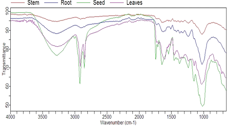
Fourier transform infra-red (FTIR) spectra of different parts of C. sinensis L.
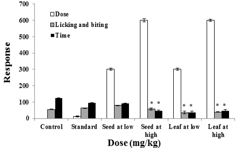
Effect of C. sinensis L. ethanolic seed and leaf extract on licking and biting response along with time duration in six mice groups in two phases categorized as (0-300s) early phase at low (300mg/kg) and high (600mg/kg) doses, respectively. Seed treated mice group at high dose showed significant decrease in licking and biting responses at high dose, while leaf group showed significant decrease at both low and high doses compared to control and standard groups.
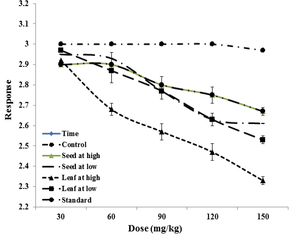
Comparative study of reduction in oedema in the hind paw of mice with passage of time among different groups. Control group showed persistent inflammation in the hind paw as no dose was given in this group. Significant reduction in the inflammation was observed in the leaf extract at high dose among the groups. Seed at high dose produced the same effect as that was observed in standard group.
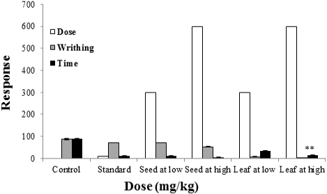
Relation between the reduction in the number of time and writhes among the groups. Control group showed high count of writhes for greater amount of time. Significant reduction in the time and number of writhes was observed in the case of leaf extract at high dose which were better than seed extract at the same dose level. The results showed that crude extract of seed and leaf produce highly significant results compared to the standard.
References
- Boussouf, L., Boutennoune, H., Kebieche, M., Adjeroud, N., Al-Qaoud, K. and Madani, K., 2017. Anti-inflammatory, analgesic and antioxidant effects of phenolic compound from Algerian Mentha rotundifolia L. leaves on experimental animals. S. Afr. J. Bot., 113: 77-83. https://doi.org/10.1016/j.sajb.2017.07.003[CrossRef]
- Chan, E.W., Soh, E.Y., Tie, P.P., and Law, Y.P, 2011. Antioxidant and antibacterial properties of green, black, and herbal teas of Camellia sinensis. Pharmacogn. Res., 3: 266. https://doi.org/10.4103/0974-8490.89748[CrossRef]
- Chen, Y., Tao, S., Zeng, F., Xie, L. and Shen, Z., 2015. Antinociceptive and anti-inflammatory activities of Schefflera octophylla extracts. J. Ethnopharm., 171: 42-50. https://doi.org/10.1016/j.jep.2015.04.050[CrossRef]
- Deraedt, R., Jouquey, S., Delevallée, F. and Flahaut, M., 1980. Release of prostaglandins E and F in an algogenic reaction and its inhibition. Eur. J. Pharm., 61: 17-24. https://doi.org/10.1016/0014-2999(80)90377-5[CrossRef]
- Farooqui, A., Khan, A., Borghetto, I., Kazmi, S.U., Rubino, S., and Paglietti, B., 2015. Synergistic antimicrobial activity of Camellia sinensis and Juglans regia against multidrug-resistant bacteria. PLoS One, 10: e0118431. https://doi.org/10.1371/journal.pone.0118431[CrossRef]
- Folashade, O., Omoregie, H., and Ochogu, P., 2012. Standardization of herbal medicines. A review. Int. J. Biodivers. Conserv., 4: 101-112. https://doi.org/10.5897/IJBC11.163[CrossRef]
- Ganeshpurkar, A., and Rai, G., 2013. Experimental evaluation of analgesic and anti-inflammatory potential of Oyster mushroom Pleurotus florida. Indian J. Pharmacol., 45: 66. https://doi.org/10.4103/0253-7613.106438[CrossRef]
- Gupta, D., and Kumar, M., 2017. Evaluation of in vitro antimicrobial potential and GC–MS analysis of Camellia sinensis and Terminalia arjuna. Biotechnol. Rep., 13: 19-25. https://doi.org/10.1016/j.btre.2016.11.002[CrossRef]
- Hamilton-Miller, J., 2001. Anti-cariogenic properties of tea (Camellia sinensis). J. med. Microbiol. 50: 299-302. https://doi.org/10.1099/0022-1317-50-4-299[CrossRef]
- Ijaz, N., Durrani, A.I., Rubab, S. and Bahadur, S., 2021. Formulation and characterization of Aloe vera gel and tomato powder containing cream. Acta Ecol. Sin., pp. 1-9. https://doi.org/10.1016/j.chnaes.2021.01.005[CrossRef]
- Iqbal, S., Parveen, N., Bahadur, S., Ahmad, T., Shuaib, M., Nizamani, M.M., Urooj, Z. and Rubab, S., 2020. Paclobutrazol mediated changes in growth and physio-biochemical traits of okra (Abelmoschus esculentus L.) grown under drought stress. Gene Rep., 21: 1-9. https://doi.org/10.1016/j.genrep.2020.100908[CrossRef]
- Ishtiaq, S., Hanif, U., Ajaib, M., Shaheen, S., Afridi, M.S.K., and Siddiqui, M.F., 2018. Pharamcognostical and physicochemical characterization of Amaranthus graecizans subsp. Silvestris: An anatomical perspective. Pak. J. Bot., 50: 307-312.
- Janakiraman, N., Sahaya, S.S., and Johnson, M., 2012. Anti–bacterial studies on Peristrophe bicalyculata (Retz.) Nees. Asian Pac. J. trop. Biomed., 2: S147-S150. https://doi.org/10.1016/S2221-1691(12)60146-4[CrossRef]
- Kerwat, K., Kerwat, M., Graf, J., and Wulf, H., 2010. Resistance to antibiotics and multiresistant pathogens. Anasthesiol. Intensivmed. Notfallmed. Schmerzther., 45: 242-243. https://doi.org/10.1055/s-0030-1253091[CrossRef]
- Liaqat, I., 2009. Fourier transform infrared spectroscopy of dental unit water line biofilm bacteria. Spectroscopy, 23: 175-189. https://doi.org/10.1155/2009/892569[CrossRef]
- Liaqat, I., Arshad, N., Arshad, M., Mirza, S.A., Ali, N.M. and Shoukat, A., 2017. Antimicrobial activity of some medicinal plants extracts against food industry isolates. Pakistan J. Zool., 49: 565-572. https://doi.org/10.17582/journal.pjz/2017.49.2.523.530[CrossRef]
- Mahmood, T., Akhtar, N., and Khan, B.A., 2010. The morphology, characteristics, and medicinal properties of Camellia sinensis tea. J. med. Pl. Res., 4: 2028-2033. https://doi.org/10.5897/JMPR10.010[CrossRef]
- Meek, I.L., Van de Laar, M.A., and Vonkeman, E.H., 2010. Non-steroidal anti-inflammatory drugs: An overview of cardiovascular risks. Pharmaceuticals, 3: 2146-2162. https://doi.org/10.3390/ph3072146[CrossRef]
- Mumtaz, T., Rizwani, G.H., and Shareef, H., 2017. Analgesic activities of crude ethanolic extract and various fractions of Adansonia digitata L. grown at the Sindh province of Pakistan. Pak. J. Pharm. Sci., 30: 1657-1663.
- Mustaffa, F., Indurkar, J., Ismail, S., Mordi, M., Ramanathan, S., and Mansor, S., 2010. Analgesic activity, toxicity study and phytochemical screening of standardized Cinnamomum iners leaves methanolic extract. Pharmacogn. Res., 2: 76. https://doi.org/10.4103/0974-8490.62952[CrossRef]
- Olila, D., and Opuda-Asibo, J., 2001. Antibacterial and antifungal activities of extracts of Zanthoxylum chalybeum and Warburgia ugandensis, Ugandan medicinal plants. Afr. Hlth. Sci., 1: 66-72.
- Paliwal, S.K., Sati, B., Faujdar, S., and Sharma, S., 2017. Studies on analgesic, anti-inflammatory activities of stem and roots of Inula cuspidata CB Clarke. J. Tradit. Complement. Med., 7: 532-537. https://doi.org/10.1016/j.jtcme.2016.08.005[CrossRef]
- Pawar, P.L., and Nabar, B.M., 2010. Effect of plant extracts formulated in different ointment bases on MDR strains. Indian J. Pharm. Sci., 72: 397. https://doi.org/10.4103/0250-474X.70494[CrossRef]
- Ponnusamy, S., Gnanaraj, W.E., Marimuthu, J., Selvakumar, V., and Nelson, J., 2010. The effect of leaves extracts of Clitoria ternatea Linn against the fish pathogens. Asian Pac. J. trop. Med., 3: 723-726. https://doi.org/10.1016/S1995-7645(10)60173-3[CrossRef]
- Radji, M., Agustama, R.A., Elya, B., and Tjampakasari, C.R., 2013. Antimicrobial activity of green tea extract against isolates of methicillin–resistant Staphylococcus aureus and multi drug resistant Pseudomonas aeruginosa. Asian Pac. J. trop. Biomed., 3: 663-667. https://doi.org/10.1016/S2221-1691(13)60133-1[CrossRef]
- Rajeswari, A., 2015. Evaluation of phytochemical constituents, Quantitative analysis and antimicrobial efficacy of potential herbs against selected microbes. Evaluation, 8: 2-4.
- Rubab, S., Bahadur, S., Hanif, U., Durrani, A.I., Sadiqa, A., Shafique, S., Zafar, U., Shuaib, M., Urooj, Z., Nizamani, M.M. and Iqbal, S., 2021. Phytochemical and antimicrobial investigation of methanolic extract/fraction of Ocimum basilicum L. Biocatal. Agric. Biotechnol., 31: 101894. https://doi.org/10.1016/j.bcab.2020.101894[CrossRef]
- Rubab, S., Hussain, I., Khan, B.A., Unar, A.A., Abbas, K.A., Khichi, Z.H., Khan, M., Khanum, S., Rehman, K.U. and Khan, H., 2017. Biomedical description of Ocimum basilicum L. J. Inf. Image Manage., 12: 57-69.
- Rubab, S., Rizwani, G.H., Bahadur, S., Shah, M., Alsamadany, H., Alzahrani, Y., Shuaib, M., Hershan, A., Hobani, Y.H. and Shah, A.A., 2020a. Determination of the GC–MS analysis of seed oil and assessment of pharmacokinetics of leaf extract of Camellia sinensis L. J. King Saud Univ. Sci., 32: 3138-3144. https://doi.org/10.1016/j.jksus.2020.08.026[CrossRef]
- Rubab, S., Rizwani, G.H., Bahadur, S., Shah, M., Alsamadany, H., Alzahrani, Y., Alghamdi, S.A., Anwar, Y., Shuaib, M., Shah, A.A. and Muhammad, I., 2020b. Neuropharmacological potential of various morphological parts of Camellia sinensis L. Saudi J. biol. Sci., 27: 567-573. https://doi.org/10.1016/j.sjbs.2019.11.025[CrossRef]
- Saha, D., Dasgupta, S., and Saha, A., 2005. Antifungal activity of some plant extracts against fungal pathogens of tea (Camellia sinensis.). Pharm. Biol., 43: 87-91. https://doi.org/10.1080/13880200590903426[CrossRef]
- Santangelo, C., Varì, R., Scazzocchio, B., Di Benedetto, R., Filesi, C., and Masella, R., 2007. Polyphenols, intracellular signalling and inflammation. Ann. Ist. Super. Sanita., 43: 394.
- Tatiya, A.U., Saluja, A.K., Kalaskar, M.G., Surana, S.J., and Patil, P.H., 2017. Evaluation of analgesic and anti-inflammatory activity of Bridelia retusa (Spreng) bark. J. Tradit. Complement. Med., 7: 441-451. https://doi.org/10.1016/j.jtcme.2016.12.009[CrossRef]
- Wollen, K.A., 2010. Alzheimer’s disease: The pros and cons of pharmaceutical, nutritional, botanical, and stimulatory therapies, with a discussion of treatment strategies from the perspective of patients and practitioners. Altern. Med. Rev., 15: 223-244.
- Xiong, L., Li, J., Li, Y., Yuan, L., Liu, S., Huang, J.A., and Liu, Z., 2013. Dynamic changes in catechin levels and catechin biosynthesis-related gene expression in albino tea plants (Camellia sinensis L.). Pl. Physiol. Biochem., 71: 132-143. https://doi.org/10.1016/j.plaphy.2013.06.019[CrossRef]
- Yashin, A.Y., Nemzer, B.V., Combet, E., and Yashin, Y.I., 2015. Determination of the chemical composition of tea by chromatographic methods: A review. J. Fd. Res., 4: 56. https://doi.org/10.5539/jfr.v4n3p56[CrossRef]
- Yougbare-ziebrou, M.N., lompo, M., Ouedraogo, N., Yaro, B., and Guissoun, I.P., 2016. Antioxidant, analgesic and anti-inflammatory activities of the leafy stems of Waltheria indica L. (Sterculiaceae). J. appl. Pharm. Sci., 6: 124-129. https://doi.org/10.7324/JAPS.2016.60219[CrossRef]
- Zulqarnain, A., Durrani, A.I., Saleem, H. and Rubab, S., 2021. Development of an ultrasonic-assisted extraction technique for the extraction of natural coloring substance chlorophyll from leaves of carica papaya. J. Oleo Sci., 70: 1367-1372. https://doi.org/10.5650/jos.ess21118[CrossRef]
Supplementary material
Services
Article usage statistics
Loading…