Research Article · Volume 13, Issue 2, 21 January 2025 · Open Access
Neurocognitive and Affective Disorders Induced by Inorganic Mercury Exposure in Male Wistar Rats: Unraveling the Role of Oxidative Stress
· · · · ·
Affiliations & Notes ⌄
1Laboratory of Biology and Health, Department of Biology, Faculty of Sciences, Ibn Tofail University, Kenitra, Morocco. *Correspondence | Sofia Azirar, Abdelghafour El Hamzaoui, Laboratory of Biology and Health, Department of Biology, Faculty of Sciences, Ibn Tofail University, Kenitra, Morocco
These results underscore the importance of refining risk assessment strategies and developing targeted preventive therapies supporting the internal antioxidant defense systems to safeguard the public health of heavy metal-induced behavioral disorders.
The findings also highlight the need for enhanced environmental and public health policies.
Despite low doses, chronic Hg exposure can accumulate and exert significant neurotoxic effects, even at levels that may be encountered in real-world settings.
Incorporating these insights into public health guidelines could help mitigate the risks of Hg-related neurobehavioral disorders.
Selected from the article's own text.
Abstract
In this study, we assessed the impact of prolonged exposure to low doses of mercury (Hg) on affective and cognitive impairments in male Wistar rats, highlighting the significance of understanding chronic exposure to environmental levels of Hg that may not immediately show overt toxicity. 24 rats received either NaCl 0.9% (control group) or mercurial chloride (HgCl2) via intraperitoneal injections at 0.25, 0.5, or 1 mg/kg for eight weeks. Behavioral evaluations included the open field (OFT), elevated plus maze (EPM), and forced swimming tests (FST) for anxiety and depression assessments, alongside the Y maze and Morris water maze (MWM) tests for memory and learning. In addition, markers of oxidative stress (OS) in the hippocampus— catalase (CAT), nitric oxide (NO), and lipid peroxidation (LPO)—were measured post-treatment. Results revealed dose-dependent increases in anxiety and depression-like behaviors, such as significant reductions in time spent in the center area (TCA) in OFT at 0.5 and 1 mg/kg compared to controls (p < 0.001) and increased immobility time in FST across all doses compared to controls (p < 0.001). Memory impairments were most pronounced at 1 mg/kg in the Y maze test (p < 0.01) and in MWM (p < 0.001). OS markers correlated with behavioral changes, with elevated LPO (p < 0.001 at 0.5 and 1 mg/kg compared to controls) and decreased CAT activity across doses (p < 0.001), implicating OS in Hg-induced neurotoxicity. Although these findings suggest a link between low-dose Hg exposure and neurobehavioral impairment, further studies are needed to clarify causative mechanisms.
Mercury (Hg), a xenobiotic heavy metal, remains a global health and environmental threat, underlined by historical public health catastrophes in Minamata, Japan (Ministry of the Environment Japan, 2015), and Iraq . Among its various forms, inorganic Hg (Hg²⁺), commonly found in industrial emissions, dental amalgams, and some pharmaceuticals, is recognized for its neurotoxic effects . This form of Hg is a pervasive contaminant that can affect many populations worldwide (Wu et al., 2024).
Of interest, organic Hg, the prevalent form of human intoxication, undergoes a progressive metabolic conversion by intestinal microflora into inorganic Hg at a low daily rate . Both elemental and organic Hg can cross the blood-brain barrier (BBB), undergoing rapid intracellular oxidation into ionic Hg²⁺, subsequently persisting within cerebral cells for years . Accumulation of Hg²⁺ in the brain has been associated with neural excitability and irritability , neuropsychiatric disorders , and neurodegenerative diseases, such as Alzheimer’s and Parkinson’s . It has also been demonstrated that Hg species can cross the placental barrier, leading to significant neurodevelopmental impairments . Recent studies also report an association between elevated Hg levels and decreased cognitive performance in otherwise healthy young adults , indicating that Hg toxicity extends to mental health even at low environmental exposure levels.
The neurotoxicity of inorganic Hg involves complex interactions within the central nervous system (CNS). The myriad ways in which Hg disrupts cellular and molecular processes, leading to oxidative stress (OS) , mitochondrial dysfunction , and inflammation , remain a subject of ongoing investigations. OS, in particular, serves as a key mechanism by which heavy metals like Hg exert their toxic effects ; ; and is increasingly involved in the onset of neurocognitive and affective disorders . Several studies have demonstrated that chronic Hg exposure can trigger reactive oxygen species (ROS) production, including superoxide and hydroxyl radicals , causing damage to lipids, proteins, and DNA molecules, leading to cellular dysfunction . Given its reduced antioxidant capacity, high oxygen consumption, and high lipid content, the brain is especially vulnerable to OS .
Despite the well-documented neurotoxic effects of high-dose Hg exposure, the impact of sub-chronic low-dose exposure remains less well understood. Therefore, in this experiment, we aim to address a specific gap in the literature by exploring the neurobehavioral effects of low-dose Hg exposure over an extended period, mimicking real-world environmental exposure levels that could pose risks to human health. In addition, this study investigates the role of OS in affective and cognitive impairments induced by prolonged, low-dose exposure to mercuric chloride (HgCl2) in adult male Wistar rats. By performing extensive behavioral and biochemical assessments, we aim to elucidate how low-dose Hg disrupts neural processes, particularly focusing on the hippocampus and its vulnerability to OS. This research addresses the following questions:1) Does prolonged low-dose Hg exposure induce measurable anxiety, depression, and memory impairments in rats? 2) How does Hg exposure at sub-chronic low doses affect markers of OS in the hippocampus? 3) Is there a dose-dependent relationship between Hg exposure levels and the severity of neurobehavioral alterations? Through this investigation, we aim to provide a nuanced understanding of Hg’s neurotoxic mechanisms at low exposure levels, contributing critical insights to risk assessments and preventive strategies for Hg-related neurobehavioral disorders.
MATERIALS AND METHODS
Animal Model and Housing Conditions
Male adult Wistar rats (approximately 60 days old, initial weight:120 ± 20 g) were obtained from Ibn Tofaïl University’s Central Animal House. The choice of age and weight aligns with prior studies assessing neurobehavioral outcomes in rats, as adulthood begins after the eighth week of postnatal life . The animals were housed in conditions of 21 ± 1°C and 40 ± 5% relative humidity, under a 12-hour light-dark cycle, with unlimited access to commercial food pellets and tap water. Ethical approval for experimental procedures was granted by the local institutional ethics committee for animal experiments. The animals were weighed weekly for dose adjustment.
Experimental Design
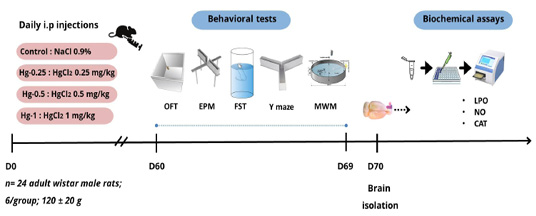
A total of 24 rats were allocated into four groups (n = 6 per group). This group size is consistent with prior studies employing similar behavioral paradigms and dosing protocols ; while also recognizing the associated limitations in statistical power. Figure 1 provides an overview of the experimental design. The groups were as follows:
Group
DRUGS AND DOSES
Control
NaCl 0.9%
Hg-0.25
HgCl2 at 0.25 mg/kg
Hg-0.5
HgCl2 at 0.5 mg/kg
Hg-1
HgCl2 at 1 mg/kg
Drug Administration
Saline solution, or HgCl2, was injected intraperitoneally (i.p.) and was obtained from Sigma Aldrich (St. Louis, MO, USA). Injections were performed daily between 4:00 pm and 4:30 pm for 8 weeks. Post-administration, rats underwent behavioral tests, followed by evaluations for OS indicators post-sacrifice.
Neurobehavioral Tests
A battery of neurobehavioral tests was performed. All equipment was properly cleaned and sterilized with a 7% alcohol solution before testing each rat to prevent any behavioral biases from residual odors.
Anxiety-Like Measurement
Open field test (OFT): Rats, when placed in new environments, tend to explore. Their behavior within this environment provides insight into their anxiety levels . Rats were individually placed in an apparatus measuring 100-L × 100-W × 40-H cm. Their behavior was monitored and recorded with emphasis on three metrics:Time Spent in the Center Area (TCA), Number of Returns to the Center (NRC), and Number of Total Squares (NTS). Anxiety levels were determined based on TCA and NRC, whereas NTS evaluated locomotor activity.
Elevated plus maze (EPM): This test gauges anxiety-like behavior in rats using the dual drive of exploration of a new space and aversion due to the elevation and brightness of the maze . Conducted a day after the OFT, each rat was placed on the central area (10× 10 cm) of the maze facing an open arm. The maze has two open arms (50 × 10 cm) and two closed arms (50 × 10 × 40 cm). Key metrics recorded during a 5-minute duration included the number of entries in open arms (EOA), time spent in open arms (TOA), and total arm entries (TAE). EOA and TOA indicate open space-induced anxiety-like behavior. TAE measures locomotor activity.
Depression-Like Measurement
Forced swimming test (FST): The FST is based on the principle that when rodents are placed in a container filled with water from which they cannot escape, they will initially display vigorous movement (interpreted as an escape behavior). Over time, they’ll exhibit periods of immobility, which are indicative of a state of despair or hopelessness ; . Rats were placed individually in a 30 cm-diameter transparent cylindrical container filled with water (23 ± 2 °C) to a height of 35 cm. Each rat’s behavior during a 5-minute FST was recorded, specifically:Immobility Time (IT) and Climbing Time. An increase in IT suggests depressive-like behavior, while increased climbing time indicates the opposite.
Cognitive Measurement
Y-maze test: The Y-maze is based on the innate curiosity of rodents. They have a natural tendency to explore new environments, especially when given a choice. A decrease in spontaneous alternation (choosing a different arm than the one previously entered) suggests a potential impairment in the rodent’s short-term spatial memory . At the onset of an 8-minute test, rats were placed on the central area of a Y-maze with three arms (A, B, and C; dimensions 61 × 35 × 12 cm3). Sequences of arm entries were recorded for the entire test duration. Spontaneous alternation behavior, an indicator of spatial working memory, is characterized by the consecutive entry into three distinct arms. It is typically measured using the following formula:
Equation 1.
Morris water maze test (MWM): The MWM is used to assess learning and memory cognitive functions . The test involved a large circular water tank (110 cm in diameter, 20 cm in height) filled with opaque water maintained at 22°C. A circular platform was hidden 0.5 cm below the water surface in the northeast section of the tank. The experiment consisted of two phases:an acquisition phase (4 trials per day over 4 days) and a probe trial conducted on the 5th day. The acquisition phase tracked each rat’s ability to locate the hidden platform within 60 seconds. If unsuccessful, they were guided to the platform and allowed a 10s stay. The 5th day’s probe trial tested spatial memory over a 60-second duration. Post completion of the behavioral tests, rats are anesthetized and euthanized by rapid decapitation, followed by the extraction and preparation of their brains for comprehensive biochemical and histological analysis.
Sample Preparation and Analysis of OS Parameters in Hippocampus Tissues
The hippocampus was meticulously dissected using a freehand technique under a dissecting microscope. Each dissected tissue was homogenized using a mechanical homogenizer in 10 volumes (50 mM W/V) of ice-cold phosphate buffer (pH 7.4) . The tissue homogenates were centrifuged at a relative centrifugal force of 1500 rpm for 10 minutes at 4 °C using a refrigerated centrifuge, and the supernatant was used for the analysis of nitric oxide (NO) and lipid peroxidation (LPO) levels and catalase (CAT) activity.
NO levels were estimated using the Griess reagent . The formation of lipid peroxides was provided by measuring the thiobarbituric-acid-reacting substances (TBARS) in cells via the method of Draper and Hadley (1990). The CAT activity was calculated using the method described by Aebi . All readings were normalized to protein content, determined using the Bradford method. CAT activity was measured in units/g of tissue, NO levels were expressed in μM/g of tissue, and LPO was quantified in nM/g of tissue ; ; .
Statistical Analysis
The data for this investigation were examined using a one-way analysis of variance (ANOVA), followed by Tukey’s HSD post-hoc test to identify statistically significant differences between the experimental groups. A repeated measure ANOVA was used for the MWM test. Results are presented as mean ± SEM. In addition to p-values, Cohen’s d-effect sizes were calculated to assess the magnitude of differences between groups. A p-value of less than 0.05 was considered indicative of statistical significance. A magnitude of d equal to or superior to 0.8 was interpreted as a large effect size . All analyses were conducted using SPSS version 29 (IBM Corp., Armonk, NY, USA), and graphs were made using Prism 8 (GraphPad Software Inc., San Diego, 189 CA, USA.)
RESULTS AND DISCUSSION
Effect of Hg on the Levels of Anxiety-Like Measured in Open Field Test
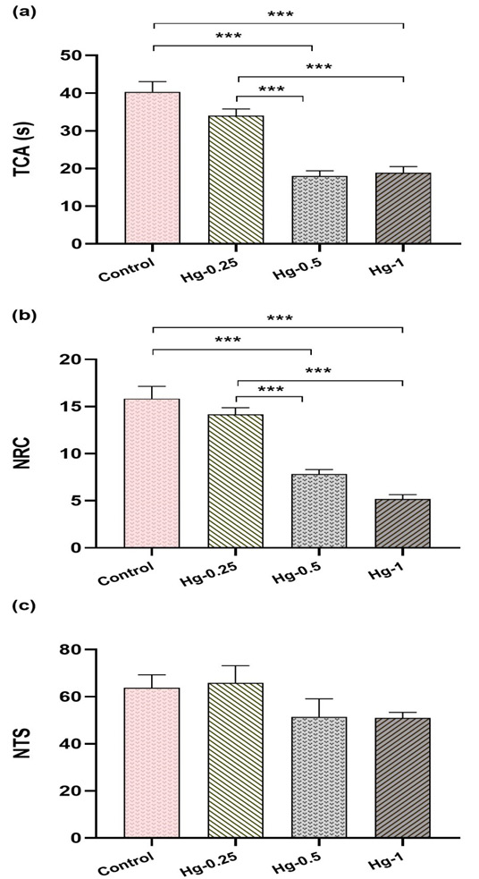
Statistical analysis revealed that the TCA and NRC parameters were significantly affected by Hg treatment (Figures 2a and 2b). Both parameters decreased in comparison to the control group (TCA:Cont/Hg-0.5:p < 0.001, d = 4.18 and Cont/Hg-1:p < 0.001, d = 3.86; NRC:Cont/Hg-0.5:p < 0.001, d = 0.64 and Cont/Hg-1:p < 0.001, d = 1.02). Additionally, a dose-dependent response was observed, with statistically significant differences observed between the groups (TCA:Hg-0.25/Hg-0.5:p < 0.001, d = 4.00 and Hg-0.25/Hg-1:p < 0.001, d = 3.53; NRC:Hg-0.25/Hg-0.5:p < 0.001, d = 0.55 and Hg-0.25/Hg-1:p < 0.001, d = 1.28). However, locomotor activity, represented by the NTS, was unaffected by any treatment (p > 0.05), with an effect size of d = 0.3, indicating a minimal change (Figure 2c). This stability in NTS suggests that the anxiety-like behavior observed is not attributable to altered locomotor activity, which strengthens the interpretation of TCA and NRC reductions as true indicators of anxiety-like behavior rather than confounded by mobility limitations.
Effect of Hg on Anxiety Levels Measured in Elevated Plus Maze Test
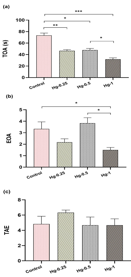
Results indicated that EOA was significantly decreased by Hg treatment in a dose-dependent manner (Figure 3b). Compared to the control group, EOA was reduced in the Hg-0.25 (p < 0.01, d = 0.78), Hg-0.5 (p < 0.05, d = 0.35), and Hg-1 (p < 0.001, d = 1.53) groups. TAE showed no significant differences among groups (p > 0.05) (Figure 3a), with negligible effect sizes (d lower to 0.1), suggesting resilience in exploratory behavior despite the presence of anxiety-like responses. This lack of significant difference in TAE supports the notion that Hg exposure primarily affects anxiety responses without diminishing general activity (Figure 3c).
Effect of Hg on Depression Levels Measured in Forced Swimming Test
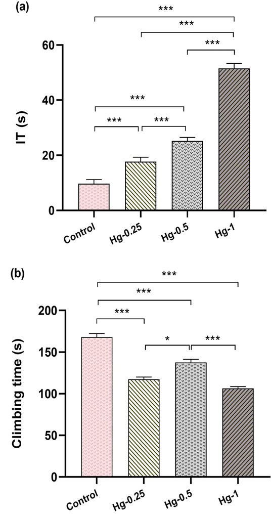
Statistical analyses for FST parameters revealed that sub-chronic Hg injection notably increased IT and decreased climbing time compared to the control group, regardless of the dose of treatment (p < 0.001) (Figure 4a). A dose-dependent effect on IT was also observed between the distinct groups (Hg-0.25/Hg-0.5:p < 0.001, d = 2.09; Hg-0.25/Hg-1:p < 0.001, d = 3.18; Hg-0.5/Hg-1:p < 0.001, d = 4.51). The climbing time reductions were particularly pronounced at 1 mg/kg, indicating that Hg exposure suppresses adaptive escape behavior, a hallmark of depressive-like states in the FST (Control vs. Hg-1:d = 2.96) further reinforcing this depressive-like response (Figure 4b).
Effect of Hg on Memory Measured in Y-maze
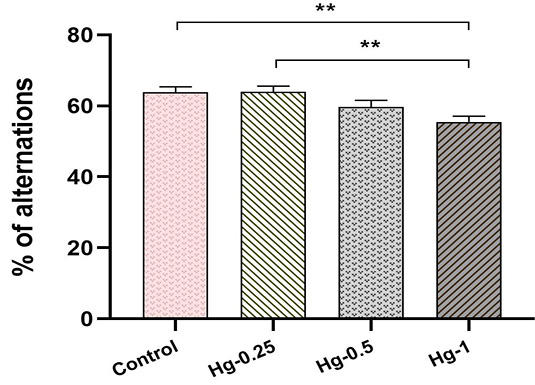
The spontaneous alternation percentage was significantly impacted by Hg treatment compared to the control group (Cont/Hg-1:p < 0.01, d = 2.20). Likewise, a dose-related significant increase in this parameter was observed in the Hg-0.25 group compared to the Hg-1 treated group (p < 0.01, d = 2.13) (Figure 5).
Effect of Hg on Memory Measured in Morris Water Maze
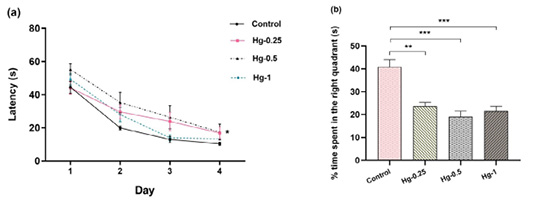
All groups showed decreased latency over four days during the acquisition phase to reach the hidden platform. Statistical analysis revealed a significant difference (p < 0.05) between the rats in the Hg-0.5-treated group and the control group (Figure 6a).
In the probe trial, the percentage of time spent in the target quadrant decreased significantly in all Hg-treated groups compared to controls (Cont/Hg-0.25:p < 0.01, d = 2.76; Cont/Hg-0.5:p < 0.001, d = 3.14; Cont/Hg-1:p < 0.001, d = 2.93) (Figure 6b).
Hg effect on Oxidative stress
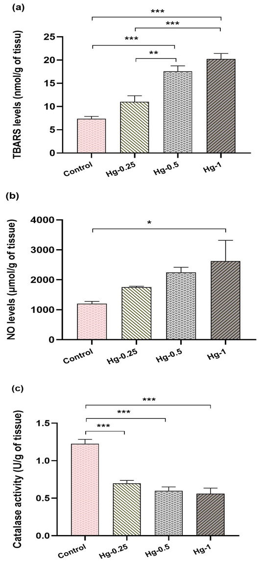
Lipid peroxidation in hippocampus: Statistical analysis showed a significant dose-dependent increase in TBARS levels in the hippocampus at 0.5 and 1 mg/kg of Hg (Cont/Hg-0.5:p < 0.001, d = 1.0; Cont/Hg-1:p < 0.001, d = 1.3), indicating enhanced LPO. No significant increase was observed at 0.25 mg/kg (Cont/Hg-0.25 :p > 0.05, d = 0.02). Moreover, significant differences were noted between the Hg-0.25/Hg-0.5 and Hg-0.25/Hg-1 groups (p < 0.01, d = 0.65; p < 0.001, d = 0.80, respectively), but not between the Hg-0.5 and Hg-1 groups (p > 0.05) (Figure 7a).
Nitric oxide concentrations in the hippocampus: Hg at 1 mg/kg significantly reduced NO levels in the hippocampus compared to controls (p < 0.05, d = 0.85). No notable differences were observed among the other groups (Figure 7b).
Catalase activity in the hippocampus: Statistical analyses revealed that sub-chronic Hg injection notably decreased CAT activity compared to the control group, regardless of the dose of treatment (p < 0.001, d ranging from 0.9 to 1.2), with no significant differences between the treatment groups (p > 0.05) (Figure 7c).
Cognitive functions, including memory, learning, attention, and executive abilities, play a pivotal role in guiding complex behaviors and decision-making processes. Concurrently, affective regulation significantly influences an individual’s emotional and social well-being. Hg-induced disturbances in these domains not only present a profound impact on the exposed organisms but also raise concerns about public health and environmental safety.
This study seeks to expand the current understanding of the neurobehavioral and biochemical effects of sub-chronic i.p. administration of gradually increasing low doses (below the lowest observed effect dose) of inorganic Hg on adult Wistar rats. Although data suggests that inorganic Hg can barely cross the BBB, chronic exposure has proven otherwise . The i.p. route was chosen to bypass the conversion of inorganic Hg to methylmercury (MeHg) in the gut , whose effects are well documented ; . Furthermore, other Hg forms, well-studied for their neurotoxic effects, convert to inorganic Hg once inside the body. Therefore, it’s possible that Hg²⁺, the oxidized form of Hg, plays a direct role in the toxic effects linked to Hg vapor (Hg0) and MeHg exposure . Experiments with primates indicate that chronic MeHg exposure increases Hg2+ levels in the CNS due to demethylation . Since Hg2+ cannot cross back the BBB easily, it accumulates in the CNS .
Although the neurotoxic effects of Hg exposure on the developing brain are well-established, effects on adults have yielded inconsistent results , possibly due to fully matured protective mechanisms such as BBB. This protocol was designed to simulate real-life environmental conditions involving continuous exposure of the adult brain to very low doses. Overall, our data reveal significant enzymatic and behavioral changes, supporting the presence of HgCl2 in the brain.
The anxiety-like behavior, as evidenced by the OFT and EPM results, indicates that rats exhibited a significant dose-dependent effect of HgCl2 on anxiety levels in response to the unfamiliar environments of the testing apparatus. The anxiogenic effect starts at a dose of 0.25 mg/kg and reaches the highest significant difference at a dose of 1 mg/kg in comparison with the control group. Of interest, the depression-like measurements in the FST revealed distinct behavioral patterns, with HgCl2-exposed rats displaying significantly increased IT, suggesting a state of despair or hopelessness, while controls exhibited significantly prolonged climbing time, indicating a more adaptive response. It is important to note that a significant difference was observed between treated groups, highlighting the dose-dependent depressogenic-like effect. In addition to these affective abnormalities, sub-chronic exposure to HgCl2 has been shown to alter cognitive functionalities, such as the working and short-term spatial memory tested in the Y maze, in which the spontaneous alternation score was significantly reduced following the treatment only with a 1 mg/kg dosage. Conversely, the spatial learning evaluated in the MWM was significantly affected by all treatment doses. These behavioral outcomes align with several prior studies associating Hg exposure with heightened anxiety-like, depression-like levels and compromised cognitive functions in rodents ; ; .
Biochemical investigations revealed significantly higher levels of LPO in the hippocampus, as confirmed by elevated TBARS concentrations in treated rats compared to controls. LPO was affected in a dose-dependent manner; an increase was observed following the 0.25 mg/kg treatment, but no statistical significance was reached. While 0.5 mg/kg and 1 mg/kg doses significantly increased LPO levels compared to the control and Hg-0.25 groups. LPO affects mitochondrial membranes , and so does Hg, altering mitochondrial permeability and leading to increased Ca2+ release , which stimulates acetylcholine (Ach) production .
A study on adult mice showed that exposure to low HgCl2 doses decreased Ach Esterase (AchE) activity . This cascade of cellular events contributes to the accumulation of ACh at the synaptic cleft, potentially leading to the desensitization of cholinergic receptors on the postsynaptic membrane . The resulting disruption in cholinergic signaling may provide an alternative mechanism underlying the observed behavioral impairments. In line with prior research , our biochemical analyses illustrate Hg’s role in augmenting OS; diminished CAT activity and increased NO levels in the hippocampus post-Hg exposure testify to perturbations in the OS-mediated signaling pathways, previously suggested as a neurotoxic mechanism of trace heavy metals ; ; ; . Here, we report on the inhibition of CAT activity; a significant decrease in this parameter was observed in comparison to controls, regardless of the treatment dose, as the effect was highly tangible even in the Hg-0.25 group treated with the lowest dose. NO levels were only sensitive to the 1 mg/kg dose, showing an increase in all treated groups but reaching statistical significance only with the highest dose. Among several brain regions, literature has proven that the hippocampus is particularly vulnerable to OS . The inhibition of CAT provides evidence that the enzymatic antioxidant defense system of the hippocampus was damaged, which can be the promoter of LPO observed above. Through a cascade of intracellular reactions, these disruptions could initiate the increase of NO . This is particularly relevant to the anxiety, depression, and impaired memory observed in treated animals, as previous data provide evidence for a causal direct link between OS and high affective and cognitive disorders ; . Hovatta et al., uncovered an intriguing aspect of anxiety genetics; the expression levels of two antioxidant enzymes, glyoxalase 1 (Glo1) and glutathione reductase 1 (Gsr), were found to be positively correlated with the anxiety state of multiple mouse strains . Recent research suggests that the combination of aggregated β-amyloid (Aβ) and a prooxidant cocktail is the most accurate method for replicating the natural progression of Alzheimer’s disease (AD), effectively mimicking hippocampal cell damage in vitro . Our findings on memory impairment and the presence of high OS markers in the hippocampus of Hg-exposed Wistar rats align with this model. This similarity highlights Hg’s potential role in accelerating neurodegenerative processes, particularly in populations with existing vulnerabilities. Expanding neurotoxicology research to focus on the long-term effects of low-dose Hg exposure could provide valuable insights into broader neurodegenerative trends.
One potential mechanism of Hg neurotoxicity involves its interference with glutamate transport. Hg increases glutamate release and inhibits its uptake by astrocytes, leading to neuronal excitotoxicity . Elevated glutamate levels in the synaptic cleft cause excess Ca2+ influx through NMDA receptors in the post-synaptic terminal, which increases NO synthase (iNOS) activity, which in turn boosts NO production in mitochondria . Additionally, microglia are extremely reactive to OS . ROS and reactive nitrogen species (RNS) can trigger microglial activation, leading them to release pro-inflammatory cytokines like TNF-α, IL-1β, and IL-6, and additional ROS/RNS , creating a feed-forward loop of inflammation and OS. This process activates the nuclear factor-kappa B (NF-κB) pathway , a crucial nuclear transcription factor, leading to the expression of genes encoding pro-inflammatory mediators . Both ROS and the neuroinflammation state can harm BBB endothelial cells, increasing permeability and thus allowing peripheral immune cells to infiltrate the CNS, which further exacerbates the inflammatory response . This mechanism is further supported by the stipulation of quick and robust activation of microglial cells following Hg treatment, mainly causing rapid ROS generation and glutathione depletion . Moreover, the comorbidity of neuroinflammation with the development of neuropsychiatric and neurodegenerative diseases is now well-established in the scientific literature .
A cross-sectional epidemiological study conducted on 920 Japanese young adults revealed that even low-level exposure to Hg, mainly from daily fish consumption, is significantly and negatively associated with cognitive performance. Interestingly, those results indicated that greater hair Hg level was significantly associated with lower gray matter volume in the left hippocampus . The accumulation of Hg in the brain could be associated with reduced cerebral blood flow, which has been proven to play a role in neurodegeneration . Accordingly, a recent meta-analysis found that exposure to low blood Hg levels was strongly correlated with levels of free thyroxine and thyroid-stimulating hormone and inversely correlated with thyroxine . Clinical investigations have also evidenced a significant association between untreated diagnosed thyroid disorders and depression and anxiety . To support our hypothesis, there is a notable connection between increased ROS generation and oxidative damage with thyroid diseases . Furthermore, thyroid dysfunction may lead to dysregulation of the hypothalamic-pituitary-adrenal axis, which is highly implicated in affective disorders and emotional responses . These interconnections may partially explain the anxiety-like and depression-like behavior observed in our tested animals.
CONCLUSIONS AND RECOMMENDATIONS
Taken together, the current findings indicate that sub-chronic, low-dose inorganic Hg exposure in adult Wistar rats significantly affects cognitive functions and emotional regulation, revealing pronounced anxiety-like and depression-like behaviors, hippocampus-dependent cognitive deficits, increased OS, elevated LPO, and decreased antioxidant enzyme activity. The 1 mg/kg dose of HgCl2, identified as the minimal dose with the maximum effect, will be the focus of further investigations. These results underscore the importance of refining risk assessment strategies and developing targeted preventive therapies supporting the internal antioxidant defense systems to safeguard the public health of heavy metal-induced behavioral disorders. The findings also highlight the need for enhanced environmental and public health policies. Despite low doses, chronic Hg exposure can accumulate and exert significant neurotoxic effects, even at levels that may be encountered in real-world settings. This supports regulatory calls to limit Hg exposure in industrial and agricultural practices and raises awareness about the risks of Hg in consumer products. Incorporating these insights into public health guidelines could help mitigate the risks of Hg-related neurobehavioral disorders.
However, our study has certain limitations, including a relatively small sample. This decision was guided by ethical considerations to minimize animal use, in accordance with principles of animal experimentation. Future studies should consider larger sample sizes and a more diverse range of animal models to strengthen the generalizability and robustness of the findings. Additionally, expanding research to include both male and female subjects may reveal sex-specific responses to Hg exposure.
ACKNOWLEDGMENTS
We are appreciative of the advice and knowledge provided by the biology department staff of Ibn Tofail University’s Faculty of Science.
NOVELTY STATEMENT
This study is, to our knowledge, the first to investigate the low dose-dependent effects of subchronic exposure to HgCl2 on anxiety-like and depression-like behaviors, as well as memory impairment and OS, in adult male Wistar rats.
AUTHOR’S CONTRIBUTIONS
Sofia azirar and Abdelghafour El Hamzaoui performed the experiments, analyzed the data and wrote the paper. Mouloud Lamtai and Mohamed Yassine El Brouzi participated in behavioral analysis and statistical significance. Aboubaker El Hessni reviewed and provided comments on the content and interpretation of the manuscript. Abdelhalem Mesfioui supervised the work, revised and approved the manuscript.
Highlights
Chronic Hg administration induces neurotoxicity in rats.
Hg administration provokes oxidative stress in rats.
Hg provokes its effects in a dose-dependent manner.
Conflict of Interest
The authors declare no conflict of interest.
Figures
Figure 1:.
Description of the experimental setup, including the exposure of the model to HgCl2 and the composition of the experimental groups exposed to either NaCl (0.9%) or HgCl2; following a 60-day intraperitoneal injection period, rats underwent behavioral testing, including the Open Field test (OFT), Elevated Plus Maze (EPM), Forced Swimming Test (FST), Y-maze and Morris Water Maze (MWM) test; Subsequently, animals were euthanized and their hippocampi were collected for the evaluation of oxidative stress markers (lipid peroxidation (LPO), nitric oxide (NO) levels, and catalase (CAT) activity).
Figure 2:.
Low dose-dependent sub-chronic effect of HgCl2 on anxiety-related behavior of male Wistar rats subjected to the open field test. (a) Total amount time spent in the center (TCA); (b) Number of returns into center area of the arena in the open-field behavior apparatus (NRC); (c) Number of total squares (NTS). Data are expressed as mean ± SEM. The significance level is 0.05 with *p < 0.05, **p < 0.01, ***p < 0.001.
Figure 3:.
Low dose-dependent sub-chronic effect of HgCl2 on anxiety-related behavior of male Wistar rats subjected to the elevated plus maze. (a) Total amount of time spent in exposed arms (TOA); (b) Number of entries in exposed arms (EOA); (c) Total number of arms entries (TEA). Data are expressed as mean ± SEM. The significance level is 0.05 with *p < 0.05, **p < 0.01, ***p < 0.001.
Figure 4:.
Low dose-dependent sub-chronic effect of HgCl2 on depression-related behavior of male Wistar rats subjected to the forced swimming test. (a) Immobility time (IT); (b) climbing time. Data are expressed as mean ± SEM. The significance level is 0.05 with *p < 0.05, **p < 0.01, ***p < 0.001.
Figure 5:.
Low dose-dependent sub-chronic effect of HgCl2 on cognitive-related behavior of male Wistar rats subjected to the Y maze. % of alternation:spontaneous alternation percentage. Data are expressed as mean ± SEM. The significance level is 0.05 with *p < 0.05, **p < 0.01, ***p < 0.001.
Figure 6:.
Low dose-dependent sub-chronic effect of HgCl2 on cognitive-related behavior of male Wistar rats subjected to the Morris water maze. (a) Latency:escape latency during the acquisition phase; (b) Time spent in the target quadrant. Data are expressed as mean ± SEM. The significance level is 0.05 with *p < 0.05, **p < 0.01, ***p < 0.001.
Figure 7:.
Low dose-dependent sub-chronic effect of HgCl2 on levels of (a) thiobarbituric acid reactive substances (TBARS) content, (b) nitric oxide (NO) and (c) catalase activity (CAT) in the hippocampus of male Wistar rats. Data are expressed as mean ± SEM. The significance level is 0.05 with *p < 0.05, **p < 0.01, ***p < 0.001.
References
Aebi H (1984). Catalase in vitro. Methods Enzymol., 105:121–126. https://doi.org/10.1016/s0076-6879(84)05016-3[CrossRef]
Angelova PR, Esteras N, Abramov AY (2021). Mitochondria and lipid peroxidation in the mechanism of neurodegeneration:Finding ways for prevention. Med. Res. Rev., 41: 770–784. https://doi.org/10.1002/med.21712[CrossRef]
Apweiler M, Saliba SW, Sun L, Streyczek J, Normann C, Hellwig S, Bräse S, Fiebich BL (2024). Modulation of neuroinflammation and oxidative stress by targeting GPR55 – new approaches in the treatment of psychiatric disorders. Mol. Psychiatry., https://doi.org/10.1038/s41380-024-02614-5[CrossRef]
Azevedo LF, Karpova N, Rocha BA, Barbosa Junior F, Gobe GC, Hornos Carneiro MF (2023). Evidence on Neurotoxicity after Intrauterine and Childhood Exposure to Organomercurials. Int. J. Environ. Res. Public. Health., 20. https://doi.org/10.3390/ijerph20021070[CrossRef]
Beauvais-Flück R, Slaveykova VI, Cosio C (2017). Cellular toxicity pathways of inorganic and methyl mercury in the green microalga Chlamydomonas reinhardtii. Sci. Rep., 7: 1–12. https://doi.org/10.1038/s41598-017-08515-8[CrossRef]
Brikat S, Lamtai M, Chakit M, Ibouzine-Dine L, Fitah I, Abouyaala O, Mesfioui A, El Hessni A (2024). Curcuma Longa Methanolic Extract and Losartan Improves Memory Impairment and Oxidative Stress induced by a High Caloric Diet in Wistar Rats. Adv. Anim. Vet. Sci., 12. https://doi.org/10.17582/journal.aavs/2024/12.4.614.623[CrossRef]
Cariccio VL, Samà A, Bramanti P, Mazzon E (2019). Mercury Involvement in Neuronal Damage and in Neurodegenerative Diseases. Biol. Trace. Elem. Res., 187: 341–356. https://doi.org/10.1007/s12011-018-1380-4[CrossRef]
Carola V, D’Olimpio F, Brunamonti E, Mangia F, Renzi P (2002). Evaluation of the elevated plus-maze and open-field tests for the assessment of anxiety-related behaviour in inbred mice. Behav. Brain. Res., 134: 49–57.
Chamoli A, Karn SK (2024). The Effects of Mercury Exposure on Neurological and Cognitive Dysfunction in Human:A Review. In:Kumar N (ed) Mercury Toxicity Mitigation:Sustainable Nexus Approach. Springer. Nature. Switzerland., Cham, 117–135.
Chung TD, Linville RM, Guo Z, Ye R, Jha R, Grifno GN, Searson PC (2022). Effects of acute and chronic oxidative stress on the blood–brain barrier in 2D and 3D in vitro models. Fluids. Barriers. CNS., 19: 33. https://doi.org/10.1186/s12987-022-00327-x[CrossRef]
Clarkson TW, Magos L (2006). The toxicology of mercury and its chemical compounds. Crit. Rev. Toxicol., 36:609–662. https://doi.org/10.1080/10408440600845619[CrossRef]
Cobley JN, Fiorello ML, Bailey DM (2018). 13 reasons why the brain is susceptible to oxidative stress. Redox. Biol., 15: 490–503. https://doi.org/10.1016/j.redox.2018.01.008[CrossRef]
Cohen J (1988). Statistical Power Analysis for the Behavioral Sciences., 2nd Editio. Routledge, New York
Crespo-Lopez ME, Augusto-Oliveira M, Lopes-Araújo A, Santos-Sacramento L, Souza-Monteiro JR, da Rocha FF, Arrifano G de P (2022). Mercury neurotoxicity in gold miners. Adv. Neurotoxicol., 7: 283–314. https://doi.org/10.1016/bs.ant.2022.04.003[CrossRef]
de Paula Arrifano G, Crespo-Lopez ME, Lopes-Araújo A, Santos-Sacramento L, Barthelemy JL, de Nazaré CGL, Freitas LGR, Augusto-Oliveira M (2023). Neurotoxicity and the Global Worst Pollutants:Astroglial Involvement in Arsenic, Lead, and Mercury Intoxication. Neurochem. Res., 48: 1047–1065. https://doi.org/10.1007/s11064-022-03725-7[CrossRef]
Draper HH, Hadley M (1990). Malondialdehyde determination as index of lipid peroxidation. Methods. Enzymol., 186: 421–431. https://doi.org/10.1016/0076-6879(90)86135-i[CrossRef]
El Brouzi MY, Lamtai M, Fath N, Rezqaoui A, Zghari O, El Hamzaoui A, Ibouzine-dine L, El Hessni A, Mesfioui A (2024). Exploring the neuroprotective role of melatonin against nickel-induced neurotoxicity in the left hippocampus. BioMetals., https://doi.org/10.1007/s10534-024-00618-w[CrossRef]
El Brouzi MY, Lamtai M, Zghari O, Ouakki S, Azizoun I, El Hessni A, Mesfioui A, Ouichou A (2021). Intrahippocampal Effects of Nickel Injection on the Affective and Cognitive Response in Wistar Rat:Potential Role of Oxidative Stress. Biol. Trace. Elem. Res., 199: 3382–3392. https://doi.org/10.1007/s12011-020-02457-5[CrossRef]
El Hamzaoui A, Lamtai M, El Brouzi MY, Azirar S, Rezqaoui A, Zghari O, El Aoufi M, Nouar R, El-Hessni A, Mesfioui A (2024). Melatonin attenuates affective disorders and cognitive deficits induced by perinatal exposure to a glyphosate-based herbicide via antioxidant pathway in adult male and female rats. Int. J. Dev. Neurosci., 84: 745-757. https://doi.org/10.1002/jdn.10374[CrossRef]
ElAli A, Rivest S (2016). Microglia Ontology and Signaling. Front. Cell. Dev. Biol., 4: 72. https://doi.org/10.3389/fcell.2016.00072[CrossRef]
El-Hamzaoui A, Lamtai M, Ibouzine–Dine L, Azirar S, El-Brouzi MY, Rezqaoui A, El-Hessni A, Mesfioui A (2024). Dose-Dependent Effects of Subchronic Exposure to Glyphosate-Based Herbicide on Behavior and Biochemical Alterations in Adult Rats. Adv. Anim. Vet. Sci., 12: 1047–1054. https://doi.org/10.17582/journal.aavs/2024/12.6.1047.1054[Full text][CrossRef]
Farina M, Aschner M, Rocha JBT (2011). Oxidative stress in MeHg-induced neurotoxicity. Toxicol. Appl. Pharmacol., 256:405–417. https://doi.org/10.1016/j.taap.2011.05.001[CrossRef]
Ghaznawi R, Zwartbol MHT, Zuithoff NPA, de Bresser J, Hendrikse J, Geerlings MI, on behalf of the UCC-SMART Study Group (2021). Reduced parenchymal cerebral blood flow is associated with greater progression of brain atrophy:The SMART-MR study. J. Cereb. Blood. Flow. Metab., 41: 1229–1239. https://doi.org/10.1177/0271678X20948614[CrossRef]
Green LC, Wagner DA, Glogowski J, Skipper PL, Wishnok JS, Tannenbaum SR (1982). Analysis of nitrate, nitrite, and [15N]nitrate in biological fluids. Anal. Biochem., 126: 131–138. https://doi.org/10.1016/0003-2697(82)90118-x[CrossRef]
Han B, Lv Z, Han X, Li S, Han B, Yang Q, Wang X, Wu P, Li J, Deng N, Zhang Z (2022). Harmful Effects of Inorganic Mercury Exposure on Kidney Cells:Mitochondrial Dynamics Disorder and Excessive Oxidative Stress. Biol. Trace. Elem. Res., 200: 1591–1597. https://doi.org/10.1007/s12011-021-02766-3[CrossRef]
Hovatta I, Tennant RS, Helton R, Marr RA, Singer O, Redwine JM, Ellison JA, Schadt EE, Verma IM, Lockhart DJ, Barlow C (2005). Glyoxalase 1 and glutathione reductase 1 regulate anxiety in mice. Nature., 438: 662–666. https://doi.org/10.1038/nature04250[CrossRef]
Hu Q, Han X, Dong G, Yan W, Wang X, Bigambo FM, Fang K, Xia Y, Chen T, Wang X (2021). Association between mercury exposure and thyroid hormones levels:A meta-analysis. Environ. Res., 196: 110928. https://doi.org/10.1016/j.envres.2021.110928[CrossRef]
Huang T-T, Leu D, Zou Y (2015). Oxidative stress and redox regulation on hippocampal-dependent cognitive functions. Arch. Biochem. Biophys., 576: 2–7. https://doi.org/10.1016/j.abb.2015.03.014[CrossRef]
Ibouzine-Dine L, Berkiks I, Lamtai M, Mallouk H, Rezqaoui A, Nassiri A, Mesfioui A, El Hessni A (2024a). Age and Gender-Dependent in Nitric Oxide Postnatal Change Activity in the Rat Brain. Adv. Anim. Vet. Sci., 12. https://doi.org/10.17582/journal.aavs/2024/12.5.915.922[CrossRef]
Ibouzine-Dine L, Berkiks I, Mallouk H, Lamtai M, El Marzouki Z, Dimaoui A, Mesfioui A, El Hessni A (2024b). Impact of Early Immune Challenge by LPS on Nitric Oxide Release Dynamics in the Neonatal Rat Brain:Triggering Long-lasting Sex- and Age-Related Affective Behavioral Alterations in Adolescence. Adv. Anim. Vet. Sci., 12. https://doi.org/10.17582/journal.aavs/2024/12.10.1976.1988[Full text][CrossRef]
Ittermann T, Völzke H, Baumeister SE, Appel K, Grabe HJ (2015). Diagnosed thyroid disorders are associated with depression and anxiety. Soc. Psychiatry. Psychiatr. Epidemiol., 50: 1417–1425. https://doi.org/10.1007/s00127-015-1043-0[CrossRef]
Jan AT, Azam M, Siddiqui K, Ali A, Choi I, Haq QMohdR (2015). Heavy Metals and Human Health:Mechanistic Insight into Toxicity and Counter Defense System of Antioxidants. Int. J. Mol. Sci., 16: 29592–29630. https://doi.org/10.3390/ijms161226183[CrossRef]
Karapetyan G, Fereshetyan K, Harutyunyan H, Yenkoyan K (2022). The synergy of β amyloid 1-42 and oxidative stress in the development of Alzheimer’s disease-like neurodegeneration of hippocampal cells. Sci. Rep., 12: 17883. https://doi.org/10.1038/s41598-022-22761-5[CrossRef]
Kern JK, Geier DA, Bjørklund G, King PG, Homme KG, Haley BE, Sykes LK, Geier MR (2014). Evidence supporting a link between dental amalgams and chronic illness, fatigue, depression, anxiety, and suicide. Neuroendocrinol. Lett., 35: 537–552
Khan IS, Dar KB, Ganie SA, Niamat, Ali Md (2022). Toxicological impact of sodium benzoate on inflammatory cytokines, oxidative stress and biochemical markers in male Wistar rats. Drug. Chem. Toxicol., 45:1345–1354. https://doi.org/10.1080/01480545.2020.1825472[CrossRef]
Kochman J, Jakubczyk K, Bargiel P, Janda-Milczarek K (2021). The influence of oxidative stress on thyroid diseases. Antioxidants, 10: 1–11. https://doi.org/10.3390/antiox10091442[CrossRef]
Lamtai M, Azirar S, Zghari O, Ouakki S, El Hessni A, Mesfioui A, Ouichou A (2021). Melatonin Ameliorates Cadmium-Induced Affective and Cognitive Impairments and Hippocampal Oxidative Stress in Rat. Biol. Trace. Elem. Res., 199: 1445–1455. https://doi.org/10.1007/s12011-020-02247-z[CrossRef]
Lamtai M, Benmhammed H, Azirar S, Rezqaoui A, Zghari O, El Hamzaoui A, El Brouzi MY, Chahirou Y, Bikri S, Mesfioui A, El Hessni A (2024). Subchronic Exposure to Mixture of Cadmium, Copper, and Nickel Induces Neurobehavioral Deficits and Hippocampal Oxidative Stress of Wistar Rats. Biol. Trace. Elem. Res., https://doi.org/10.1007/s12011-024-04166-9[CrossRef]
Lamtai M, Zghari O, Ouakki S, Marmouzi I, Mesfioui A, El Hessni A, Ouichou A (2020). Chronic copper exposure leads to hippocampus oxidative stress and impaired learning and memory in male and female rats. Toxicol. Res., 36: 359–366. https://doi.org/10.1007/s43188-020-00043-4[CrossRef]
Li H, Lin X, Zhao J, Cui L, Wang L, Gao Y, Li B, Chen C, Li YF (2019). Intestinal Methylation and Demethylation of Mercury. Bull. Environ. Contam. Toxicol., 102: 597–604. https://doi.org/10.1007/s00128-018-2512-4[CrossRef]
Maccani JZJ, Koestler DC, Lester B, Houseman EA, Armstrong DA, Kelsey KT, Marsit CJ (2015). Placental DNA Methylation Related to Both Infant Toenail Mercury and Adverse Neurobehavioral Outcomes. Environ. Health. Perspect., 123: 723–729. https://doi.org/10.1289/ehp.1408561[CrossRef]
Malqui H, Anarghou H, Ouardi FZ, Ouasmi N, Najimi M, Chigr F (2018). Continuous Exposure to Inorganic Mercury Affects Neurobehavioral and Physiological Parameters in Mice. J. Mol. Neurosci., 66: 291–305. https://doi.org/10.1007/s12031-018-1176-1[CrossRef]
Mello-Carpes PB, Barros W, Borges S, Alves N, Rizzetti D, Peçanha FM, Vassallo DV, Wiggers GA, Izquierdo I (2013). Chronic exposure to low mercury chloride concentration induces object recognition and aversive memories deficits in rats. Int. J. Dev. Neurosci., 31: 468–472. https://doi.org/10.1016/j.ijdevneu.2013.05.009[CrossRef]
Ministry of the Environment Japan (2015). Lessons from Minamata Disease and Mercury Management in Japan. Jpn. Minist. Environ., 64
Mohammad Abu-Taweel G, Al-Fifi Z (2021). Protective effects of curcumin towards anxiety and depression-like behaviors induced mercury chloride. Saudi. J. Biol. Sci., 28: 125–134. https://doi.org/10.1016/j.sjbs.2020.09.011[CrossRef]
Morgan MJ, Liu Z (2011). Crosstalk of reactive oxygen species and NF-κB signaling. Cell. Res., 21: 103–115. https://doi.org/10.1038/cr.2010.178[CrossRef]
Morris R (1984). Developments of a water-maze procedure for studying spatial learning in the rat. J. Neurosci. Methods., 11: 47–60. https://doi.org/10.1016/0165-0270(84)90007-4[CrossRef]
Murphy MP, Bayir H, Belousov V, Chang CJ, Davies KJA, Davies MJ, Dick TP, Finkel T, Forman HJ, Janssen-Heininger Y, Gems D, Kagan VE, Kalyanaraman B, Larsson N-G, Milne GL, Nyström T, Poulsen HE, Radi R, Van Remmen H, Schumacker PT, Thornalley PJ, Toyokuni S, Winterbourn CC, Yin H, Halliwell B (2022). Guidelines for measuring reactive oxygen species and oxidative damage in cells and in vivo. Nat. Metab., 4: 651–662. https://doi.org/10.1038/s42255-022-00591-z[CrossRef]
Nassiri A, Lamtai M, Berkiks I, Benmhammed H, Coulibaly SM, Chakit M, Ibouzine-dine L, Mesfioui A, El-Hessni A (2024). Age and Sex Dependent Effects of Maternal Deprivation on Anxiety-Like and Depressive-Like Behaviors and Oxidative Stress in the Prefrontal Cortex of Rats. Adv. Anim. Vet. Sci., 12. https://doi.org/10.17582/journal.aavs/2024/12.3.457.466[CrossRef]
Ni M, Li X, Yin Z, Sidoryk-Weogonekgrzynowicz M, Jiang H, Farina M, Rocha JBT, Syversen T, Aschner M (2011). Comparative study on the response of rat primary astrocytes and microglia to methylmercury toxicity. Glia., 59: 810–820. https://doi.org/10.1002/glia.21153[CrossRef]
Novo JP, Martins B, Raposo RS, Pereira FC, Oriá RB, Malva JO, Fontes-Ribeiro C (2021). Cellular and Molecular Mechanisms Mediating Methylmercury Neurotoxicity and Neuroinflammation. Int. J. Mol. Sci., 22. https://doi.org/10.3390/ijms22063101[CrossRef]
Paduraru E, Iacob D, Rarinca V, Rusu A, Jijie R, Ilie O-D, Ciobica A, Nicoara M, Doroftei B (2022). Comprehensive Review Regarding Mercury Poisoning and Its Complex Involvement in Alzheimer’s Disease. Int. J. Mol. Sci., 23. https://doi.org/10.3390/ijms23041992[CrossRef]
Panzenhagen AC, dos Santos Petry F, Alves-Teixeira A, Santos L, Carazza-Kessler FG, Gelain DP, Moreira JCF (2024). Biomarkers of methylmercury neurotoxicity and neurodevelopmental features:A systematic review. Food. Chem. Toxicol., 191: 114851. https://doi.org/10.1016/j.fct.2024.114851[CrossRef]
Pellow S, Chopin P, File SE, Briley M (1985). Validation of open:closed arm entries in an elevated plus-maze as a measure of anxiety in the rat. J. Neurosci. Methods., 14: 149–167. https://doi.org/10.1016/0165-0270(85)90031-7[CrossRef]
Pollard KM, Cauvi DM, Toomey CB, Hultman P, Kono DH (2019). Mercury-induced inflammation and autoimmunity. Biochim. Biophys. Acta - Gen. Subj., 1863: 129299. https://doi.org/10.1016/j.bbagen.2019.02.001[CrossRef]
Porsolt RD, Bertin A, Jalfre M (1977). Behavioral despair in mice:a primary screening test for antidepressants. Arch. Int. Pharmacodyn. Ther., 229: 327–336
Pyatha S, Kim H, Lee D, Kim K (2022). Association between Heavy Metal Exposure and Parkinson’s Disease:A Review of the Mechanisms Related to Oxidative Stress. Antioxidants., 11. https://doi.org/10.3390/antiox11122467[CrossRef]
Reddy GR, Devi BC, Chetty CS (2007). Developmental lead neurotoxicity:Alterations in brain cholinergic system. NeuroToxicology., 28: 402–407. https://doi.org/10.1016/j.neuro.2006.03.018[CrossRef]
Rhaimi S, Brikat S, Lamtai M, Ouhssine M (2023). Acute Oral Toxicity and Neurobehavioral Effects of Salvia officinalis Essential Oil in Female Wistar Rats. Adv. Anim. Vet. Sci., 11. https://doi.org/10.17582/journal.aavs/2023/11.4.654.662[Full text][CrossRef]
Rooney JPK (2007). The role of thiols, dithiols, nutritional factors and interacting ligands in the toxicology of mercury. Toxicology., 234: 145–156. https://doi.org/10.1016/j.tox.2007.02.016[CrossRef]
Sengupta P (2013). The Laboratory Rat:Relating Its Age With Human’s. Int. J. Prev. Med., 4:624–630
Sierksma ASR, van den Hove DLA, Pfau F, Philippens M, Bruno O, Fedele E, Ricciarelli R, Steinbusch HWM, Vanmierlo T, Prickaerts J (2014). Improvement of spatial memory function in APPswe/PS1dE9 mice after chronic inhibition of phosphodiesterase type 4D. Neuropharmacology., 77: 120–130. https://doi.org/10.1016/j.neuropharm.2013.09.015[CrossRef]
Simpson DSA, Oliver PL (2020). ROS Generation in Microglia:Understanding Oxidative Stress and Inflammation in Neurodegenerative Disease. Antioxidants., 9. https://doi.org/10.3390/antiox9080743[CrossRef]
Skerfving SB, Copplestone JF (1976). Poisoning caused by the consumption of organomercury-dressed seed in Iraq. Bull. World. Health. Organ., 54: 101–112
Skok M, Gergalova G, Lykhmus O, Kalashnyk O, Koval L, Uspenska K (2016). Nicotinic acetylcholine receptors in mitochondria:subunit composition, function and signaling. Neurotransmitter., https://doi.org/10.14800/nt.1290[CrossRef]
Songur A, Sarsilmaz M, Sogut S, Ozyurt B, Ozyurt H, Zararsiz I, Turkoglu AO (2004). Hypothalamic superoxide dismutase, xanthine oxidase, nitric oxide, and malondialdehyde in rats fed with fish omega-3 fatty acids. Prog. Neuropsychopharmacol. Biol. Psychiatry., 28: 693–698. https://doi.org/10.1016/j.pnpbp.2004.05.006[CrossRef]
Steckling N, Bose-O’Reilly S, Pinheiro P, Plass D, Shoko D, Drasch G, Bernaudat L, Siebert U, Hornberg C (2014). The burden of chronic mercury intoxication in artisanal small-scale gold mining in Zimbabwe:Data availability and preliminary estimates. Environ. Health. Glob. Access. Sci. Source., 13. https://doi.org/10.1186/1476-069X-13-111[CrossRef]
Stojsavljević A, Lakićević N, Pavlović S (2023). Mercury and Autism Spectrum Disorder:Exploring the Link through Comprehensive Review and Meta-Analysis. Biomedicines., 11. https://doi.org/10.3390/biomedicines11123344[CrossRef]
Tafet GE, Nemeroff CB (2020). Pharmacological Treatment of Anxiety Disorders:The Role of the HPA Axis. Front. Psychiatry., 11. https://doi.org/10.3389/fpsyt.2020.00443[CrossRef]
Takeuchi H, Shiota Y, Yaoi K, Taki Y, Nouchi R, Yokoyama R, Kotozaki Y, Nakagawa S, Sekiguchi A, Iizuka K, Hanawa S, Araki T, Miyauchi CM, Sakaki K, Nozawa T, Ikeda S, Yokota S, Magistro D, Sassa Y, Kawashima R (2022). Mercury levels in hair are associated with reduced neurobehavioral performance and altered brain structures in young adults. Commun. Biol., 5:529. https://doi.org/10.1038/s42003-022-03464-z[CrossRef]
Teixeira FB, Fernandes RM, Farias-Junior PMA, da Costa NMM, Fernandes LMP, Santana LNS, Silva-Junior AF, Silva MCF, Maia CSF, Lima RR (2014). Evaluation of the Effects of Chronic Intoxication with Inorganic Mercury on Memory and Motor Control in Rats. Int. J. Environ. Res. Public. Health., 11: 9171–9185
Teleanu DM, Niculescu A-G, Lungu II, Radu CI, Vladâcenco O, Roza E, Costăchescu B, Grumezescu AM, Teleanu RI (2022). An Overview of Oxidative Stress, Neuroinflammation, and Neurodegenerative Diseases. Int. J. Mol. Sci., 23. https://doi.org/10.3390/ijms23115938[CrossRef]
Wu Y-S, Osman AI, Hosny M, Elgarahy AM, Eltaweil AS, Rooney DW, Chen Z, Rahim NS, Sekar M, Gopinath SCB, Mat Rani NNI, Batumalaie K, Yap P-S (2024). The Toxicity of Mercury and Its Chemical Compounds:Molecular Mechanisms and Environmental and Human Health Implications:A Comprehensive Review. ACS. Omega., 9: 5100–5126. https://doi.org/10.1021/acsomega.3c07047[CrossRef]
Zghari O, Azirar S, Lamtai M, Hessni A El, Ouichou A, Mesfioui A (2023a). Intrahippocampal dose-dependent effects of aluminum injection on affective and cognitive response in male Wistar rat:potential role of oxidative stress. Egypt. J. Basic. Appl. Sci., 10: 460–475. https://doi.org/10.1080/2314808X.2023.2229623[CrossRef]
Zghari O, Lamtai M, Azirar S, El-BrouZi MY, Benmhammed H, El-hessni A, Ouichou A, Mesfioui A (2023b). Neuroprotective Effects of Melatonin Against Neurotoxicity Induced by Intrahippocampal Injection of Aluminum in Male Wistar Rats:Possible Involvement of Oxidative Stress Pathway. Adv. Anim. Vet. Sci., 11: 711–719
Zhang W, Xiao D, Mao Q, Xia H (2023). Role of neuroinflammation in neurodegeneration development. Signal. Transduct. Target. Ther., 8: 267. https://doi.org/10.1038/s41392-023-01486-5[CrossRef]
Zhang Y, Kiryu H (2023). Identification of oxidative stress-related genes differentially expressed in Alzheimer’s disease and construction of a hub gene-based diagnostic model. Sci. Rep., 13: 6817. https://doi.org/10.1038/s41598-023-34021-1[CrossRef]
Zhao Y, Zhou C, Guo X, Hu G, Li G, Zhuang Y, Cao H, Li L, Xing C, Zhang C, Yang F, Liu P (2021). Exposed to Mercury-Induced Oxidative Stress, Changes of Intestinal Microflora, and Association between them in Mice. Biol. Trace. Elem. Res., 199: 1900–1907. https://doi.org/10.1007/s12011-020-02300-x[CrossRef]
Neurocognitive and Affective Disorders Induced by Inorganic Mercury Exposure in Male Wistar Rats: Unraveling the Role of Oxidative Stress · ResearchersLinks