Research Article · Volume 14, Issue 3, 20 August 2026 · Open Access
Morphometric and Histopathologic Evaluation of a Cryptorchid Testis in Cat
· · · · ·
Affiliations & Notes ⌄
1Department of Medicine and Surgery, Faculty of Veterinary Medicine, Chattogram Veterinary and Animal Sciences University, Chattogram, Bangladesh
2Department of Anatomy and Histology, Faculty of Veterinary Medicine, Chattogram Veterinary and Animal Sciences University, Chattogram, Bangladesh
3Department of Anatomy and Histology, Faculty of Veterinary, Animal and Biomedical Sciences, Sylhet Agricultural University, Sylhet, Bangladesh
4Department of Microbiology and Immunology, Faculty of Veterinary, Animal and Biomedical Sciences, Sylhet Agricultural University, Sylhet, Bangladesh. *Correspondence | Kazi Asma Akther, Department of Medicine and Surgery, Faculty of Veterinary Medicine, Chattogram Veterinary and Animal Sciences University, Chattogram, Bangladesh
The cat under study had a morphologicallydistinct features in cryptorchid testis from a normal testis, with decreased dimensions in terms of length, width, and diameter.
The study’s results will aid in more sophisticated morphometrical analysis in the future, even though there isn’t a published report on the comparison of normal and cryptorchid testis morphometrics.
The average diameter and length of seminiferous tubules in cryptorchid testis were found to be 35.37 µm and 146.26 µm smaller than those of normal testis, respectively, according to the histomorphometrical study.
It is concluded that, retained testis was found structurally and functionally impaired and imperfect in regard to reproduction.
Selected from the article's own text.
Abstract
Cryptorchidism is considered as a heritable reproductive disease in cats causing failure of testis to descend mostly at birth or shortly after birth. Persian cats were considered as most susceptible breed for cryptorchidism, therefore this study was conducted to investigate the morphometric, histopathologic alterations of retained testis and hormonal alterations of a cat affected with unilateral cryptorchidism. For confirmation of cryptorchidism, physical examinations, palpations and X-ray imaging were carried out consistently. The organ was obtained by laparotomy and biometrical study was conducted with naked eye examination, measuring scale and tissue processing- following standard protocol. Microscopic study was conducted, compared with normal and tissue alterations were photographed and recorded. This study revealed, that the specified cat’s hormonal activity differed from that of a normal cat, and there were distinct gross morphometrical (reduced length - 10.43%, width - 40.98% and diameter - 36% of testis with epididymis) and histomorphometrical (reduced % of average tubular length and % of average diameter were 46.52% and 31.49% respectively) alterations in the cryptorchid testis. Different remarkable changes including, disarray of testicular cells, histological degenerations, and alterations in the external appearance of the affected testis, a hormonal assay revealed a lower level of testosterone in the blood (0.33 ng/mL) when compared to animals with normal testicular development were observed. It is concluded that, retained testis was found structurally and functionally impaired and imperfect in regard to reproduction.
The position of the testes in prenatal life is in the abdominal cavity, somewhat caudal to the corresponding kidneys. The right testicle is a little cranial in location in comparison to left one . The testes normally descend into the scrotum just immediately prior to birth and they can also freely traverse up and down in the inguinal canal before puberty . The testicles descend into the scrotum in three stages: (a) translocation of the testicles within the inguinal canal; (b) trans-inguinal testicular migration; and (c) inguino-scrotal testicular migration, which is the process of moving the testicles from the outer ring of the inguinal canal into the scrotum ; ; . The inability of one or both testes to descend from the abdomen into the scrotum within the proper time frame for an individual species after birth is known as cryptorchidism. Another term for cryptorchidism is maldescensus testis . Many factors, including genetic, epigenetic, and environmental, are now recognized as contributing to cryptorchidism. Environmental factors significance hasn’t been established yet . It’s widely acknowledged that a genetic abnormality is typically the cause of cryptorchidism . Cryptorchidism in cat is thought to be inherited as actual etiology is unknown and a polygenic route of inheritance has been proposed to be suggested ; . It’s arguably the most prevalent congenital reproductive abnormalities in animals ; . Normally, the condition is detected at birth or within a very short time after birth. The undescended testicle may be retained in the inguinal canal, in the abdominal cavity, or alongside the penis and prepuce at the base of the scrotum, and sometimes it may be located in subcutaneous tissues in the groin region, in between the inguinal canal and the scrotum . The percentages of the cryptorchid testis in the pre-scrotal, intra-abdominal, and inguinal canal areas were 49%, 33%, and 14%, respectively . Multiple research investigations demonstrate that the occurrence of cryptorchidism in dogs is between 1–10%, whereas in cats it is around 2%. Smaller breeds such as Chihuahuas, Poodles, Yorkshire Terriers, and Pomeranians are approximately 2.7 times more probable to be affected than larger breeds ; ; . Within the feline population, Persians are more likely to be affected than any other breed, with a 1.3-6.2% incidence in purebred or pedigreed cats .There are two types of cryptorchidism: unilateral and bilateral. The former is thought to be more commonly seen (78–90%) and affects both the right and left testicles equally, while other studies indicate that the right testis is more likely to be affected. It is stated that the cryptorchid testis produces testosterone, but the thermal suppression has prevented spermatogenesis from taking place. Because of this, although they may exhibit reproductive behavior because they have reproductive hormones, animals with bilateral cryptorchidism are often sterile. In contrast, animals with unilateral cryptorchidism, in which only one testis is still functional, may exhibit fertility . Furthermore, according to some other findings, the cryptorchid testis is 13.6 times more susceptible than the normal testicle to developing neoplastic tissue. The reduced size of the retained testicle makes it challenging to diagnose internal cryptorchidism just by palpation; thus, an X-ray or ultrasonographic examination is typically thought to be useful in this situation . Because there is a chance of testicular torsion and the development of testicular neoplasia, as well as because the testicles of cats with cryptorchidism generate testosterone, the treatment option for cats with this condition is cryptorchidectomy . Laparotomy using a caudal midline approach is advised for the surgical management of intra-abdominal cryptorchidism, according to the literature . Laparoscopy and laparoscopy-assisted cryptorchidectomy were noted but least research have been found which focused the morphometric, histopathologic alterations of retained testis and hormonal alterations of a cat affected with unilateral cryptochidism. Therefore, understanding the tomcat’s gross morphometrical and histological characteristics as well as the variations between normal and cryptorchid testicles were the goal of this investigation. The hormonal action in cryptorchid animals was also demonstrated. The study may help with more in-depth investigation into animal cryptorchidism as well as its impact on reproduction.
Materials and Methods
Study animal
The study animal was a Persian tomcat of around one year of age, which was brought to Veterinary clinics for treatment.
Study period and location
The study was conducted from February 2023 to October 2023. Tissue processing, microscopic study, photography and recording were accomplished at the post graduate laboratory, Department of Anatomy and Histology and Department of Pathology and Parasitology in Chittagong Veterinary and Animal Sciences University, Chittagong.
Clinical examination and confirmation and confirmation of cryptorchid condition
The clinical examination was conducted by expert’s veterinarian and recorded. For confirmation of cryptorchidism, abdominal palpation and X-rays were performed.
Laparotomy and sample (testes) collection
The study cat with unilateral abdominal cryptorchidism underwent a laparotomy procedure following standard protocol to remove both cryptorchid and normal testis, afterwards collected and shifted to the laboratory for tissue processing.
Physical/naked eye examination
In order to evaluate any significant gross morhological changes, the obtained testes (normal and cryptorchid) were examined with naked eyes.
Measurement of organ length, width and diameter
The length, width, and diameter of the normal and cryptorchid testes were measured by measuring scale manually for comparative study.
Microscopic study, recording and analyzing of photomicrograph
After processing of tissue, a microscopic study was performed by using a Biological Digital Microscope with 10X, 20X, and 40X magnification for assessing histological changes in cryptorchid testis compared to normal testis. The recorded photomicrographs were analyzed with the help of ImageJ.exe software for measuring average tubular length and diameter of both normal and cryptorchid testis.
Results
Clinical findings
The studied cat was showing reproductive behavior after being neutered (only one testicle removed) as another testicle was retained intra-abdominally and secreting testosterone. Abdominal palpation and X-rays were performed by the attending veterinarian to confirm cryptorchidism.
Gross morphometrical study
Naked eye examination
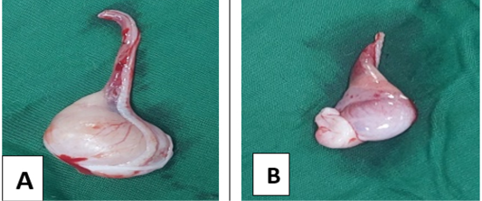
The external appearance of cryptorchid testicles differs markedly from normal in naked eye observation in terms of shape, size, weight and color. The cryptorchid testis was atrophied, congested and considerably smaller in size than the normal testis (Figure 1).
Measurement of length, width and diameter of testes
The cryptorchid testis was smaller in size than the normal testis. In terms of length, width, and diameter the percentages of disparities were found 10.43%, 40.98%, and 36% respectively. In addition, the study showed that, the head, body, and tail of the epididymis of a cryptorchid testis were found smaller than normal testis (Table 1).
Histomorphometrical study
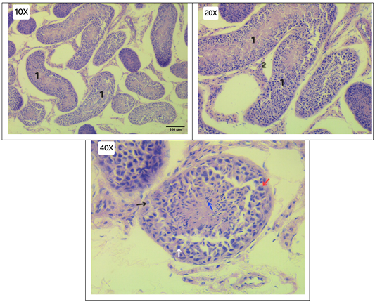
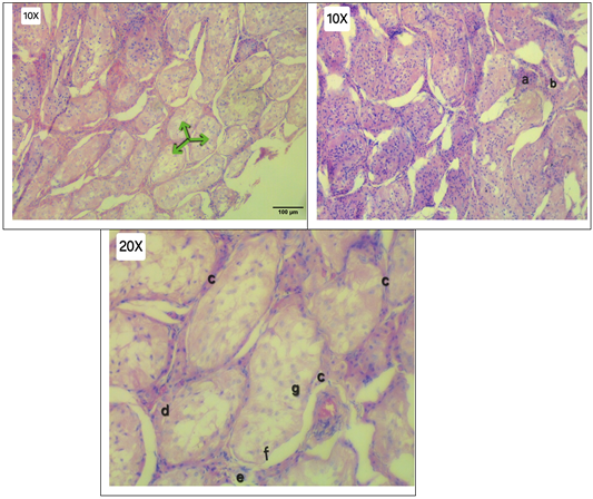
To assess the histoarchiectural alterations, processed tissue sections of both normal and cryptorchid testes were studied under microscope using different magnification, compared, photographed and recorded Figures 2 and 3. Well organized tubules and spermatogenic lineage were observed in normal testis whereas seminiferous tubules in cryptorchid testis were found atrophied (reduced length and diameter), compacted and highly irregular in shape with disorganised spermatogenic lineage, as more spermatogonia were closer to the basement membrane, and other germ cells were also found somewhat disorganized in relation to their normal position, indicating irregular and abnormal spermatogenesis. The average length and diameter of the seminiferous tubules in the cryptorchid testis were found to be 46.52% and 31.49%
Table 1: Morphometrical findings of normal and cryptorchid testis.
Parameter
Normal testis (Right)
Cryptorchid testis (Left)
Difference
Percentage of difference
Measurement of testis with epididymis
Length
19.18 mm
17.88 mm
2 mm
10.43%
Width
14.3 mm
8.44 mm
5.86 mm
40.98%
Diameter
5 cm
3.2 cm
1.8 cm
36%
Measurement of epididymis
Head
Length
6.96 mm
6.48 mm
0.48 mm
6.89%
Width
5.42 mm
2.88 mm
2.54 mm
46.86%
Body
Length
13.48 mm
10.06 mm
3.42 mm
25.82%
Width
1.67 mm
1.45 mm
0.22 mm
13.17%
Tail
Length
5.85 mm
5.69 mm
0.16 mm
2.74%
Width
5.16 mm
3.07 mm
2.09 mm
40.50%
Table 2: Histomorphometrical findings of normal and cryptorchid testis.
Parameter
Normal testis (10X)
Cryptorchid testis (10X)
Difference
Percentage of difference
Measurement of Seminiferous tubules
Avg. Length (µm)
314.39
168.13
146.26
46.52%
Avg. Diameter(µm)
112.31
76.94
35.37
31.49%
smaller than in the normal testis, respectively (Table 2). Multilayered and comparatively thicker basement membrane was found in cryptorchid testis due to leydig cell hyperplasia, vacuolated leydig cells and deformed sertoli cells. Moreover, peri-tubular fibrosis and interstitial mastocytosis were present in the cryptorchid testis but absent in the normal one.
Hormonal assay
Blood testosterone hormone level in the tomcat’s blood was measured and found 0.33 ng/mL of testosterone.
Discussion
Persian cats are the most likely breed to be affected by cryptorchidism, which is also coherent with this study. A number of studies have shown that unilateral cryptorchidism is more common, with a greater frequency of 84.2% affecting the right testicle compared to the left ; which is inconsistent with the findings as the study animal was infected with left abdominal cryptorchidism. On the other hand, several other researchers reported that the prevalence of left and right cryptorchidism is identical . The exact cause of these variations is yet unknown in the incidence of unilateral (right or left) and/or bilateral cryptorchidism; however, heredity may be responsible for these variations. Young animals that have suffered typically do not exhibit symptoms of the illness until a related pathology, such as neoplasia or torsion, manifests in the affected testicle. As a result of their lack of clinical symptoms, owners frequently are unaware of the illness which also supported by the investigation of previous research . The diagnosis made on the examined cat was made using X-ray imaging technology and thorough palpation, both of which are supported by earlier research ; ; . The cat under study had a morphologicallydistinct features in cryptorchid testis from a normal testis, with decreased dimensions in terms of length, width, and diameter. Though the precise reason for the reduction in the size (length, width and diameter) of the cryptorchid testis is yet unknown, it could be brought on by hormonal imbalances, inadequate blood flow, and malnutrition. The study’s results will aid in more sophisticated morphometrical analysis in the future, even though there isn’t a published report on the comparison of normal and cryptorchid testis morphometrics. Histological structures have shown reduced spermatogenesis, fewer or no sertoli cells, leydig cell hyperplasia and disorganized germ cells that shed into the lumen, which are consistent with the findings of previously published report ; . Histological observations also reveal atrophied germinal epithelia, reduced and/ or disorganized germ cells, thickening of the basement membrane, and interstitial mastocytosis which were also in the findings of ; ; . The average diameter and length of seminiferous tubules in cryptorchid testis were found to be 35.37 µm and 146.26 µm smaller than those of normal testis, respectively, according to the histomorphometrical study. The results of this study will be useful for future, more complex histomorphometrical investigations, even though there isn’t a published report that compares the histomorphometrics of the normal and cryptorchid testis. The results of earlier studies ; supported the reduced tubular length, diameter, and abnormal architecture in semeiniferous tubules of cryptorchid testis compared to normal testis, although they were inconsistent with the findings of Fell (1923); which reported the diameter and shape of cryptorchid testis were similar to normal testis. However, no exact histomorphometrical study was present. These alterations of the testis may cause problem in productive and reproductive performance of the cat . The results of earlier investigations ; also validate the findings of this study such as altered anatomical and histological structures of the cryptorchid testis. The results of earlier research showed that the average blood testosterone level in normal cat was 3.42±0.49 ng/ml, whereas the blood testosterone level of the study was 0.33 ng/mL, supporting the lower-than-normal blood testosterone level in the studied tom cat. It may be inferred that although the testis of cryptorchid animals can release testosterone hormone, even in small amounts, and exhibit reproductive behavior, it is unable to carry out the regular process of spermatogenesis. As a result, cryptorchid animals may exhibit sterility . It is concluded that, retained testis was found structurally and functionally impaired and imperfect in regard to reproduction.
Limitations
This study was limited to a single case of unilateral cryptorchidism in a Persian cat, excluding statistical analysis and limiting the generalizability of the findings to the broader feline poplulation. Gross and histopathological evaluations were restricted to descriptive measurements and routine HandE staining without quantitative lesion scoring or advanced techniques such as special staining, immunohistochemistry, and ultrastructural analysis etc. due to time and resources constriction. Further studies with larger sample sizes and comprehensive morphological and molecular approaches are needed to validate these findings.
Conclusion
Based on the findings of the study such as reduced gross morphometrical testicular development and histomorphological testicular tissue alterations along with lower testosterone hormone production; It is concluded that, retained testis was found structurally and functionally impaired and imperfect in regard to reproduction.
Acknowledgements
Our gratitude goes out to the vets who contributed their skills in surgery and biopsies for the study. We also appreciate the sample preparation done by the personnels of Department of Anatomy and Histology and Department of Pathology and Parasitology, CVASU, for photomicrograph of histological slides.
Novelty Statement
This study provides a detailed comparative assessment of a unilateral abdominal cryptorchid testis and the contralateral normal testis in a Persian cat, integrating gross morphometry, histomorphometry, histopathological evaluation, and serum testosterone assessment. To the best of our knowledge, quantitative comparison of testicular and seminiferous tubular dimensions between normal and cryptorchid testes in the cat has been insufficiently documented. The study demonstrates marked reductions in testicular dimensions and seminiferous tubular length and diameter, together with distinctive histopathological alterations and reduced serum testosterone concentration in the retained testis. These findings provide useful baseline morphometric and histomorphometric information for understanding the structural and functional consequences of feline cryptorchidism and may support further investigations into its reproductive implications.
The authors received no financial assistance for the research, authorship, and/or publication of this article.
Ethical considerations
The owner of the animal used in this study provided verbal and written informed consent for all procedure(s) that were carried out. In order to publish this work, no extra informed consent was needed because neither persons nor animals are identifiable.
Generative AI and AI assisted technology statement
The authors declare that no generative AI and AI assisted technology was used in the creation of this manuscript.
Conflict of interest
The authors have declared no conflict of interest.
Figures
Figure 1:.
Gross morphology of normal testis (A) and cryptorchid (B) testis of cat.
Figure 2:.
Photomicrographs of H and E stained histological slides of normal testis showing normal architecture with normal and regular spermatogenesis-Seminiferous tubules (1), Leydig cell (2), Sertoli cell (Black arrow), Spermatogonia ( Red arrow), Spermatocyte (White arrow), Spermatozoa (Blue arrow). 10X, 20X and 40X indicates magnification of slides, 100 µm indicates measurement of scale bar used for measuring tubular length and diameter.
Figure 3:.
Photomicrographs of H and E stained histological slides of cryptorchid testis of cat exhibiting atrophic and degenerated seminiferous tubules (green arrow), interstitial mastocytosis (a), vacuolated leydig cells (b), thicker basement membrane (c), more spermatogonia closer to basement membrane (d), peritubular fibrosis by fibrous connective tissue (e), deformed sertoli cell (f), disorganized spermatogenic lineage (g). * Because of the blurry image, a 40X magnification photomicrograph of the histology slide of the cryptorchid testis could not be taken. 10X and 20X indicate magnification of slides. 100 µm indicates measurement of scale bar used for measuring tubular length and diameter.
References
Ali OJ, Ali TG, Raouf GM, Dana OI (2022). Clinical and histological aspects of cryptorchidism in dogs and cats. Al-Anbar J. Vet. Sci., 15(1): 34-42. https://doi.org/10.37940/AJVS.2022.15.1.5[CrossRef]
Amann RP, Veeramachaneni DNR (2018). Cryptorchidism and associated problems in animals. Anim. Reprod., 3(2): 108-120.
Babaei H, Azari O, Kheirandish R, Abshenas J, Mohammadi N (2010). Zinc therapy improves deleterious effects of experimental unilateral cryptorchidism: histopathological evaluation of testes. Iran. J. Vet. Surg., 5(1-2): 77-88.
Brückner M (2015). Laparoscopic-assisted cryptorchidectomy in a cat. Tierärz. Praxis Ausgabe K: Kleintiere/Heimtiere, 43(4): 248-252. https://doi.org/10.15654/TPK-140628[CrossRef]
Chaudhary AK, Purohit GN, Choudhary S (2024). Diseases of the reproductive system of male dogs and cats. In: Introduction to diseases, diagnosis, and management of dogs and cats. pp. 163-179. Academic Press. https://doi.org/10.1016/B978-0-443-18548-9.00011-1[CrossRef]
Christensen BW (2012). Disorders of sexual development in dogs and cats. Vet. Clin. Small Anim. Pract., 42(3): 515-526. https://doi.org/10.1016/j.cvsm.2012.01.008[CrossRef]
Diagone KV, Feliciano MA, Pacheco MR, Vicente WR (2012). Histology and morphometry of the testes of adult domestic cats (Felis catus). J. Feline Med. Surg., 14(2): 124-130. https://doi.org/10.1177/1098612X11431531[CrossRef]
Fell HB (1923). A histological study of the testis in cases of pseudo‐intersexuality and cryptorchism with special reference to the interstitial cells. Quart. J. Exp. Physiol. Trans. Integr., 13(2): 145-158. https://doi.org/10.1113/expphysiol.1923.sp000285[CrossRef]
Felumlee AE, Reichle JK, Hecht S, Penninck D, Zekas L, Dietze YA, Lowry J (2012). Use of ultrasound to locate retained testes in dogs and cats. Vet. Radiol. Ultras., 53(5): 581-585. https://doi.org/10.1111/j.1740-8261.2011.01943.x[CrossRef]
Goericke-Pesch S, Wehrend A (2013). Cryptorchidism in dogs and cats-definition, prevalence, diagnosis and therapy.
Gradil C, McCarthy R (2023). Cryptorchidism. Small animal soft tissue surgery, pp. 720-725. https://doi.org/10.1002/9781119693741.ch59[CrossRef]
Gubbels EJ, Scholten J, Janss L, Rothuizen J (2009). Relationship of cryptorchidism with sex ratios and litter sizes in 12 dog breeds. Anim. Reprod. Sci., 113(1-4): 187-195.
Hill FI, Tse MP, Mills SW, Sandy JR, Elsohaby I, Barrs VR (2024). Histopathological changes in testicular lesions in cats. J. Feline Med. Surg., 26(9): https://doi.org/10.1177/1098612X241264124[CrossRef]
Karasu A, Kuşcu Y, Kayıkcı C, Gençcelep M, Kavas B, Ekin M (2024). Evaluation of cryptorchidism in cats and dogs. Turk. J. Vet. Res., 8(1): 81-87. https://doi.org/10.47748/tjvr.1440967[CrossRef]
Khan FA, Gartley CJ, Khanam A (2018). Canine cryptorchidism: An update. Reprod. Domest. Anim., 53(6): 1263-1270. https://doi.org/10.1111/rda.13231[CrossRef]
Little S (2011). Feline reproduction: Problems and clinical challenges. J. Feline Med. Surg., 13(7): 508-515. https://doi.org/10.1016/j.jfms.2011.05.008[CrossRef]
Miller NA, Van Lue SJ, Rawlings CA (2004). Use of laparoscopic-assisted cryptorchidectomy in dogs and cats. J. Am. Vet. Med. Assoc., 224(6): 875-878. https://doi.org/10.2460/javma.2004.224.875[CrossRef]
Millis DL, Hauptman JG, Johnson CA (1992). Cryptorchidism and monorchism in cats: 25 cases (1980-1989). J. Am. Vet. Med. Assoc., 200(8): 1128-1130. https://doi.org/10.2460/javma.1992.200.08.1128[CrossRef]
Moon JH, Yoo DY, Jo YK, Kim GA, Jung HY, Choi JH, Jang G (2014). Unilateral cryptorchidism induces morphological changes of testes and hyperplasia of Sertoli cells in a dog. Lab. Anim. Res., 30(4): 185-189. https://doi.org/10.5625/lar.2014.30.4.185[CrossRef]
Mosallanejad B, Gooraninejad S, Fatemi TSR, Ghasemi GA (2019). A survey on seasonal changes of serum testosterone concentration in male cats of Ahvaz region. Vet. Clin. Pathol. (Vet. J. Tabriz), 13(2): 109-119.
Richardson EF, Mullen H (1993). Cryptorchidism in cats. The Compendium on continuing education for the practicing veterinarian (USA). https://www.cabidigitallibrary.org/doi/full/10.5555/19942211090
Romagnoli SE (1991). Canine cryptorchidism. The veterinary clinics of North America. Small Anim. Pract., 21(3): 533-544. https://doi.org/10.1016/S0195-5616(91)50059-0[CrossRef]
Slaoui M, Fiette L (2010). Histopathology procedures: from tissue sampling to histopathological evaluation. In: Drug safety evaluation: Methods and protocols (pp. 69-82). Totowa, NJ: Humana Press. https://doi.org/10.1007/978-1-60761-849-2_4[CrossRef]
Spangenberg C (2021). Canine cryptorchidism: A concise review of its origin, diagnosis and treatment caroline spangenberg. BEMS Rep., 7(1): 1-3. https://doi.org/10.5530/bems.7.1.1[CrossRef]
Villalobos-Gomez J, Del-Angel-Caraza J, Tapia-Araya A, Brandao F, Hernández-López CA, Martínez-Gomariz F, Properzi R (2023). A retrospective study of laparoscopic cryptorchidectomy in 19 cats with intra-abdominal testes. Animals, 13: 181. https://doi.org/10.3390/ani13010181[CrossRef]
Yates D, Hayes G, Heffernan M, Beynon R (2003). Incidence of cryptorchidism in dogs and cats. Vet. Rec., 152(16): 502-504. https://doi.org/10.1136/vr.152.16.502[CrossRef]