Research Article · Volume 13, Issue 7, 1 June 2025 · Open Access
Morphological and Histochemical Adaptations of the Urinary System in the male Euphrates Jerboa (Scarturus euphraticus)
· · · ·
Affiliations & Notes ⌄
- 1Department of Anatomy and Histology, College of Veterinary Medicine, University of Al-Qadisiyah, Iraq
- 2Department of Anatomy, Histology and Embryology, Faculty of Veterinary Medicine, University of Kufa. Iraq. *Correspondence | Nabeel Abd Murad Almamoori, Department of Anatomy and Histology, College of Veterinary Medicine, University of Al-Qadisiyah, Iraq
- 3Email: nabeel.almamorri@qu.edu.iq, nabeelmurad9@gmail.com
Article Info ⌄
- Journal:
- Advances in Animal and Veterinary Sciences
- Published:
- 1 June 2025

From the article
- Anatomically, our findings studied three parts of the urinary system of the Euphrates Jerboa including left and right kidneys, ureters and urinary bladders.
- The mean thickness of cortex and medulla of right kidney was measured (4.26±0.02) mm and (6.18±0.02) mm respectively, whereas, the mean thickness of cortex and medulla of left kidney accounted for (4.21±0.01) mm and (6.20±0.02) mm, respectively (Table 1).
- Also, the kidney displayed uni-pyramid (uni-papillate kidneys) consisted mainly of tubules that extended from the cortical layer the kidney to the calyces, and opened to renal pelvis before it passes through the ureter to the bladder (Figure 1).
- The urinary bladder demonstrated four layer; mucosa, submucosa, muscularis and serosa.
Selected from the article's own text.
Abstract
INTRODUCTION
Rodentia is a well-distributed small animal class comprising various species like rats, mice, and the Dipodidae family . Dipodidae constitute the subfamilies: Allactaginae, Cardiocraniinae, Dipodinae, and Euchoreutinae . The Euphrates jerboa (Scarturus euphraticus) is a small desert rodent found in parts of the Middle East. While its name comes from Iraq’s Euphrates River, this species also inhabits neighboring countries including Syria, southeastern Turkey, western Iran, and possibly Jordan. These animals live in dry, sandy environments, particularly near river valleys and desert plains . Mice and rats are two main vertebrate experimental models for laboratory studies . The urinary system of rodents, Dipodidae inclusive, exhibits great diverse adaptations to the habitats, ranging from deserts through forests to human-populated spaces ; . The Euphrates jerboa (Scarturus euphraticus) is especially intriguing due to its unique adaptation to desert conditions and its capacity to endure with very little water intake . This system, made up of the kidneys, ureters, and bladder, is critically important for fluid and electrolyte maintenance and efficient water conservation inside the animal . Compared to kidneys in humid environments, kidneys in these conditions are not very good at conserving water. Rather than preserving electrolytes and water, they are designed to eliminate excess of them .
Rodent kidneys may be small but anatomically are nearly identical to those of larger mammals, including humans . Their bean-like shape, positioned against the spine in the slightly below the middle posterior, is maintainable in all species . Structurally, these kidneys have a cortical and medullary layer, of which the basic structural components are nephrons, the functional units of the kidney ; ; Kose F et al., 2025). Each nephron has a renal corpuscle, a tuft of capillaries (glomerulus) fed by afferent arterioles and drained via efferent arterioles and supported by mesangial cells . The glomerulus is enclosed in Bowman’s capsule, lined by podocyte foot processes . This filtration unit is where the filtration of blood begins and leads to the reabsorption of necessary substances in the removal of unwanted ones like urea and creatinine .
Directly from this the tubules of the nephron will extend along with fluid from the cortex through the medulla to renal the pelvis and ureter . The primary function of the proximal convoluted tubule within the cortex is reabsorption of glucose, amino acids, and electrolytes through simple cuboidal epithelium possessing a brush border .
The loop of Henle of the nephron drained from the thin descending limb to the thick ascending limb. The major functions of these segments involve the complex processes of filtering materials, reabsorbing nutrients, emptying urine, and thus maintaining the chemical balance in the body .
The ascending limb is structurally differentiated into proximal and distal parts. The proximal part is lined by simple squamous epithelium, and the distal part is lined by simple cuboidal epithelium . The tubule continues from the distal straight to the distal convoluted tubule within the cortex of the kidney, where further calcium reabsorption and secretion of phosphate take place, under hormonal control .
The collecting tubule is the last component of the nephron to drain into the collecting duct system. It is lined by simple cuboidal epithelium and comprises two types of cells: principle cells and intercalated cells . It is primarily responsible for water and salt reabsorption, excretion of nitrogenous waste products received from the collecting tubules, and further concentration of urine .
Several studies reported the rodent’s kidneys continued with the ureters and bladder similar to other species ; . They have their own particular structural adaptations that fit their functions. The ureters are retroperitoneal tubes connecting both kidneys with the bladder. Its thick muscular walls enable peristaltic contractions, propelling urine toward the bladder (Low et al., 2006). The ureter’s inner lining, the mucosa, comprises transitional epithelium that stretches and recoils according to activity of the muscular layer . The muscular layer itself comprises both longitudinal and circular smooth muscle fibers, orchestrating the peristaltic waves . Finally, there is an outer layer of adventitia, which consists of connective tissue and also houses the blood supply to the ureter . The rodent bladder is a pear-shaped structure in the pelvic cavity that temporarily stores urine . Urine produced in the kidneys is stored before elimination via the urethra. Similar to the ureters, the bladder is lined with a mucosa made of transitional epithelium capable of responding to varying amounts of urine . This lining rests on a muscular layer of smooth muscle capable of contracting during voiding to expel urine, Finally, an adventitial layer of connective tissue surrounds the bladder .
This study determined morphology and histochemistry of the urinary system (i.e. kidneys, ureters, and bladder) in the male Euphrates jerboa, to further understand the very unique modifications seen in its urinary system given that the organism is living in an arid environment with very limited water availability.
MATERIALS AND METHODS
Animal Ethics
All animals of the Euphrates Jerboa (Scarturus euphraticus) were first acclimatized and health checked before euthanasia, and they ranged in weight from around 200-250g. Euthanasia was performed and the animals were then dissected, as per the Animal Ethics Guidelines of the College of Veterinary Medicine/University of Al-Qadisiyah (The Scientific Research Ethics Committee formed by administrative order No. 2326 on May 29, 2024, from the College of Veterinary Medicine/University of Al-Qadisiyah, has sanctioned and accepted this study plan based on details regarding the submitted research project as above since it is in conformity with the relevant regulations and guidelines).
Morphometric Parameters
The kidneys were carefully excised from the abdominal cavity and dissected from the surrounding tissues after dissecting and removing the intestines. Weighing, measuring length, width, circumference, and thickness were done for each kidney. The kidneys were split longitudinally, thereby exposing their internal structure with the help of digital calipers. Thickness measurements of the cortex and medulla were taken, as well as their distance from the edge. The ureters were detected, and their lengths and diameters were recorded. The urinary bladder was located and examined both internally and externally. All findings were photographed using an HTC digital camera .
Histologic Procedure
Kidney, ureter, and urinary bladder tissues were harvested and promptly fixed in 10% neutral buffered formalin (NBF) for approximately 48 hours. Following fixation, the samples were washed for two hours and then dehydrated through ascending graded series of ethanol alcohol (50%, 70%, and 100%). Consequently, the samples were cleared in xylene (two changes, 2 minutes each) and embedded in paraffin. Sections of 4–6 μm thickness were cut and mounted on slides . These sections were stained with hematoxylin and eosin, periodic acid-Schiff (PAS), and Masson’s trichrome stain. Light microscopy (Olympus, Japan) was used to examine the kidney, ureter, and urinary bladder tissue sections, and photographs were captured at various magnifications (4x, 10x, 20x, 40x, and 100x oil immersion lens).
Statistical Analysis
The measurements of the anatomical and histological kidneys, ureters and urinary bladders were analyzed using Paired Samples Test (T. Test) using IBM SPSS Statistics 23.0. Accordingly, means and standard errors of the kidneys, ureters and urinary bladders for all parts of urinary system were recorded and analysed. .
RESULTS
Anatomically, our findings studied three parts of the urinary system of the Euphrates Jerboa including left and right kidneys, ureters and urinary bladders. Both right and left kidneys were bean-shaped and located in sub-lumbar of the dorsal surface of abdomen at the upper lumbar vertebra, however, right kidney was more progressed cranially in comparison with left kidney. Unusually, the kidneys were not surrounded by adipose tissue, and surrounded by a delicate fibrous connective tissue capsule.
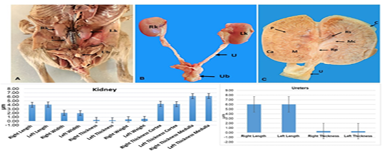
So, the right and left kidneys were brown to red in colour, and each kidney was had two poles (cranial and caudal), two surfaces (dorsal and ventral), and two borders (lateral and medial). The medial border was concave with an apparent indentation area (the hilus) where the ureter, renal artery, and vein enter or exit from or to the kidney, but the lateral border was convex (Figure 1). The mean weight of right and left kidney accounted for (0.46±0.07) grams (g) and (0.56±0.07) g respectively (Table 1). Also, the mean length, width and thickness of right kidney was (4.04±0.09) mm, (2.04±0.08) mm and (0.28±0.04) mm respectively (Table 1), as the mean length, width and thickness of left kidney was accounted for (4.10±0.12) mm, (1.98±0.10) mm and (0.56±0.07) mm respectively (Table 1).
Similarly, the internal surface of kidneys was showed two main regions; externally, thin outer renal cortex was dark brown in color, while the inner renal layer medulla was lighter then renal cortex in color and larger distance compare to the renal cortex (Figure 1). The mean thickness of cortex and medulla of right kidney was measured (4.26±0.02) mm and (6.18±0.02) mm respectively, whereas, the mean thickness of cortex and medulla of left kidney accounted for (4.21±0.01) mm and (6.20±0.02) mm, respectively (Table 1).
Table 1: Morphometric measurement of the kidney and ureter in the male Euphrates Jerboa.
| Kidney | ||||
| No. | Parameters | Mean (N=5) | Std. Deviation | Std. Error Mean |
| 1 | Right Length | 4.04 mm | 0.21 | 0.09 |
| 2 | Left Length | 4.10 mm | 0.27 | 0.12 |
| 3 | Right Width | 2.04 mm | 0.18 | 0.08 |
| 4 | Left Width | 1.98 mm | 0.23 | 0.10 |
| 5 | Right Thickness | 0.28 mm | 0.08 | 0.04 |
| 6 | Left Thickness | 0.22 mm | 0.08 | 0.04 |
| 7 | Right Weight | 0.46gm | 0.17 | 0.07 |
| 8 | Left Weight | 0.56gm | 0.15 | 0.07 |
| 9 | Right Thickness Cortex | 4.26 mm | 0.05 | 0.02 |
| 10 | Left Thickness Cortex | 4.21 mm | 0.03 | 0.01 |
| 11 | Right Thickness Medulla | 6.18 mm | 0.03 | 0.02 |
| 12 | Left Thickness Medulla | 6.20 mm | 0.04 | 0.02 |
| Ureter | ||||
| 1 | Right Length | 6.04 mm | 0.18 | 0.08 |
| 2 | Left Length | 6.04 mm | 0.11 | 0.05 |
| 3 | Right diameter | 0.34 mm | 0.039 | 0.017 |
| 4 | Left diameter | 0.32 mm | 0.039 | 0.017 |
Importantly, renal medulla showed large number irregular shape of the pyramids constituted the main interstitium of renal medulla had small columns sizes distributed between medulla pyramids. Also, the kidney displayed uni-pyramid (uni-papillate kidneys) consisted mainly of tubules that extended from the cortical layer the kidney to the calyces, and opened to renal pelvis before it passes through the ureter to the bladder (Figure 1).
Moreover, right and left ureters were extended from hilus (medial border) for each kidney through the hilus and runs caudally on either side of the midline in the dorsal of the abdomen (abdominal section of the ureter). The ureter displayed two regions including abdominal and pelvic regions, with thin muscular wall holding by a mesentery fold. The pelvic portion of the ureter entered the bladder from the craniodorsal aspect (Figure 1). Subsequently, the mean length of right ureter was (6.04±0.08) mm and diameter was (0.34±0.17) mm (Table 1). While left ureter was measured (6.04±0.05) mm and diameter (0.32±0.17) mm. (Table 1).
Furthermore, the urinary bladder was white in color and appeared as hollow sac, and compressed small pear-shaped, was located in the pelvic cavity of the animal, and the rounded cranial portion of the bladder expanding in the midline of the caudal abdomen, and opened to the urethra, (Figure 1).
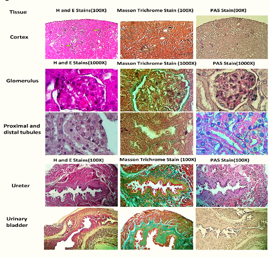
Histologically, kidney was consistent with anatomical results, it consisted of an external delicate capsule, narrow renal cortex and internal renal medulla. There is no clear boundary separated between the cortex and the medulla, however, renal cortex demonstrated many renal corpuscles which were absent in the renal medulla (Figure 2). So, the mean thickness of capsule, cortex and medulla were recorded (0.14 ±0.244) μm, (4.38±0.131) μm and (5.48 ±0.135) μm respectively. Microscopically, renal cortex formed of many nephrons interposed between interstitium of cortex, blood vessels as well as renal tubules including the renal the proximal convoluted tubules, the distal convoluted tubules, and the collecting tubules (Figure 2).
The renal corpuscles enveloped by Bowman’s capsule contained the glomerulus, which were spherical renal corpuscle in shape. Bowman’s capsule exposed two double-walled layers; visceral and parietal layers. Bowman’s capsule lined by podocytes (epithelial cells) around the glomerulus. The capsular (urinary) space was clear between Bowman’s capsule and glomerulus, so the most renal corpuscles had small area distance between the renal corpuscles visceral of the Bowman’s capsule and glomerulus. In addition to podocytes that surround the capillaries, there are other specialized cells in the glomerulus, called mesangial cells that are also attached to the capillaries (Figure 2). The mean diameter of glomeruli was accounted for (0.46±0.244) μm.
Additionally, the proximal convoluted tubules were lined by cuboidal epithelial cells based on the basement membrane. So, the epithelial cells were had large central nucleus, ground eosinophilic cytoplasm, and an apical surface with brush borders extended toward lumen of tubules as microvilli. Whereas, the apical surface of the distal convoluted tubule was covered by low cuboidal epithelial cells with short microvilli, and the ground cytoplasm was obviously less eosinophilic compare to the proximal tubules (Figure 2), however, these distal convoluted tubules were increased in lumen toward renal medulla and collecting tubules. Subsequently, the mean diameter of proximal and distal convoluted tubules was accounted for (0.46±0.244) μm and (0.14±0.244) μm in respectively. Moreover, medullary rays were obviously extended from renal medulla toward the cortex including collecting tubules, ascending straight segments of the distal tubules, and descending straight segments of the proximal and distal tubules. Also, the renal medulla noticeably contained straight parts of the tubules including thick and thin descending segments and thin and thick ascending segments with large lumen toward renal pelvis (Figure 2).
In addition, the ureter lumen appeared a star-shaped lumen and histologically comprised with three tunica including; tunica mucosa, muscularis, and thin adventitia. The tunica mucosa displayed a thick transitional epithelium based on the basement membrane. The muscular layer presented three layers of muscle fibers directions including internal longitudinal, middle circular and outer longitudinal layer. Externally, tunica adventitia was outer layer consisted of the fibrous connective tissue (Figure 2).
The urinary bladder demonstrated four layer; mucosa, submucosa, muscularis and serosa. The transitional epithelium lined by lumen of urinary bladder and divided into folds, and extended toward lamina propria. Muscular layers revealed three layers of muscle fibers directions including internal longitudinal, middle circular and outer longitudinal layer through connective tissue. While, serosa exhibited adipose connective tissue and blood supply (Figure 2).

DISCUSSION
Anatomical findings showed the Euphrates Jerboa kidneys are bean-shaped, located in the sub-lumbar region, with the right kidney positioned more cranially than the left. They are smaller than mice and rats, and have a delicate layer capsule ; . The left kidney has longer morphological differences from the right. The kidney similar bean-shaped morphologies are observed in various species ; . Our observation found that perirenal fat in jerboas requires more consideration under their desert habitat. Subsequently, different figure shape observed in the left kidney and a lack of surrounding adipose tissue in Euphrates Jerboas may be attributed to arid environmental conditions acting upon physiological adaptations of these animals.
Moreover, our findings confirmed that kidneys of the Euphrates jerboa were typically mammalian in structure having two poles (superior and inferior), with a couple of surfaces (anterior and posterior) and borders (medial and lateral) along with a hilum ; . While the right and left kidneys of the Euphrates jerboa differed slightly in size and weight, these differences were not significance. Even the weight of the kidneys themselves was small relative to the animal’s overall body weight. In contrast, mice and rats have large weight of the kidneys compare whole body weight ; .
According to our findings, the kidney’s internal structure is divided into medullary and cortical regions. Each renal pyramid in the kidneys of this species empties into a single renal pelvis, making them unipapillary. The medulla and cortex are histologically organized similarly to those of other mammals. There are numerous renal corpuscles in the cortex; each corpuscle has a glomerulus that stains positive for PAS and is supported by mesangial cells. Each renal corpuscle is surrounded by Bowman’s capsule lined with podocytes. Proximal and distal convoluted tubules interpose through the cortical interstitium toward the medulla, which has descending and ascending limbs of the collecting ducts. Proximal and distal convoluted tubules, and descending and ascending limbs of the collecting ducts lined by low to high cuboidal epithelium stained lighter with PAS stain, and stained with thin basement membrane stained by mass trichrome stain. This general histological architecture is maintained throughout several mammalian species, which include mice, rats, rabbits, and cats, dogs, and goats ; . Ureters, and urinary bladder showed same gross appearance to other species, though, urinary bladder was compressed and small in Euphrates jerboa ; . We thought that the relatively smaller capacity of their bladder, as compared to mesic rodents, could be related to the strategies of water acquisition. Therefore, our study suspects that If jerboas mostly depend on metabolic water, then it could be expected that low amounts of urine volumes produced will make such small bladders suffice. Or, perhaps a smaller bladder with a highly concentrated urine output could indicate a frequent excretion approach to lose as little water as possible due to waste excretion in an arid environment. So, the ureter renal connected the hilus to urinary bladder, and described as abdominal and pelvically divided, having craniodorsal entry into the bladder. As, urinary bladder, a pear-shaped white hollow sac was located in the pelvic cavity, its cranial portion extending into the abdomen and opening into the urethra. These finding are consistent with other mammalians .
Moreover, histological examination revealed a star-shaped lumen of the ureter, and three layers compose its wall- tunica mucosa (with transitional epithelium) stained with PAS, muscularis (with three muscle layers), and tunica adventitia. Connective tissue staining using Masson trichrome exhibited high intensity in the muscularis mucosa for connective tissue, and this was also clear staining for the connective tissue positioned between muscles and the adventitia. In the case of the urinary bladder, four layers constitute the bladder wall, namely the mucosa (lined with transitional epithelium) stained with PAS, the submucosa, muscularis (with three muscle layers) and the serosa. The staining of the connective tissue using Masson trichrome was also highly positive for the connective tissue in the muscularis mucosa, and for connective tissue between muscles and serosa.
General histological architecture structure of the ureter, and urinary bladder of the Euphrates jerboa showed high similarity with other species and agreed with other studies ; ; .
CONCLUSIONS AND RECOMMENDATIONS
In conclusion, the urinary system of the Euphrates jerboa includes typical mammalian features but also reflects some peculiar adaptations. There are some relative size differences compared to body weight: a relatively small kidney organ in particular on the left- kidney surface displays slightly different shapes. There is no subcutaneous fat around the kidneys, which might represent adaptations to living in an arid environment. The ureters and urinary bladder resemble their counterparts in other mammals in their gross form as well as in their histological structure; wonder, bladder that is rather small and compressed. These findings seem to suggest a compromise between mammalian conserved features of the urinary system and the particular adaptations specific to the habitat of the Euphrates jerboa.
ACKNOWLEDGMENTS
This work was funded and supported by the department of anatomy and histology/College of Veterinary Medicine/the University of Qadisiyah. Furthermore, we appreciate the collaboration of technician staff in the department of anatomy and histology/College of Veterinary Medicine, University of Qadisiyah.
NOVELTY STATEMENT
This research provides a detailed morphological and histological description of the urinary system of the Euphrates jerboa (Scarturus euphraticus), with emphasis on several distinctive features compared to the general mammalian model. Notably, the jerboa’s kidneys lack the typical surrounding adipose tissue and are instead encapsulated by a thin fibrous connective tissue layer. Furthermore, the urinary bladder appears small and compressed relative to other rodents and mammals. These specific anatomical and histological characteristics of the Euphrates jerboa’s urinary system are potentially adaptations to the arid environmental conditions of its habitat. Finally, these findings constitute novel data on the urinary system of the Euphrates jerboa, representing a first for Iraq, and may serve as a foundation for future research.
AUTHOR’S CONTRIBUTIONS
All researchers contributed equally.
Conflict of Interest
None.
Figures

A general anatomical illustrative image and graphical data for the urinary system (kidneys and ureters, and urinary bladder). A: This section indicates the opened view of the abdominal cavity; hence, it contains the cut kidneys (Rk and Lk for Right and Left Kidney respectively), ureters (U), and possibly the renal vessels. The numbers appear to be reference points or labels for the structures. B: A closer isolate picture of kidneys and ureters showing their comparative position and size. The ureters (U) are seen more clearly, extended from the renal hila. Urinary bladder (Ub) C: Cross-section through a kidney showing its layers or regions. ‘C’ (cortex), ‘M’ (medulla), ‘Rp’ (renal pelvis), and others indicate special anatomical structures within the kidney. Diagrams revealed the different parameter measurements for the left and right kidneys and ureters.

Light microscopy images of kidney and ureter tissues stained by hematoxylin and eosin (H&E), Masson’s trichrome, and periodic Acid-Schiff (PAS) were displayed in the figure. The images were arranged in the grid. The higher magnification (1000x) view of the kidney cortex shows glomeruli, proximal tubules, and distal tubules, whereas the lower magnification (100x) view shows the ureter and urinary bladder.
References
- AL-Mamari SA (2023). Anatomy of the Kidney. In Urogenital Trauma: A Practical Guide, Springer:3-16. https://doi.org/10.1007/978-981-99-6171-9_1[CrossRef]
- AL-Samawy ER (2012). Morphological and Histological study of the kidneys on the Albino rats. al-Anbar J. Vet. Sci., 5: 115-119.
- Almhanna H, Yassin A, AL-Mamoori N, Kilroy D, Kumar A (2024). Structural and network analysis of ADAM9 and ADAM10 and its transcript expression in the male reproductive tract of Bos Taurus. Ger. J. Vet. Res., 4:126-139 https://doi.org/10.51585/gjvr.2024.3.0104.[CrossRef]
- Bachmann S, Kriz W (1998). Histology, Cytology, Ultrastructure Nephron and Collecting Duct Structure in the Kidney, Rat. In Urinary System, Springer: 3-36. https://doi.org/10.1007/978-3-642-80335-2_1[CrossRef]
- Bagga A, Srivastava R, Gupta AK, Jana M, Bal C, Thergaonkar R, Khandelwal P (2016). Renal Anatomy and Physiology. Paediatr. Nephrol., 19: https://doi.org/10.5005/jp/books/12792_2[CrossRef]
- Braun EJ (1998). Comparative renal function in reptiles, birds, and mammals. In Seminars in Avian and Exotic Pet Medicine, Elsevier: 62-71. https://doi.org/10.1016/S1055-937X(98)80044-3[CrossRef]
- Bryman A, Cramer D (2004). Quantitative data analysis with SPSS 12 and 13: A guide for social scientists. Routledge, 394 1st Edition (Book). https://doi.org/10.4324/9780203498187[CrossRef]
- Costa G (2012). Behavioral adaptations of desert animals, Springer Science and Business Media
- Crisler R, Johnston NA, Sivula C, Budelsky CL (2020). Functional anatomy and physiology. In The Laboratory Rat, Elsevier: 91-132. https://doi.org/10.1016/B978-0-12-814338-4.00004-0[CrossRef]
- Dahms, Piechota, Dahiya, Lue, Tanagho (1998). Composition and biomechanical properties of the bladder acellular matrix graft: Comparative analysis in rat, pig and human. BJU Int., 82: 411-419. https://doi.org/10.1046/j.1464-410X.1998.00748.x[CrossRef]
- Dantzler WH (1989). Comparative physiology of the vertebrate kidney, Springer. Zoophysiology, 22: X-198-208. https://doi.org/10.1007/978-3-642-83521-6[CrossRef]
- Darawiroj D, Choisunirachon N (2019). Morphological assessment of cat kidneys using computed tomography. Anatomia, histologia, embryologia. 48: 358-365. https://doi.org/10.1111/ahe.12448[CrossRef]
- Delaney MA, Kowalewska J, Treuting PM (2018). Urinary system. In Comparative anatomy and histology, Elsevier: 275-301. https://doi.org/10.1016/B978-0-12-802900-8.00016-6[CrossRef]
- Donnelly TM, Bergin I, Ihrig M (2015). Biology and diseases of other rodents. In Laboratory animal medicine, Elsevier, 285-349. https://doi.org/10.1016/B978-0-12-409527-4.00007-9[CrossRef]
- Elger M, Kriz W (1992). The renal glomerulus–the structural basis of ultrafiltration. In Oxford Textbook of Clinical Nephrology, Oxford Medical Publications Oxford: 129.
- Elsayed AH, EL-Gammal SM (2024). Comparative Study on the Gross Anatomy of Some Abdominal Organs in Albino Rat (Rattus norvegicus) and Albino Mouse (Mus musculus). Alex. J. Vet. Sci., 80(0): 1-17. https://doi.org/10.5455/ajvs.183386[CrossRef]
- Frazier KS, Seely JC, Hard GC, Betton G, Burnett R, Nakatsuji S, Nishikawa A, Durchfeld-Meyer B, Bube A (2012). Proliferative and nonproliferative lesions of the rat and mouse urinary system. Toxicol. path., 40: 14S-86S. https://doi.org/10.1177/0192623312438736[CrossRef]
- Goldstein DL (2022). Renal and extrarenal regulation of body fluid composition. In Sturkie’s avian physiology, Elsevier: 411- 443. https://doi.org/10.1016/B978-0-12-819770-7.00034-7[CrossRef]
- Iwanaga R, Orlicky DJ, Arnett J, Guess MK, Hurt KJ, Connell KA (2016). Comparative histology of mouse, rat, and human pelvic ligaments. Int. Urogynecol. J., 27: 1697-1704. https://doi.org/10.1007/s00192-016-3008-6[CrossRef]
- Jokinen MP, Seely JC (2018). Urinary bladder, ureter, and urethra. In Boorman’s Pathology of the Rat, Elsevier, 167-188. https://doi.org/10.1016/B978-0-12-391448-4.00012-5[CrossRef]
- Kose F, Orhan I, Beyaz F, Poscu OT, Alan A, Duzler A (2025). A Comparative Study on Kidney Morphology of Anatolian Ground Squirrels, Rabbits, and Rats. MRT. 1-13. https://doi.org/10.1002/jemt.24797[CrossRef]
- Kirmiz, John P (1962). Adaptation to Desert Environment; A Study on the Jerboa, Rat and Man. London: Butterworths., 29.
- Kriz W, Kaissling B (1992). Structural organization of the mammalian kidney. The kidney: Physiol. Pathophysiol., 3: 587-654.
- Lebedev VS, Shenbrot GI, Krystufek B, Mahmoudi A, Melnikova MN, Solovyeva EN, Lisenkova AA, Undrakhbayar E, Rogovin KA, Surov AV (2022). Phylogenetic relations and range history of jerboas of the Allactaginae subfamily (Dipodidae, Rodentia). Sci. Rep., 12: 842. https://doi.org/10.1038/s41598-022-04779-x[CrossRef]
- Lescay HA, Jiang J, Leslie SW, Tuma F (2024). Anatomy, abdomen and pelvis ureter. In StatPearls internet, StatPearls Publishing.
- Low P, Molnar K, Kriska G (2016). Dissection of the Rat (Rattus norvegicus), Springer, 325-399. https://doi.org/10.1007/978-3-319-25172-1_12[CrossRef]
- Metheny N (2012). Fluid and electrolyte balance, Jones and Bartlett Publishers.
- Mohamed AA (2014). Morphological anatomical and histological study of the normal kidneys in the House Mice (Mus Musculus). Muthanna J. Pure Sci., 2: 83-81.
- Moinuddin Z, Dhanda R (2015). Anatomy of the kidney and ureter. Anaesth. Intensive Care Med., 16: 247-252. https://doi.org/10.1016/j.mpaic.2015.04.001[CrossRef]
- Oliveira P, Colaco A, LF De La Cruz P, Lopes C (2006). Experimental bladder carcinogenesis–rodent models. Exp. Oncol., 28(1): 2-11.
- Parker GA, Picut CA (2016). Atlas of histology of the juvenile rat, Academic Press.
- Pattanaik B, Gayki S, Khutwad J, Puranik S (2023). Excretion in Animals. Principal, Progressive Education Society’s Modern College of Science, Shivajinagar, Pune-5.
- Rajab JM, Abbood AS, Mohsin RA (2024). Comparative histological study of the kidneys in two types of mammals. Pharma. Innov. J., 13(11): 86-91. https://doi.org/10.22271/tpi.2024.v13.i11b.25915[CrossRef]
- Ragab G, Seif M, Hagag U (2010). Radiologic and ultrasonographic studies of kidneys in goat. J. Vet. Med. Res., 20: 30-37. https://doi.org/10.21608/jvmr.2020.77577[CrossRef]
- Rouiller C (1969). General anatomy and histology of the kidney. In The kidney, Elsevier, 61-156. https://doi.org/10.1016/B978-1-4832-2825-9.50008-5[CrossRef]
- Salmon A (2013). Glomerular structure and function. Landmark Papers in Nephrology, 1. https://doi.org/10.1093/med/9780199699254.003.0001[CrossRef]
- Shehata RA (1977). Comparative study of the urinary bladder and the intramural portion of the ureter. Cells Tissues Organs. 98: 380-395. https://doi.org/10.1159/000144817[CrossRef]
- Tripathi R (2014). Integrated management of rodent pests, in Integrated Pest Management, Elsevier: 419-459. https://doi.org/10.1016/B978-0-12-398529-3.00022-1[CrossRef]
- Treuting PM, Dintzis SM, Montine KS (2017). Comparative anatomy and histology: a mouse, rat, and human atlas, Academic Press.
- Treuting PM, Kowalewska J (2012). Urinary system. In Comparative Anatomy and Histology, Elsevier. 229-251. https://doi.org/10.1016/B978-0-12-381361-9.00016-0[CrossRef]
- Wolf JR JS, Humphrey PA, Rayala HJ, Gardner SM, Mackey RB, Clayman RV (1996). Comparative ureteral microanatomy. J. Endourol., 10: 527-531. https://doi.org/10.1089/end.1996.10.527[CrossRef]
Services
Article usage statistics
Loading…