
To elucidate the mechanism of neuropeptide Y (npy) gene in feeding in gibel carp, we cloned and identified the npy cDNA sequence. Furthermore, we examined the npy mRNA distribution in 21 tissues and investigated the effect of its expression at different nutritional levels. Herein, we found that gibel carp npy has an Open Reading Frame (ORF) containing 291 bp. Moreover, the expression of npy was detected in all tested tissues, with the hypothalamus showing the highest expression in gibel carp. The npy mRNA expression in the hypothalamus significantly decreased 1 and 3 h after feeding (P < 0.01). Furthermore, its expression in the hypothalamus was significantly increased after fasting for 7 days and declined 2 days after refeeding (at 11-day and 14 -day; P < 0.01), suggesting that npy is an orexigenic peptide in gibel carp. In conclusion, these results indicated that npy is a conserved peptide within vertebrates and might exert orexigenic role in gibel carp.
Research Article · Volume 51, Issue 3, 31 March 2019 · Open Access
Molecular Cloning and Function Characterization in Feeding of Neuropeptide Y in Gibel Carp, Carassius auratus gibelio
· · · · · · · ·
Affiliations & Notes ⌄
- 1College of Animal Sciences, Southwest University, Chongqing 402460, People’s Republic of China 2Department of Aquaculture, College of Life Sciences, Neijiang Normal University, Neijiang, Sichuan, 641000, People’s Republic of China 3Tibet Academy of Agricultural and Animal Husbandry Sciences, Lhasa, Tibet 850002, People’s Republic of China , Luo Lei and Xingxing Deng have contributed equally to this work. , * Corresponding authors: 442589528@qq.com
- 2421807518@qq.com
Article Info ⌄
- Journal:
- Pakistan Journal of Zoology
- Published:
- 31 March 2019

From the article
- The results indicated that the gibel carp npy gene is highly conserved within teleost species.
- To explore the gene’s expression profile in gibel carp, npy mRNA was investigated in peripheral and central tissues by qPCR.
- The npy mRNA expression of fasted fish at day 7 was significantly higher compared with the fed group (P < 0.01).
- Furthermore, fasting caused its mRNA expression to change in gibel carp, suggesting that npy may have orexigenic activity in gibel carp.
Selected from the article's own text.
Abstract
Introduction
Feeding is a complicated behavior involving food ingestion and appetitive behavior, which reflects the motivation for food intake ; . Furthermore, it is an important process in mammals and fish . Feeding is ultimately regulated by receiving and transducing endocrine signals from the central nervous system and periphery tissues ; ; ; . To date, many appetite-regulating peptides have been identified in fish including cocaine, leptin , ghrelin (GRLN) , peptide YY (PYY) , neuropeptide Y (NPY) ; . Although lots of research on food intake in fish has progressed in the last few years, very little attention has been paid to the endocrine regulation mechanism of food intake. How to strengthen the production performance of fish through appetite regulation is still a hot research area.
Since neuropeptide Y (NPY) was first extracted from a mammalian brain in 1982 , many studies have found that the npy mRNA is expressed in the central nervous system, peripheral nervous system and peripheral tissues of mammals and teleosts, with the highest expression in the brain ; ; ; . NPY is a highly conserved neuroendocrine peptide , which is involved in the regulation of multiple physiological functions, such as circadian rhythms , body temperature , food intake , blood pressure and sexual behavior ; . In general, it is a multifunctional complicated hormone that governs various physiological processes in several animals.
In recent years, several researchers have reported that NPY and its related peptides are important in feeding in mammals and fish ; . In Schizothorax prenanti, the npy expression level in fed fish was lower than in fasting fish . A similar result was found in other fish, such as Cyprinus carpio , Danio rerio and Seriola quinqueradiata , suggesting that NPY plays an orexigenic role in teleost fish. However, the npy expression level was elevated after feeding in Atlantic salmon . Differences in these studies could be due to fish species, age, size, and also to the physiological differences among different fish species ; ; . Therefore, further study is needed to validate the molecular mechanisms and biological function of NPY in feeding in fish.
Gibel carp (Carassius auratus gibelio), one of the major farmed fish species in China, has been subjected to major breeding efforts with high yield. So far, in gibel carp, most studies on the appetite and feeding modes have emphasized the effectiveness of the diet , but only little attention was given to the molecular regulation mechanism of food intake. Research on the biological function of NPY in feeding in gibel carp has not been described.
Here, we identified the gibel carp npy mRNA sequence and analyzed its expression patterns in different tissues. Furthermore, we analyzed the change in npy mRNA expression level between postprandial and fasting status using real-time quantitative PCR (RT-qPCR). This study provides the foundation for further investigation into the molecular mechanisms regulating food intake in gibel carp.
Materials and methods
Ethics statement
Based on the Regulations for the Administration of Affairs Concerning Experimental Animals (Ministry of Science and Technology, China, revised in June 2004), all the research that involved animals was approved by the Institutional Animal Care and Use Committee in the College of Animal Science, Southwest University, Chongqing, China. The feeding conditions, including dissolved oxygen and temperature were under strict control.
Fish
All the gibel carp were purchased from a local nursery of fish in Chongqing, China, and then transported to the laboratory in the Aquaculture Department at Southwest University. Before the experiments, the fish were in a circulating aquarium system at 23 ± 2°C for 14 days and fed a commercial diet (6% fat and 40% crude protein, Haida, Guangzhou, China) once a day (17:00).
According to an overview about regulation of food intake in fish , npy is highly expressed in the brain. Therefore, for the cloning study, brain samples were collected from six fish with a mean body weight of 293.5 ± 17.56 g(1:1 gender ratio). For the tissue distribution study, 21 tissues were collected from six fish with a mean body weight of 316.33 ± 11.7 g (1:1 gender ratio; for more information about the 21 tissues see section 2.3).
To conduct the postprandial research, seven groups of weight-matched fish with a mean body weight of 308.7.36 ± 17.5 g (n = 15/group) were allowed to acclimate to the tank conditions (110 × 44 × 44 cm3). In terms of the feeding activity and behavior, no difference was observed between the tanks. The fish were sampled before the feeding (−3 h and -1 h), at commencement of feeding (0 h) and after feeding (+1 h and +3 h). For the unfed control groups, two unfed groups were sampled at +1 and +3 h. Six individuals were sampled for every sampling point and group.
For the long fasting study, fish with a mean body weight of 458.33 ± 20.1 g (n = 30 × 6, three tanks of unfed fish and three tanks of fed fish) were sampled. After 2 weeks of feeding, three groups’ fish were endured fasting, while other three groups’ fish were fed as before. The fasted group’s fish were sampled severally on day 1, 3, 5 and 7. Six fish in the fed group were sampled at every sampling point. The fasted group was fed again on day 9 on a regular basis, and six fish were sampled on day 9, 11 and 14.
Tissue collection and RNA extraction
Before being sacrificed, fish were anesthetized with tricaine methanesulfonate (MS-222, 0.02%). Then, 21 tissues (telencephalon, hypothalamus, mesencephalon, cerebellum, myelencephalon, liver, spleen, heart, eye, skin, foregut, gill, white muscle, hindgut, midgut, red muscle, trunk kidney and gonad) were removed immediately and stored in liquid nitrogen. RNA was extracted from these tissues with TRIzol A+ reagent, according to the manufacturer’s instructions (TaKaRa, Dalian, China). The concentration of all the RNA samples was determined by a photometer (Thermo Scientific, Dreieich, Germany) at 280 nm and 260 nm. Until further use, the total RNA was stored at –80°C.
Molecular cloning
RNA (around 2 µg) was used for first strand synthesis of cDNA with a Prime Script RT reagent Kit (TaKaRa, Dalian, China). The primers for npy fragment amplification were designed based on npy sequences of other fish species found in the NCBI GenBank.
SMART RACE cDNA Construction Kit (Clontech, USA) was used to synthesize the entire brain’s first-strand cDNA with 3′ or 5′ adaptors for cDNA ends (RACE) PCR rapid amplification. 3′ and 5′ RACE PCR were conducted to obtain the sequence of the full-length gibel carp npy. The RACE PCR products were obtained from the nested RACE PCR for npy’s 3′ and 5′ RACE. Based on the npy fragment, the primers for the gibel carp npy gene were designed. The PCR conditions were: 5 cycles of 94°C for 30 s and 72°C for 180 s; 5 cycles of 94°C for 30 s, 70°C for 30 s and 72°C for 180 s; 20 cycles of 94°C for 30 s, 68°C for 30 s and 72°C for 180 s. The PCR product was separated by agarose gel electrophoresis (1.5%). Next, the target bands were extracted by a Universal DNA Purification Kit (TIANGEN, Beijing, China). Subsequently, the PCR fragments were ligated to a pMD®19-T plasmid (TaKaRa, Dalian, China). The ligated plasmid was transformed into Escherichia coli DH5α. Colonies with inserts were cultured in LB medium at 37°C overnight. OMEGA Mini Plasmid Kit (TaKaRa, Dalian, China) was used to extract the plasmids from the colonies, which were then sequenced by an automated sequencing study.
Structural analysis
Multiple sequence alignments were generated using the clustalx1.83. The cleavage site of the signal peptide was estimated using the SignalP Ver. 4.0 program (http://www.cbs.dtu.dk/services/SignalP/). A phylogenetic tree based on the amino acid sequences was constructed by the neighbor-joining method of the Clustal W (http://www.ddbj.nig.ac.jp/search/clustalw-e.html) and MEGA 5.1 programs (http://www.megasoftware.net/index.html) .
Real-time quantitative PCR
Real-time quantitative PCR (qPCR) was used to evaluate the tissue distribution, postprandial variations and fasting effect on npy mRNA. According to the sequence information gained from cloning (Table I), gene-specific primers were designed for qPCR. SYBR Green (TaKaRa) was used to conduct the qPCR on npy with QuantStudio™ 3 Real-Time PCR (ABI) (TaKaRa). All the real-time qPCR reactions were carried out in triplicate. Every PCR reaction (total volume, 25 µL) included 9.5 µL of RNase-free H2O, 2.5 µL of cDNA, 0.5 µL of each primer and 12.5 µL of 2×SYBR Premix Ex TaqTM. To analyze the samples, the 2-ΔΔCt method was used to determine the mRNA abundance. The amplification reaction conditions were: 95°C for 30 s, followed by 45 cycles of 95°C for 5 s and 60°C for 30 s. In this research, 18S and β-actin were used as housekeeping control genes.
Statistical analysis
The normality test was performed before the Student’s t-test or one-way analysis of variance with SPSS 18.0 software (SPSS Inc., Chicago, IL, USA), to ensure that the t-test is suitable for data comparison. Data were expressed as means ± SEM. P < 0.05 was considered statistically significant.
Table I.- Primers used for sequences.
| Primer name | Sequence (5′→3′) | Application |
| NPY-1F | ACCCACCGAGCAAGAAGT | NPY cloning |
| NPY-1R | TCACTATCCTGACGATGGCT | NPY cloning |
| NPY3′-F1 | TGTCAGCACTGGACCTTTTGCCATACCT | NPY 3′RACE outer |
| NPY3′-F2 | ACCCTTCCGTTAGAGTTCCCAAGCAGAC | NPY 3′RACE inner |
| NPY5′-F1 | CTGCTTGGGAACTCTAACGGAAGGGTAT | NPY 5′RACE outer |
| NPY5′-F2 | ATAACACGGCAGAGGTATGGCAAAAGGT | NPY 5′RACE inner |
| NPY-F | CCTTCCTCTTGTTCGTCTGCT | Real time PCR |
| NPY-R | TGCCATACCTCTGCCGTGT | Real time PCR |
| β-Actin-F | CGAGCTGTCTTCCCATCCA | Real time PCR |
| β-Actin-R | TCACCAACGTAGCTGTCTTTCTG | Real time PCR |
| 18s F | ACCACCCACAGAATCGAGAAA | Real time PCR |
| 18s R | GCCTGCGGCTTAATTTGACT | Real time PCR |
Results
Sequence analysis of the gibel carp npy gene
The complete Open Reading Frame (ORF) of the gibel carp npy mRNA is 291 bp, encoding 96 amino acids including a 28-amino-acid signal peptide and a 36-amino-acid mature peptide (Fig. 1; GenBank accession No. MF805788).
Gibel carp NPY has a lower similarity to mammalian NPY (74% with Mus musculus) than to cyprinid NPY (98% with Carassius auratus and 99% with Schizothorax prenanti; Fig. 2). The phylogenetic analysis revealed that the gibel carp NPY was grouped with the Cyprinid fish cluster (Fig. 3).
Tissue distribution of npy in gibel carp
We investigated the expression level of npy mRNA in 21 tissues of gibel carp. As shown in Figure 4, the highest expression was found in the hypothalamus, eye and myelencephalon. The lowest expression of npy mRNA was observed in the liver, heart, gill, pituitary and hindgut.
The effect of food on npy mRNA expression in gibel carp
As shown in Figure 5, after 1 and 3 h of feeding, the npy mRNA expression in the hypothalamus was significantly lower in the fed group compared with the unfed group (P < 0.01). The npy mRNA expression of unfed group at +1h and +3h was significantly higher than the npy mRNA expression at -3h,-1h and 0h (P < 0.05).
The effect of long fasting on npy mRNA expression in gibel carp
As shown in Figure 6, on day 7, a significant change was detected between the fed group and the fasting group (P < 0.01). After refeeding, the expression of npy in the refed groups at 9, 11 and 14 days was significantly lower than that in the fed groups (P < 0.01). There was no significant change in npy mRNA expression among the fed groups (P > 0.05; Fig. 6).
Discussion
In our research, we isolated from gibel carp a 291-bp ORF of npy, encoding 96 amino acids. Using multiple sequences alignment and phylogeny evolution analysis, we found that the sequence of this gene is highly homologous with other species, especially cyprinid fish. The results indicated that the gibel carp npy gene is highly conserved within teleost species.
To explore the gene’s expression profile in gibel carp, npy mRNA was investigated in peripheral and central tissues by qPCR. The results showed that npy mRNA was highly expressed in the brain, especially in the hypothalamus, which is consistent with reports on other fish species, such as S. prenanti and Atlantic salmon . The hypothalamic neuronal circuitry is critical for the regulation of feeding behavior in vertebrates ; ; ; ; . Thus, it can be inferred from the results that npy may play a role in feeding regulation. Interestingly, we also found a high expression level in the eye. Similar result was also reported in the Sparus aurata L. . We speculated that npy may be involved in modulating neurotransmitter release. However, research on npy function in the eye is still inadequate and remains to be explored. In addition, npy was detected in the heart, foregut, ovary and testis. These results indicate that npy may be involved in a wide range of biological functions in gibel carp such as cardiac function , gastrointestinal processes ; ; and sexual behavior ; .
Previous studies have demonstrated that npy is an important appetite-regulating hormone in mammals and fish . The obtained data of the current study showed that 1 and 3 h after feeding, the npy mRNA expression in the hypothalamus was significantly lower in the fed group compared with the unfed group (P < 0.01). This result is consistent with previous studies on other fish. In goldfish, the brain npy mRNA levels sharply decreased at +1h and +3h after feeding . In S. prenanti, the expression level of npy in fed fish was significantly lower than that in unfed fish at 0.5, 1.5, 3 and 9 h after feeding . The npy mRNA expression of fasted fish at day 7 was significantly higher compared with the fed group (P < 0.01). On days 9, 11 and 14, the npy mRNA expression was significantly lower in the refed group than in the fed group (Fig. 6; P < 0.01). These results are consistent with previous research on other species. In zebrafish, the npy mRNA expression in the hypothalamus was significantly increased after 7 days of fasting . In Chinook salmon and coho salmon, npy mRNA expression in the hypothalamus increased after fasting for 2–3 weeks . In Brazilian flounder, the npy mRNA expression in the brain after 2 weeks of fasting was higher than in fed fish . In summary, these results indicated that npy has an orexigenic role in gibel carp. Nevertheless, fasting did not affect the npy mRNA expression in Atlantic salmon , suggesting that not all fish are alike in the endocrine regulation of feeding.
Conclusion
In our study, the npy gene was first identified from gibel carp. We found that it is highly expressed in the brain, especially in the hypothalamus. Furthermore, fasting caused its mRNA expression to change in gibel carp, suggesting that npy may have orexigenic activity in gibel carp. Our findings offer an essential foundation for future research on the regulating mechanisms and functions that underlie the role of npy in feeding in teleosts.
Acknowledgments
This study was supported by the Fundamental Research Funds for the Central Universities (No. XDJK2018C053) and the Youth Foundation of Southwest University Rongchang Campus (No. 132030/20700946) and the Ecological Fishery Industry Technology System of Chongqing (No. 134030/40800115).
Statement of conflict of interest
The authors declare no conflict of interest.
Figures
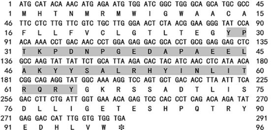
The cDNA coding sequence of Carassius auratus gibelio npy. The shaded boldface letters stand for the mature peptide. * Indicates the stop codon.
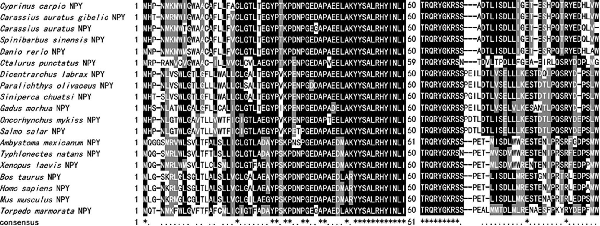
Amino acid sequence alignment of NPY. The shadowed regions are conserved in the listed peptides. * Indicates identical amino acids.
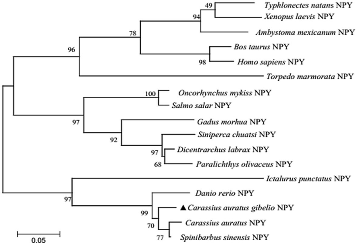
The phylogenetic tree of vertebrate NPY. Scale bar indicates the substitution rate per residue. Numbers at nodes indicate the bootstrap value, as percentages, obtained per 1000 replicates. GenBank accession numbers: Typhlonectes natans (AAD48033), Xenopus laevis (NP_001081300), Ambystoma mexicanum (AAT66407), Bos taurus (AAR37328), Homo sapiens (AAA59944), Torpedo marmorata (AAA49281), Oncorhynchus mykiss (AAB25269), Salmo salar (BAH24101), Gadus morhua (AAX19943), Siniperca chuatsi (ABS83815), Dicentrarchus labrax (CAB64932), Paralichthys olivaceus (BAB62409), Ictalurus punctatus (AAF71617), Danio rerio (NP_571149), Carassius auratus (AAA49186), Spinibarbus sinensis (ABE73783).
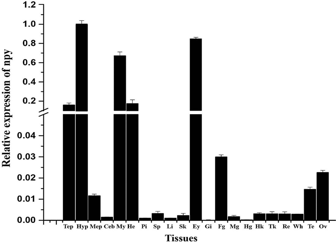
Relative expression of npy mRNA in tissues of gibel carp. Error bars represent standard error of the mean. Tep, telencephalon; Hyp, hypothalamus; Mep, mesencephalon; Ceb, cerebellum; Myp, myelencephalon; Pi, pituitary; Hk, head kidney; Tk, trunk kidney; Li, liver; Fg, foregut; Mg, midgut; Hg, hindgut; He, heart; Sp, spleen; Gi, gill; Te, testis; Ov, ovary. Re, red muscle; Wh, white muscle; Sk, skin; Ey, eye.
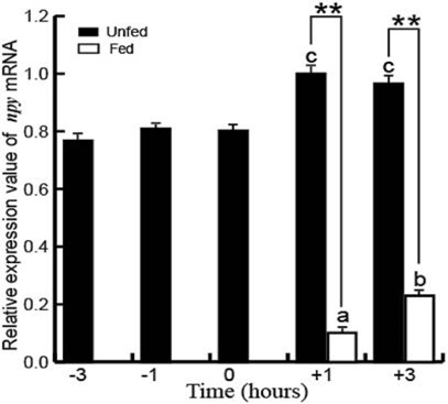
Postprandial changes in npy mRNA expression in the hypothalamus of gibel carp. The preprandial (-3, -1 and 0 h) and postprandial groups (+3 and +1 h) that differed significantly are indicated by different letters above the bars. Asterisks represent significant differences between the fed and unfed groups at the same time point. **P < 0.01.
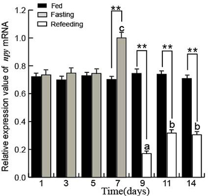
Effects of fasting and refeeding on npy mRNA expression in the hypothalamus of gibel carp. Bars with different letters represent significant differences between experimental groups. Asterisks represent significant differences between the groups at the same time point. **P < 0.01.
References
- Begg, D.P. and Woods, S.C., 2013. The endocrinology of food intake. Nature Rev. Endocrinol., 9: 584-597. https://doi.org/10.1038/nrendo.2013.136[CrossRef]
- Berthoud, H.R., 2002. Multiple neural systems controlling food intake and body weight. Neurosci. Biobehav. Rev., 26: 393-428. https://doi.org/10.1016/S0149-7634(02)00014-3[CrossRef]
- Bouali, S.M., Fournier, A., Stpierre, S. and Jolicoeur, F.B., 1995. Influence of ambient temperature on the effects of NPY on body temperature and food intake. Pharmacol. Biochem. Behav., 50: 473-475. https://doi.org/10.1016/0091-3057(94)00266-L[CrossRef]
- Campos, V.F., Collares, T., Deschamps, J.C., Seixas, F.K., Dellagostin, O.A., Lanes, C.F., Sandrini, J., Marins, L.F., Okamoto, M. and Sampaio, L.A., 2010. Identification, tissue distribution and evaluation of brain neuropeptide Y gene expression in the Brazilian flounder Paralichthys orbignyanus. J. Biosci., 35: 405-413. https://doi.org/10.1007/s12038-010-0046-y[CrossRef]
- Carr, J.A., Brown, C.L., Mansouri, R. and Venkatesan, S., 2002. Neuropeptides and amphibian prey-catching behavior. Comp. Biochem. Physiol. Part B, 132: 151-162. https://doi.org/10.1016/S1096-4959(01)00545-0[CrossRef]
- Chen, R. and Li, W., 2005. cDNA cloning and mRNA expression of neuropeptide Y in orange spotted grouper, Epinephelus coioides. Comp. Biochem. Physiol. Part B: Biochem. Mol. Biol., 142: 79-89. https://doi.org/10.1016/j.cbpc.2005.06.003[CrossRef]
- Chen, T., Chen, S., Ren, C., Hu, C., Tang, D. and Yan, A., 2016. Two isoforms of leptin in the White-clouds Mountain minnow (Tanichthys albonubes): Differential regulation by estrogen despite similar response to fasting. Gen. Comp. Endocrinol., 225: 174-184. https://doi.org/10.1016/j.ygcen.2015.08.002[CrossRef]
- Clark, J.T., Kalra, P.S., Crowley, W.R. and Kalra, S.P., 1984. Neuropeptide Y and human pancreatic polypeptide stimulate feeding behavior in rats. Endocrinology, 115: 427-429. https://doi.org/10.1210/endo-115-1-427[CrossRef]
- Clark, J.T., Kalra, P.S. and Kalra, S.P., 1985. Neuropeptide Y stimulates feeding but inhibits sexual behavior in rats. Obesity, 117: 2435.
- Dahlöf, C., Dahlöf, P. and Lundberg, J.M., 1985. Neuropeptide Y (NPY): Enhancement of blood pressure increase upon alpha-adrenoceptor activation and direct pressor effects in pithed rats. Eur. J. Pharmacol., 109: 289-292. https://doi.org/10.1016/0014-2999(85)90433-9[CrossRef]
- Dalmolin, C., Almeida, D.V., Figueiredo, M.A. and Marins, L.F., 2015. Food intake and appetite control in a GH-transgenic zebrafish. Fish Physiol. Biochem., 41: 1131-1141. https://doi.org/10.1007/s10695-015-0074-5[CrossRef]
- Fuentes, E.N., Kling, P., Einarsdottir, I.E., Alvarez, M., Valdés, J.A., Molina, A. and Björnsson, B.T., 2012. Plasma leptin and growth hormone levels in the fine flounder (Paralichthys adspersus) increase gradually during fasting and decline rapidly after refeeding. Gen. Comp. Endocrinol., 177: 120-127. https://doi.org/10.1016/j.ygcen.2012.02.019[CrossRef]
- Gaikwad, A., Biju, K.C., Muthal, P.L., Saha, S. and Subhedar, N., 2005. Role of neuropeptide Y in the regulation of gonadotropin releasing hormone system in the forebrain of Clarias batrachus (Linn.): Immunocytochemistry and high performance liquid chromatography-electrospray ionization-mass spectrometric analysis. Neuroscience, 133: 267-279. https://doi.org/10.1016/j.neuroscience.2004.12.052[CrossRef]
- Holzer, P., Reichmann, F. and Farzi, A., 2012. Neuropeptide Y, peptide YY and pancreatic polypeptide in the gut–brain axis. Neuropeptides, 46: 261-274. https://doi.org/10.1016/j.npep.2012.08.005[CrossRef]
- Hosomi, N., Furutani, T., Takahashi, N., Masumoto, T. and Fukada, H., 2014. Yellowtail neuropeptide Y: Molecular cloning, tissue distribution, and response to fasting. Fish. Sci., 80: 483-492. https://doi.org/10.1007/s12562-014-0711-4[CrossRef]
- Hoyle, C.H., 1999. Neuropeptide families and their receptors: Evolutionary perspectives. Brain Res., 848: 1. https://doi.org/10.1016/S0006-8993(99)01975-7[CrossRef]
- Inaba, A., Komori, Y., Muroi, Y., Kinoshita, K.I. and Ishii, T., 2016. Neuropeptide Y signaling in the dorsal raphe nucleus inhibits male sexual behavior in mice. Neuroscience, 320: 140-148. https://doi.org/10.1016/j.neuroscience.2016.01.069[CrossRef]
- Keen-Rhinehart, E., Ondek, K. and Schneider, J.E., 2013. Neuroendocrine regulation of appetitive ingestive behavior. Front. Neurosci., 7: 213. https://doi.org/10.3389/fnins.2013.00213[CrossRef]
- Kehoe, A.S. and Volkoff, H., 2007. Cloning and characterization of neuropeptide Y (NPY) and cocaine and amphetamine regulated transcript (CART) in Atlantic cod (Gadus morhua). Comp. Biochem. Physiol. Part A: Mol. Integr. Physiol., 146: 451-461. https://doi.org/10.1016/j.cbpa.2006.12.026[CrossRef]
- Kumar, S., Tamura, K. and Nei, M., 2004. MEGA3: Integrated software for molecular evolutionary genetics analysis and sequence alignment. Brief. Bioinform., 5: 150-163. https://doi.org/10.1093/bib/5.2.150[CrossRef]
- MacDonald, E. and Volkoff, H., 2009. Cloning, distribution and effects of season and nutritional status on the expression of neuropeptide Y (NPY), cocaine and amphetamine regulated transcript (CART) and cholecystokinin (CCK) in winter flounder (Pseudopleuronectes americanus). Horm. Behav., 56: 58-65. https://doi.org/10.1016/j.yhbeh.2009.03.002[CrossRef]
- Malmström, R.E., 2001. Existence of both neuropeptide Y, Y1 and Y2 receptors in pig spleen: Evidence using subtype-selective antagonists in vivo. Life Sci., 69: 1999-2005. https://doi.org/10.1016/S0024-3205(01)01286-3[CrossRef]
- Montpetit, C.J., Chatalov, V., Yuk, J., Rasaratnam, I. and Youson, J.H., 2005. Expression of neuropeptide Y family peptides in the brain and gut during Stages of the life cycle of a parasitic lamprey (Petromyzon marinus) and a nonparasitic lamprey (Ichthyomyzon gagei). Annls N.Y. Acad. Sci., 1040: 140-149. https://doi.org/10.1196/annals.1327.018[CrossRef]
- Murashita, K., Kurokawa, T., Ebbesson, L.O., Stefansson, S.O. and Rønnestad, I., 2009. Characterization, tissue distribution, and regulation of agouti-related protein (AgRP), cocaine- and amphetamine-regulated transcript (CART) and neuropeptide Y (NPY) in Atlantic salmon (Salmo salar). Gen. Comp. Endocrinol., 162: 160-171. https://doi.org/10.1016/j.ygcen.2009.03.015[CrossRef]
- Narnaware, Y.K., Peyon, P.P., Lin, X. and Peter, R.E., 2000. Regulation of food intake by neuropeptide Y in goldfish. Am. J. Physiol. Regul. Integr. Comp. Physiol., 279: R1025-1034.
- Nuria, D.P. and Thrur, B.B., 2001. Regulation of food intake by neuropeptides and hormones. In: Food intake in fish (eds. D. Houlihan, T. Boujard and M. Jobling). John Wiley & Sons, Inc., New Jersey, United States. https://doi.org/10.1002/9780470999516.ch12[CrossRef]
- Peng, C., Gallin, W., Peter, R.E., Blomqvist, A.G. and Larhammar, D., 1994. Neuropeptide-Y gene expression in the goldfish brain: Distribution and regulation by ovarian steroids. Endocrinology, 134: 1095. https://doi.org/10.1210/endo.134.3.8119148[CrossRef]
- Pirone, A., Lenzi, C., Marroni, P., Betti, L., Mascia, G., Giannaccini, G., Lucacchini, A. and Fabiani, O., 2008. Neuropeptide Y in the brain and retina of the adult teleost gilthead seabream (Sparus aurata L.). Anat. Histol. Embryol., 37: 231-240. https://doi.org/10.1111/j.1439-0264.2007.00836.x[CrossRef]
- Saper, C.B., Chou, T.C. and Elmquist, J.K., 2002. The need to feed: Homeostatic and hedonic control of eating. Neuron, 36: 199. https://doi.org/10.1016/S0896-6273(02)00969-8[CrossRef]
- Silverstein, J.T., Breininger, J., Baskin, D.G. and Plisetskaya, E.M., 1998. Neuropeptide Y-like gene expression in the salmon brain increases with fasting. Gen. comp. Endocrinol., 110: 157-165. https://doi.org/10.1006/gcen.1998.7058[CrossRef]
- Simpson, E.H. and Balsam, P.D., 2016. Behavioral neuroscience of motivation. Springer International Publishing. https://doi.org/10.1007/978-3-319-26935-1[CrossRef]
- Tang, Y., Li, H., Li, J., Yu, F. and Yu, J., 2014. Characterization and expression analysis of two distinct neuropeptide Ya paralogues in Jian carp (Cyprinus carpio var. Jian). Fish Physiol. Biochem., 40: 1709-1719. https://doi.org/10.1007/s10695-014-9961-4[CrossRef]
- Tatemoto, K., Carlquist, M. and Mutt, V., 1982. Neuropeptide Y- - A novel brain peptide with structural similarities to peptide YY and pancreatic polypeptide. Nature, 296: 659-660. https://doi.org/10.1038/296659a0[CrossRef]
- Thompson, J.D., Higgins, D.G. and Gibson, T.J., 1994. CLUSTAL W: Improving the sensitivity of progressive multiple sequence alignment through sequence weighting, position-specific gap penalties and weight matrix choice. Nucl. Acids Res., 22: 4673-4680. https://doi.org/10.1093/nar/22.22.4673[CrossRef]
- Thorsell, A. and Mathé, A.A., 2017. Neuropeptide Y in alcohol addiction and affective disorders. Front. Endocrinol., 8: 178. https://doi.org/10.3389/fendo.2017.00178[CrossRef]
- Valassi, E., Scacchi, M. and Cavagnini, F., 2008. Neuroendocrine control of food intake. Nutr. Metab. Cardiovasc. Dis., 18: 158-168. https://doi.org/10.1016/j.numecd.2007.06.004[CrossRef]
- Valen, R., Jordal, A.E., Murashita, K. and Rønnestad, I., 2011. Postprandial effects on appetite-related neuropeptide expression in the brain of Atlantic salmon, Salmo salar. Gen. Comp. Endocrinol., 171: 359. https://doi.org/10.1016/j.ygcen.2011.02.027[CrossRef]
- Volkoff, H., 2016. The neuroendocrine regulation of food intake in fish: A review of current knowledge. Front. Neurosci., 10: 540. https://doi.org/10.3389/fnins.2016.00540[CrossRef]
- Volkoff, H., Canosa, L.F., Unniappan, S., Cerdá-Reverter, J.M., Bernier, N.J., Kelly, S.P. and Peter, R.E., 2005. Neuropeptides and the control of food intake in fish. Gen. Comp. Endocrinol., 142: 3-19. https://doi.org/10.1016/j.ygcen.2004.11.001[CrossRef]
- Volkoff, H., Sabioni, R., Coutinho, L.L. and Cyrino, J.E., 2017. Appetite regulating factors in pacu (Piaractus mesopotamicus): Tissue distribution and effects of food quantity and quality on gene expression. Comp. Biochem. Physiol. Part A: Mol. Integr. Physiol., 203: 241-254. https://doi.org/10.1016/j.cbpa.2016.09.022[CrossRef]
- Wei, R., Zhou, C., Yuan, D., Wang, T., Lin, F., Chen, H., Wu, H., Xin, Z., Yang, S. and Wang, Y., 2014. Characterization, tissue distribution and regulation of neuropeptideY in Schizothorax prenanti. J. Fish Biol., 85: 278-291. https://doi.org/10.1111/jfb.12413[CrossRef]
- Woods, S.C. and Begg, D.P., 2016. Regulation of the motivation to eat. Curr. Topics Behav. Neurosci., 27: 15-34. https://doi.org/10.1007/7854_2015_381[CrossRef]
- Xiang, H., 1994. Comparative aspects of the role of neuropeptide Y in the regulation of the vertebrate heart. Cardioscience, 5: 209.
- Xu, C., Li, X.F., Tian, H.Y., Jiang, G.Z. and Liu, W.B., 2015. Feeding rates affect growth, intestinal digestive and absorptive capabilities and endocrine functions of juvenile blunt snout bream Megalobrama amblycephala. Fish Physiol. Biochem., 42: 689-700. https://doi.org/10.1007/s10695-015-0169-z[CrossRef]
- Yin, Z., Xue, M., Zheng, Y., Wang, X. and Wu, X., 2017. Effect of yeast extract as feeding stimulants on diet preference of Gibel carp. Feed Indust., 38: 4-8.
- Yokobori, E., Azuma, M., Nishiguchi, R., Kang, K.S., Kamijo, M., Uchiyama, M. and Matsuda, K., 2012. Neuropeptide Y stimulates food intake in the Zebrafish, Danio rerio. J. Neuroendocrinol., 24: 766. https://doi.org/10.1111/j.1365-2826.2012.02281.x[CrossRef]
- Zhang, L., Zhang, M.L., Guo, F.F., Sun, X.R., Gao, S.L., Luo, X.U. and Pathophysiology, D.O., 2017. Effects of orexins on the food intake of rats and its mechanism. Progr. Mod. Biomed., 17: 28-31.
- Zhang, Y., Proenca, R., Maffei, M., Barone, M., Leopold, L. and Friedman, J.M., 1994. Positional cloning of the mouse obese gene and its human homologue. Nature, 372: 425-432. https://doi.org/10.1038/372425a0[CrossRef]
- Zhou, C., Zheng, J., Lei, L., Yuan, D., Zhu, C., Ye, H., Zhang, C., Wang, D., Yang, M. and Wu, J., 2016. Evidence that ghrelin may be associated with the food intake of gibel carp (Carassius auratus gibelio). Fish Physiol. Biochem., 42: 1-10. https://doi.org/10.1007/s10695-016-0246-y[CrossRef]
Services
Article usage statistics
Loading…