
Due to the emerging problem of multidrug resistant (MDR) pathogens, there is a demand of novel biological active compounds to replace currently used antibiotics in order to evade resistance. Currently, the pathogenic organisms of concern are methicillin and vancomycin resistant strains of Staphylococcus aureus, Acinetobacter spp., extended spectrum β-lactamases (ESBLs) producing strains of Escherichia coli, Klebsiella spp., Pseudomonas spp. and Proteus spp. In this study, a collection of nineteen actinomycetes strains isolated from Cholistan Desert, Pakistan, were screened biologically and biochemically for their potential bioactive secondary metabolites against MDR bacterial pathogens. The identification of strains was done by morphological, physiological and biochemical characterization and by 16SrRNA gene sequencing. For biological screening different methods including cross streak, agar plug and well diffusion method were performed. Seven Streptomyces strains including NWMD-6, NWMD-7, NWMD-10, NWMD-11, NWMD-12, NWMD-15 and NWMD-16 were found to have significant antimicrobial activity against MDR bacterial pathogens. Secondary metabolite profiling of each methanolic extract by TLC and HPLC-UV signified the presence of unique and diverse metabolic compounds. The study predicts that the seactinomycetes strains are a potential resource to cope up with evolving multi drug resistant pathogens. Furthermore, these secondary metabolites can be identified by comparative studies and these actinomycetes can be grown on larger scale to obtain high yield of potentially useful secondary metabolites.
Research Article · Volume 33, Issue 2, 20 November 2018 · Open Access
Molecular Characterization and Secondary Metabolite Profiling of the Actinomycetes Strains Active against Various MDR Bacterial Pathogens
· ·
Affiliations & Notes ⌄
- 1Department of Microbiology and Molecular Genetics, University of the Punjab, Lahore-54590, Pakistan , *Corresponding author: Imran Sajid imran.mmg@pu.edu.pk
Article Info ⌄
- Journal:
- Punjab University Journal of Zoology
- Published:
- 20 November 2018
From the article
- In melanin production, almost 52.67% of strains showed melanin production.
- The results of sugar utilization suggested that the best sugar used for utilization of carbon source is glucose because it allowed the growth of every strain.
- That suggests that Streptomyces strains have ability to utilize common sugars and their metabolic pathway do not allow degrading and utilizing complex sugars.
- In biological screening almost 80% strains showed the antimicrobial activity against Gram positive organisms whereas the results against Gram negative organisms were not that promising.
Selected from the article's own text.
Abstract
Introduction
The discovery of antibiotics for treatment of infectious diseases is greatest success in medicine century but due to extensive use of antibiotics, resistance to drugs is increasing ; . This causes the application of currently used antimicrobial agents to be quickly diminished resulting in the favor of multi drug resistant pathogens ; . Drug resistant pathogens comprises of methicillin-resistant Staphylococcus aureus (MRSA), vancomycin-resistant Staphylococcus aureus (VRSA), extended spectrum β-lactamases (ESBL) producing bacteria such as E. coli, Klebsiella sp. and Pseudomonas aeruginosa ; .
Due to emerging problem of multidrug resistance there is an increasing demand of novel biological active secondary metabolites for drugs formation . Microbial secondary metabolites are a significant source of current as well as new drugs. Some genera of bacteria, such as Streptomyces, Myxobacteria, Bacilli, Pseudomonas, and Burkholderia, are known to exhibit versatile secondary metabolism that has proven to be useful in the production of structurally varied compounds with novel biological activities such as antibiotics . Actinomycetes, specifically those belongs to the genus Streptomyces, are known to an economic source of variety of secondary compounds with diverse chemical molecules . Actinomycetes are Gram-positive bacteria that have high GC content above 55% and most of them produce mycelium . Nowadays, Streptomyces are considered as antibiotics store room or library. It have been reported that about 23,000 existing natural biological product are produced by microorganisms out of which 16,500 compounds have antibiotic activities. More than 10,000 of such antibiotics are isolated from actinomycetes. Among actinomycetes, about 7,600 antibiotics are isolated from Streptomyces species and 2470 from rare actinomycetes. Literature revealed that at least 4607 patents have been issued on actinomycete associated products ; ; .
Streptomyces produces many therapeutic agents which include anti-bacterials such as tetracyclines, antifungals such as amphotericin, and anticancer drugs such as Adriamycin and the immune-suppressants such as tacrolimus ; . In past several decades, a huge amount of Streptomyces spp. have been screened from soil, so probabilities of isolating a unique Streptomyces strain with prevailing antimicrobial activity from terrestrial areas have reduced . So researchers are hunting to explore Streptomycetes from unique places such as deep sea, forests, mountains, desert and marine areas .
In this study, Streptomyces strain previously isolated from Cholistan Desert were characterized and screened for their antimicrobial activity. A significant aim of the study has been to discover useful secondary metabolites to solve problems regarding resistant microbes. The morphological, microscopic, physiological and genetic characterization of the selected strains was performed. The strains were cultivated on smaller scale to obtain crude bioactive secondary metabolites with ethyl acetate. The antimicrobial activity of strains was analyzed by cross streak, agar plug and well diffusion methods against MDR bacterial pathogens. In chemical profiling the crude extracts were examined using TLC and HPLC-UV analysis.
Materials and Methods
Sample collection
Actinomycetes strains were obtained from Culture Collection (Cholistan Desert strains) of the Department of Microbiology and Molecular Genetics (MMG), University of the Punjab, Lahore, Pakistan.
Morphological, biochemical and physiological characterization
Identification of actinomycetes strains was done on the basis of morphological, biochemical and genetic characterization. In morphological characterization actinomycetes were observed both macroscopically and microscopically. For physiological and biochemical characterization of streptomycetes, two methods which have been employed in International Streptomyces Project (ISP), the formation of melanin and utilization of different sugars as carbon source were used. Whereas numerous other physiological tests used for characterization included determination of growth temperature range, utilization of organic acids, utilization of oxalates, hydrolysis of urea, formation of organic acids, hydrolysis of Esculin/Arbutin etc.
Genetic characterization (16S rRNA gene sequencing)
For genetic characterization genomic DNA of the selected strains were isolated by FavorPrep™ tissue Genomic DNA extraction Mini Kit. PCR amplification of 16S rRNA gene was done by using 27F forward primer (5’-AGAGTTTGATCCTGGCTCAG-3’) and 1522R reverse primer (5’-AAGGAGGTGATCCA(AG)CCGCA-3’). The amplified product was extracted from gel by using Favor Prep Micro Elute Gel Extraction Kit. The PCR amplified 16S rRNA gene was sequenced on an automated sequencer by dye terminator chemistry. After BLAST, the sequences were submitted to GenBank to get accession numbers of submitted sequences of 16S rRNA gene.
Cultivation and solvent extraction
The cell free extract of active actinomycetes strains were prepared by inoculating strains in 200 ml of GYM broth and incubated at 28°C on a rotatory shaker for 7 days. After incubation 200ml ethyl acetate was added in every flask. The cells of actinomycetes were broken by sonication by the sonicator (Selecta Ultrasons Medi-II) for about 30 min. The broken cells were transferred to a separating funnel and were agitated properly for 20 min, later the separating funnel was placed uninterrupted for some time to make two separated layers visible. Then the upper organic was it upper or lower layer was separated from aqueous layer with care. After that the solvent was removed by using the rotary vacuum evaporator. The dried crude extract was suspended in 5ml of methanol, which was kept in clean test tubes (Heidolph Laborota 4000 efficient) at 4°C. These methanolic extracts were used for the screening of actinomycetes by agar well method and further used for chemical screening including TLC and HPLC analysis.
Biological screening (determination of antimicrobial activity)
For antimicrobial screening of Streptomyces, following three typical methods including cross streak method, agar plug method and well diffusion method were used:
Cross streak method
Cross streak method is rapid test for screening of antimicrobial activity of actinomycetes. Muller Hinton agar (MH agar) was prepared, autoclaved and poured into petri plates. The strain of Streptomyces was inoculated in the form of major streak in the center of MH agar plate and incubated at 28oC for 5 days. After 5-7 days incubation, pathogenic test strains (Gram-positive and Gram-negative) were inoculated perpendicular to the main streak and the plate was incubated at 37oC for 24 h. After completion of second incubation the antimicrobial activity of steptomycetes was observed by inhibition of test strains around the main streak .
Agar plug method
In agar plug method, 24 h broth culture of test strains Gram-positive organisms (MRSA and Bacillus), Gram-negative (Klebsiella, Acinetobacter, Escherichia coli, Pseudomonas and Proteus) were swabbed on the MH agar plate by means of sterile swabs. Five days cultures of streptomycetes strains on GYM agar were taken and agar plugs of moderate size were cut and placed upside down on the already swabbed plate of test organism and then incubated at 37°C for 24 h. Due to diffusion of antimicrobial compound from plug to medium, antimicrobial activity was easily detected by the formation of clear zone of inhibition around agar plug .
Well diffusion method
Well diffusion method is standard and convenient method as compared to agar plug method for the screening of the biologically active compounds. Supernatant was made by taking the culture growth in centrifuge tubes and centrifuged for 2 min. Methanolic crude extracts was made by the procedure mentioned in cultivation and solvent extraction section. In well diffusion, Gram-positive organisms (MRSA and Bacillus), Gram-negative (Klebsiella, Acinetobacter, Escherichia coli, Pseudomonas and Proteus) were swabbed on the MH agar plate by means of sterile swabs. Then by using sterile corkborer wells were made in the agar. About 50μl of the supernatant or methanolic extracts was filled on each well. The plates were placed on bench top for 1 h at room temperature, and then incubated at 37°C for 24 h. After incubation plates were visualized for clear zones around wells. The appearance of clear zones indicated that Streptomyces had ability to inhibit the growth of test strains. The zones of inhibition were measured in millimeters.
Chemical screening
For chemical screening two methods thin layer chromatography (TLC) and high-performance liquid chromatography (HPLC) were used for the recognition of “promising” strains .
Thin layer chromatography
The TLC is a simple chemical method for the profiling of components of crude methanolic extract. On TLC plate a line 1 inch above from base was marked by lead pencil and samples names were labeled on it. Then methanolic extracts of streptomycetes samples were spotted drop wise on their respective places by using capillary tube. Then allowed the plate to be air dried and were placed in TLC tank with CH2Cl2/10%MeOH solvent system making sure and allowed the solvent to move up on the TLC plate. After development of plates, they were air dried and observed under UV at 254 nm and 366 nm and bands were marked. Then the plates were individually stained with spraying reagents Anisaldehyde/H2SO4 and Ehrlisch’s reagents. The distinct bands of various colors appeared in every crude extract were scanned and recorded.
High performance liquid chromatography analysis
The crude extracts were analysed on the HPLC (Sykum HPLC system) using the software clarity withC18column from phenomenex with 30cm length. For mobile phase methanol was used and flow rate was set at 1ml/min. The crude extract was mixed in HPLC grade methanol and 20μl was injected by using a micro-syringe. Samples were allowed to run for 15 min and detector detects UV absorbance at 254nm. The peaks formed by software was analysed properly and evaluated by using retention time (tR) with standard UV absorption data of secondary metabolites.
Table I: Morphological characteristics of streptomycetes strains after incubation at 28oC for 7 days on GYM agar.
| Strains | Colony characteristics | Color of mycelium | Soluble pigments | ||||
| Shape | Size (mm) | Margin | Texture | Substrate | Aerial | ||
| NWMD-1 | Irregular | 4 | Undulate | Hard | Brown Yellow | Grey Yellow | No |
| NWMD-2 | Irregular | 2.5 | Undulate | Hard | Cream | White | No |
| NWMD-3 | Irregular | 1.4 | Undulate | Hard | Black Yellow | Grey White | No |
| NWMD-4 | Irregular | 1 | Undulate | Hard | Brown Yellow | Grey White | No |
| NWMD-5 | Irregular | 3.3 | Undulate | Hard | Pink Orange | White Pink | No |
| NWMD-6 | Irregular | 3.6 | Undulate | Hard | Grey White | Yellow | No |
| NWMD-7 | Irregular | 4.5 | Undulate | Hard | Orange | Yellow Orange | No |
| NWMD-8 | Irregular | 3.5 | Undulate | Hard | Cream | Grey Yellow | No |
| NWMD-9 | Irregular | 3 | Undulate | Hard | Dark Brown | Grey White | No |
| NWMD-10 | Circular | 4.5 | Entire | Soft | Greenish Brown | Greenish Grey | No |
| NWMD-11 | Irregular | 3 | Undulate | Hard | Brown Yellow | Brown White | No |
| NWMD-12 | Irregular | 2 | Undulate | Soft | Orange Pink | Peach White | Yes |
| NWMD-13 | Irregular | 4 | Undulate | Soft | Cream yellow | Brown Grey | No |
| NWMD-14 | Irregular | 4 | Undulate | Hard | Orange Yellow | Pink White | No |
| NWMD-15 | Irregular | 3.5 | Undulate | Hard | Yellow Orange | Orange Pink | No |
| NWMD-16 | Irregular | 3 | Undulate | Hard | Pink Orange | Pink White | No |
| NWMD-17 | Irregular | 3.2 | Undulate | Hard | Cream yellow | Grey Yellow | No |
| NWMD-18 | Irregular | 3.5 | Undulate | Hard | Brown Yellow | Grey White | No |
| NWMD-19 | Irregular | 2 | Undulate | Soft | Cream | Brown white | Yes |
Results
A total of 19 streptomycetes strains obtained from the culture collection of the department of Microbiology and Molecular Genetics (MMG) University of the Punjab, Lahore, Pakistan were named as NWMD-1, NWMD-2, NWMD-3, and up to NWMD-19.
Morphological, biochemical and physiological characteristics and 16S rRNA gene sequencing
For morphological characterization streptomycetes colonies were observed macroscopically, most of them were irregular in shape and embedded in solid agar, size was ranging from 1-4 mm, margins were mostly undulate and texture was hard except for the strains NWMD-10, NWMD-12, NWMD-13 and NWMD-19. Substrate and aerial mycelium showed color that range from white to grey, brown yellow, pink and orange. The morphological characteristics of selected streptomycetes strains are summarized in Table I and shown in Figure 1. In Gram staining, all of them were gram positive and exhibited irregular hyphae (Figure 2). In case of physiological and biochemical, in melanin production 10 strains out of 19 including NWMD-1, NWMD-5, NWMD-12, NWMD-14, NWMD-16, NWMD-17 and NWMD-18 showed prominent melanin production whereas NWMD-7, NWMD-9 and NWMD-15 showed moderate melanin production and NWMD-2, NWMD-3, NWMD-4, NWMD-6, NWMD-8, NWMD-10, NWMD-11, NWMD-13 and NWMD-19 showed negative results. In sugar utilization test, the strainsNWMD-7, NWMD-13, NWMD-15, NWMD-16 and NWMD-17 showed growth on all nine sugars, strains NWMD-2 and NWMD-19 showed growth only on D-lactose and D-glucose whereas NWMD-3 showed growth only on D-fructose and D-glucose. NWMD-1, NWMD-4, NWMD-5, NWMD-6, NWMD-8, NWMD-9, NWMD-10, NWMD-11, NWMD-12, NWMD-14 and NWMD-18 showed growth on some sugars (Table II). For genetic characterization 16S rRNA gene sequence data was aligned with reported gene sequence in GenBank. Strain NWMD-6 (GenBank accession number KX455499) showed 99% similarity to Streptomyces pseudo griseolus whereas Strain NWMD-7 (GenBank accession number KX455500) showed 99% similarity to Streptomyces geysiriensis).The number of nucleotide sequence, maximum resemblance with Streptomyces species and GenBank accession numbers are mentioned in Table III.
Biological screening
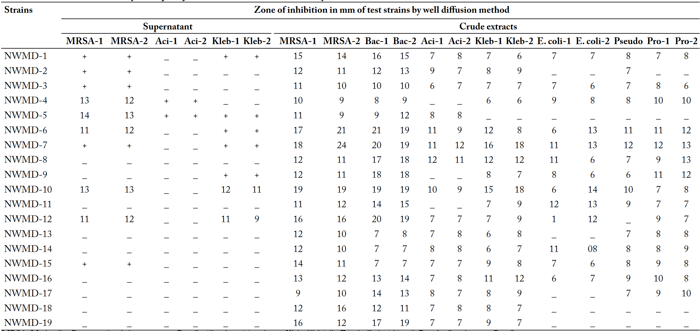
For biological screening test organisms used were well characterized strains of MRSA, Bacillus, Acinetobacter, Klebsiella, Escherichia coli, Pseudomonas and Proteus species. By using cross streak method strains were found to be more bioactive against Gram-positive organisms as compared to Gram-negative. The best activity was shown by strain NWMD-7 and NWMD-10 while strain NWMD-4, NWMD-5, NWMD-6, NWMD-10 and NWMD-15 also showed growth of inhibition against Gram-positive organisms. Results for Cross-streak method are shown in Figure 3 and Table III. In agar plug method, best activity against MRSA was exhibited by streptomycetes strains NWMD-6, NWMD-10, NWMD-14 and NWMD-15 whereas NWMD-2 and NWMD-13 exhibited no activity. Against Acinetobacter best activity was exhibited by NWMD12, NWMD-14 and NWMD-15, against Klebsiella best activity was exhibited by NWMD-4 and for Bacillus best activity was exhibited by NWMD-6, NWMD-14 and NWMD-15. Results of agar plug method are shown in Figure 3 and Table IV. In well diffusion method, by using supernatant the best activity was observed against MRSA. Best activity against MRSA was exhibited by strain NWMD-4, NWMD-5, NWMD-6, NWMD-10 and NWMD-12. For Acinetobacter best activity was exhibited by NWMD-4 and NWMD-5, for Klebsiella best activity was exhibited by NWMD-10 and NWMD-12. By using methanolic extracts best activity against MRSA was exhibited by strain NWMD-7, NWMD-10 and NWMD-12. For Bacillus best activity was exhibited by the strains NWMD-6, NWMD-7, NWMD-10 and NWMD-12, for Acinetobacter best activity was exhibited by the strain NWMD-10 whereas NWMD-4, NWMD-9 and NWMD-11 exhibited no activity, for Klebsiella best activity was exhibited by the strains NWMD-7 and NWMD-10 whereas NWMD-5 exhibited no activity, for Pseudomonas best activity was exhibited by the strain NWMD-7 whereas NWMD-5, NWMD-11. NWMD-18 and NWMD-19 exhibited no activity. Against Proteus best activity was exhibited by the strains NWMD-7 and NWMD-9 whereas NWMD-5, NWMD-18 and NWMD-19 exhibited no activity, and for Escherichia coli best activity was exhibited by the strains NWMD-11 whereas NWMD-2, NWMD-5, NWMD-13, NWMD-17 NWMD-18 and NWMD-19 exhibited no activity. Results of well diffusion method are shown in Figure 3 and Table V.
Table II: Physiological and biochemical characteristics of selected streptomycetes strains: melanin formation, utilization of sugars,utilization of organic acid, urea hydrolysis, utilization of calcium oxalate, hydrolysis of esculin and formation of organic acid.
| Strains | Mela nin form ation | Sugars | Organic acid utilization | Esc ulin | Org anic acid prod uction | |||||||||
| Gl | S | M | L | F | X | Ga | A | I | Sodium malo nate | Trisod ium citrate | ||||
| NWMD-1 | ++ | +++ | - | +++ | +++ | + | + | - | + | - | - | - | ++ | - |
| NWMD-2 | - | ++ | + | - | ++ | - | - | - | - | - | - | - | + | - |
| NWMD-3 | - | + | - | - | - | ++ | - | - | - | - | - | - | ++ | - |
| NWMD-4 | - | +++ | ++ | +++ | +++ | +++ | - | + | + | - | ++ | - | ++ | - |
| NWMD-5 | ++ | +++ | +++ | +++ | ++ | - | + | + | - | + | - | - | + | - |
| NWMD-6 | - | +++ | - | +++ | +++ | ++ | - | + | - | + | - | - | ++ | - |
| NWMD-7 | + | +++ | + | +++ | +++ | ++ | + | + | + | + | - | + | + | - |
| NWMD-8 | - | + | - | + | +++ | ++ | - | - | - | - | - | - | +++ | - |
| NWMD-9 | + | ++ | +++ | + | + | + | + | - | - | - | - | + | +++ | - |
| NWMD-10 | - | +++ | + | +++ | +++ | +++ | + | - | - | - | - | - | + | - |
| NWMD-11 | - | +++ | + | +++ | +++ | ++ | + | + | - | - | +++ | ++ | ++ | - |
| NWMD-12 | +++ | +++ | +++ | +++ | +++ | +++ | + | - | - | - | - | - | + | - |
| NWMD-13 | - | +++ | +++ | +++ | +++ | +++ | + | + | +++ | + | +++ | +++ | +++ | - |
| NWMD-14 | +++ | +++ | +++ | +++ | +++ | + | + | + | ++ | _ | - | - | + | - |
| NWMD-15 | + | +++ | +++ | +++ | +++ | + | + | + | +++ | + | - | + | ++ | - |
| NWMD-16 | +++ | +++ | +++ | +++ | +++ | + | + | + | + | + | - | +++ | + | - |
| NWMD-17 | ++ | +++ | +++ | +++ | +++ | ++ | + | + | + | - | + | ++ | ++ | - |
| NWMD-18 | +++ | +++ | ++ | ++ | +++ | ++ | - | - | - | - | - | - | + | - |
| NWMD-19 | - | + | - | - | + | - | - | - | - | - | - | - | - | - |
+++, excellent results; ++, good results; +, moderate results; –, negative results. Gl, D-glucose; S, sucrose; M, D-maltose; L, D-lactose; F, D-fructose; X, D- xylose, Ga, D-galactose; A, L-arabinose; I, meso-inositol
Table III: 16S rRNAgene sequencing.
| Isolates | Nucleotide length (bp) | %age homology | Organism | Accession No. |
| ND-6 | 766 | 99% | Streptomyces pseudogriseolus | KX455499 |
| ND-7 | 342 | 99% | Streptomyces geysiriensis | KX455500 |
Chemical profiling (metabolic fingerprinting of the selected strains)
The crude extract of Streptomyces strains were examined by thin layer chromatography (TLC). Plates were observed under short UV (254nm) and long UV (366nm) and sprayed with 2 reagents (anisaldehye / H2SO4 and Ehrlisch reagent) for determination of bands pattern of crude extracts. The crude extracts showed a great diversity of compounds after staining, the color produced after staining were yellow, purple, red, dark brown, green and pink. The highest diversity of compounds was observed in crude extracts of strain NWMD-6, NWMD-7, NWMD-10, NWMD-12, NWMD-13, NWMD-14 and NWMD-16. The pattern of bands observed on TLC plates under UV and after staining with anisaldehye / H2SO4 and Ehrlisch reagent is shown in Figure 4.
In HPLC analysis, each of the crude extract exhibited different number of peaks with different retention times (Figure 5). The crude extract of strain NWMD-7 exhibited major peak at retention time 2.344 min with area of 19517.641 (85.9%) and five small peaks at retention time (tR) 1.54, 1.86, 2.86, 3.16 and 3.45 respectively. The crude extract of strainNWMD-10 exhibited major peak at retention time 2.32 min with area of 21525.376 (77.8%) and five small peaks at retention time (tR) 1.48, 1.80, 3.15, 3.45 and 3.86. The crude extract of strainNWMD-12 exhibited major peak at retention time 2.388 min with area of 18241.980 (77.9%) and three small peaks retention time (tR) 1.84, 3.49 and 3.84. likewise different strains exhibited different peaks at different retention time but most of peaks were ranging from 2.3 to 2.6 min.
Discussion
By keeping in view the fact, that streptomycetes of different climate and unexplored region may yield some distinct and potent secondary metabolites , many researchers have screened actinomycetes strains isolated from natural habitats like soil , marine , desert ; , plants and sediments that have ability to produce biologically active compounds against infectious diseases .
In this study a total of 19 Streptomyces strains isolated from desert soil were examined for their antimicrobial activity against various MDR bacterial pathogens. The strains were characterized on the basis of morphology and microscopy, the results strongly suggested that these strains are the member of genus Streptomyces (Table I; Figures 1, 2). Subsequently, the strains were characterized by physiological, biochemical and genetic characterization. In physiological and biochemical characterization tests used for identification and characterization employed in International Streptomyces Project (ISP) are formation of melanin and utilization of different sugars as a carbon source. In melanin production, almost 52.67% of strains showed melanin production. The results of sugar utilization suggested that the best sugar used for utilization of carbon source is glucose because it allowed the growth of every strain. The most common sugars such as D-glucose, sucrose, D-maltose and D-fructose are utilized by most of the strains whereas complex sugars like D-xylose, D-lactose, D-galactose, and meso-inositol are utilized by some strains with limited growth. That suggests that Streptomyces strains have ability to utilize common sugars and their metabolic pathway do not allow degrading and utilizing complex sugars. The 16S rRNA gene sequencing of two bioactive strains (NWMD-6 and NWMD-7) proved that these strains are different species of genus Streptomyces. The assigned GenBank accession numbers are mentioned in Table III.
The main objective of this study was to screen out extraordinary and rare actinomycetes species having ability to produce undiscovered biologically active metabolites. While screening of bioactive strains for drug discovery, numerous promising strains possibly be eliminated due to incompetent procedures, too high selectivity and by missing tests. So variety of tests with low selectivity and broad range of antibacterial activity was used . For antimicrobial screening of Streptomyces strains various biological and chemical screening strategies were used. In biological screening almost 80% strains showed the antimicrobial activity against Gram positive organisms whereas the results against Gram negative organisms were not that promising. Similar types of results that extracts are more active against Gram positive organisms, were reported in other antibacterial studies by Walsh et al. (2003), Hozzein et al. (2011) and Salehghamari et al. (2015) that may be due to presence of outer membrane in Gram negative organisms that prevent the entry of antimicrobial agent to enter. Streptomyces strains with bigger than 10mm zone of inhibition was taken as active metabolites. All of the strains were found to be active against MRSA whereas only 3 were found to be active against Acinetobacter and Pseudomonas only 4 were found to be active against Klebsiella and only 6 were found to be active against E.coli and Proteus.
In chemical screening, TLC and HPLC analysis help in recognizing metabolic fingerprints of methanolic crude extract to observe the similarities and differences between different actinomycetes isolates grown under same conditions . In TLC, the best results were shown by anisaldehye/H2SO4because of its ability to stain numerous compounds with distinctive colors. The crude extracts gave several yellow, purple, red, dark brown, green and pink colors after staining with reagents. Formation of different colored bands under UV and by staining revealed the presence of variety of compounds in the crude extracts which might have the antimicrobial activity. HPLC of crude extracts provide an additional picture of metabolic fingerprints specific for each strain.
Table IV: Antimicrobial activity of Sreptomycetes strains determined by cross streak method and agar plug method.
| Strains | Zone of inhibition in mm of test strains | |||||||||
| Cross streak method | Agar plug method | |||||||||
| Gram positive | Gram negative | MRSA-1 | MRSA-2 | Aci-1 | Aci-2 | Kleb-1 | Kleb-2 | Bac-1 | Bac-2 | |
| NWMD-1 | _ | _ | + | + | + | + | + | - | + | + |
| NWMD-2 | _ | _ | _ | _ | - | + | - | - | + | + |
| NWMD-3 | _ | _ | + | + | - | + | + | - | + | + |
| NWMD-4 | 11 | _ | + | + | + | + | + | + | + | + |
| NWMD-5 | 5 | _ | 11 | 9 | - | - | + | + | + | + |
| NWMD-6 | 7 | _ | 15 | 17 | - | - | - | - | 16 | 19 |
| NWMD-7 | 17 | _ | 11 | 12 | - | - | + | + | 14 | 12 |
| NWMD-8 | _ | _ | + | + | - | - | - | - | + | + |
| NWMD-9 | _ | _ | + | + | - | - | - | - | + | + |
| NWMD-10 | 9 | _ | 14 | 13 | - | - | - | - | 13 | 12 |
| NWMD-11 | 17 | _ | 13 | 12 | - | - | - | - | + | + |
| NWMD-12 | _ | _ | 12 | 14 | 13 | 11 | - | - | + | + |
| NWMD-13 | _ | _ | - | - | - | - | - | + | + | |
| NWMD-14 | _ | _ | 19 | 20 | 13 | 12 | + | + | 19 | 16 |
| NWMD-15 | 13 | _ | 18 | 19 | 14 | 12 | - | - | 16 | 15 |
| NWMD-16 | _ | _ | + | + | + | + | - | - | + | + |
| NWMD-17 | _ | _ | + | + | - | - | - | - | + | + |
| NWMD-18 | _ | _ | + | + | + | + | + | + | + | + |
| NWMD-19 | _ | _ | + | + | - | - | - | - | + | + |
MRSA, methicillin resistant Staphylococcus aureus; Aci, Acinetobacter; Kleb, Klebsiella; Bac, Bacillus.
Table V: Antimicrobial activity of Sreptomyces crude extracts determined by well diffusion method.
MRSA, Methicillin Resistant Staphylococcus aureus; Bac, Bacillus; Aci, Acinetobacter; Kleb, Klebsiella; E. coli, Escherichia coli; Pseudo, Pseudomonas; Pro, Proteus species.
The study reveals that due to the emerging antibiotic resistance in most common pathogens, screening and isolation of potentially active secondary metabolites is necessary. By extensive analysis of the results obtained from biological screening and chemical screening we can conclude that Streptomyces strains from unexplored area (Cholistan Desert soil) including strains NWMD-6, NWMD-7, NWMD-10, NWMD-11, NWMD-12, NWMD-15 and NWMD-16 are promising source of bioactive secondary metabolites against MDR pathogens. These Streptomyces strains can be grown on larger scale to obtain high yield of potentially useful secondary metabolites and further compound identification by spectroscopic studies.
Conflicts of interest
The authors declare no conflicts of interest.
Figures
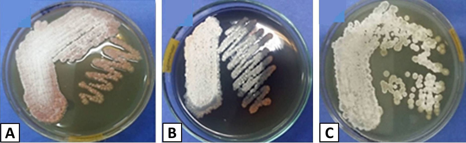
Pure cultures ofstreptomycetesstrains on GYM agar grown at 28oC for 5 to 7 days. A, NWMD-5; B, NWMD-12; C, NWMD-18.
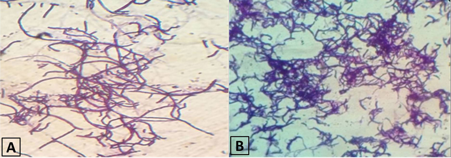
Microscopic appearance of streptomycetes strains. A, NWMD-1; B, NWMD-15.
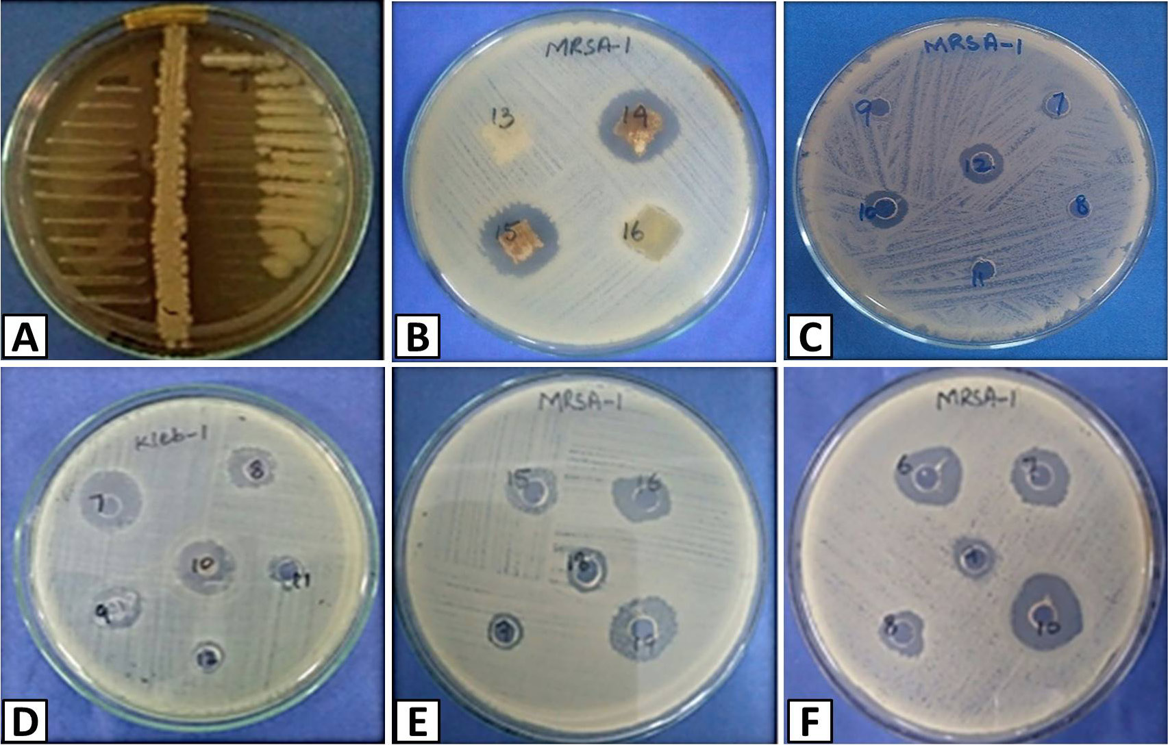
Antimicrobial activity of ofstreptomycetes strains (A) Streptomycetes strain NWMD-7 by cross streak method. On left side gram negative is streaked and on right side gram positive is streaked perpendicular to streptomycetesgrowth (B) Streptomycetes strain NWMD-13, NWMD-14, NWMD-15 and NWMD-16 against MRSA-1 by agar plug method. (C) Supernatant of streptomycetes strain NWMD-7, NWMD-8, NWMD-9, NWMD-10, NWMD-11, and NWMD-12 against MRSA-1 by well diffusion method (D) Methanolic extracts of streptomycetesstrains NWMD-7, NWMD-8, NWMD-9, NWMD-10, NWMD-11and NWMD-12 against Klebsiella by well diffusion method (E) Methanolic extracts of streptomycetes strains NWMD-15, NWMD-16, NWMD-17, NWMD-18 and NWMD-19 against MRSA by well diffusion method (F) Methanolic extracts of streptomycetes strains NWMD-6, NWMD-7, NWMD-8, NWMD-9 and NWMD-10 against MRSA by well diffusion method.
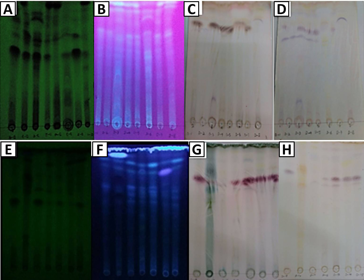
Chemical screening using TLC. A, TLC plate under UV at short wavelength (254 nm) of strain NWMD-1 to NWMD-8; B, TLC plate under UV at long wavelength (366 nm)of strain NWMD-1 to NWMD-8; C, TLC plate after treatment with Anisaldehye / H2SO4of strain ND-1 to ND-8; D, TLC plate after treatment with Ehrlisch reagent of strain NWMD-1 to NWMD-8; E, TLC plate under UV at short wavelength (254 nm) of strain NWMD-9 to NWMD-15; F, TLC plate under UV at long wavelength (366 nm)of strain NWMD-9 to NWMD-15; G, TLC plate after treatment with Anisaldehye / H2SO4of strain NWMD-9 to NWMD-15; H, TLC plate after treatment with Ehrlisch reagent of strain NWMD-1 to NWMD-8.
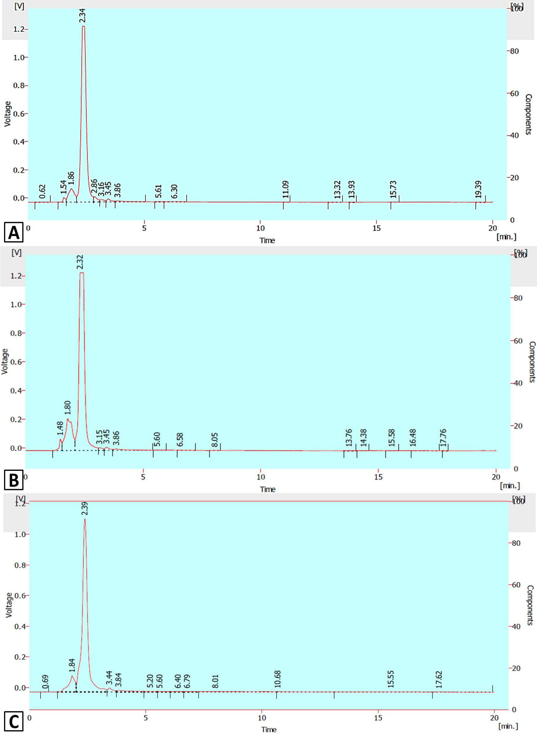
HPLC-UV chromatogram of Streptomyces strains. A, NWMD-7; B, NWMD-10; C, NWMD-12.
References
- Aftab, U., Qureshi, Z.U.A. and Sajid, I., 2016. In-vitro antitumor activity and metabolic finger printing of the actinomycetes isolated from various ecological niches in Pakistan. Pakistan J. Zool., 48: 1291-1305.
- Anwar, S., Ali, B., Qamar, F. and Sajid, I., 2014. Insecticidal activity of actinomycetes isolated from salt range, Pakistan against mosquitoes and red flour beetle. Pakistan J. Zool., 46: 83-92.
- Aouiche, A., Bijani, C., Zitouni, A., Mathieu, F. and Sabaou, N., 2014. Antimicrobial activity of saquayamycins produced by Streptomyces spp. PAL114 isolated from a Saharan soil. J. med. Mycol., 24: e17-e23.
- Balachandran, C., Duraipandiyan, V., Arun, Y., Sangeetha, B., Emi, N., Al-Dhabi, N.A., Ignacimuthu, S., Inaguma, Y., Okamoto, A. and Perumal, P.T., 2016. Isolation and characterization of 2-hydroxy-9, 10-anthraquinone from Streptomyces olivochromogenes (ERINLG-261) with antimicrobial and antiproliferative properties. Rev. Brasil. Farmacogn., 26: 285-295. https://doi.org/10.1016/j.bjp.2015.12.003[CrossRef]
- Balouiri, M., Sadiki, M. and Ibnsouda, S.K., 2015. Methods for in vitro evaluating antimicrobial activity: A review. J. Pharmaceut. Analys., 6: 71-79. https://doi.org/10.1016/j.jpha.2015.11.005[CrossRef]
- Barka, E.A., Vatsa, P., Sanchez, L., Gaveau-Vaillant, N., Jacquard, C., Klenk, H.P., Clément, C., Ouhdouch, Y. and van Wezel, G.P., 2016. Taxonomy, physiology, and natural products of Actinobacteria. Microbiol. mol. Biol. Rev., 80: 1-43. https://doi.org/10.1128/MMBR.00019-15[CrossRef]
- Bérdy, J., 2012. Thoughts and facts about antibiotics: Where we are now and where we are heading. J. Antibiot., 65: 385-395. https://doi.org/10.1038/ja.2012.54[CrossRef]
- Claverías, F.P., Undabarrena, A.N., González, M., Seeger, M. and Cámara, B.P., 2015. Culturable diversity and antimicrobial activity of actinobacteria from marine sediments in Valparaíso bay, Chile. Front. Microbiol., 6: 737. https://doi.org/10.3389/fmicb.2015.00737[CrossRef]
- Duraipandiyan, V., AL-Dhabi, N.A. and Ignacimuthu, S., 2016. New antimicrobial anthraquinone 6, 6 1-bis (1, 5, 7-trihydroxy-3-hydroxymethylanthraquinone) isolated from Streptomyces sp. isolate ERI-26. Saudi J. biol. Sci., 23:731-735. https://doi.org/10.1016/j.sjbs.2016.02.008[CrossRef]
- Hiltunen, T., Virta, M. and Laine, A.L., 2017. Antibiotic resistance in the wild: An eco-evolutionary perspective. Phil. Trans. R. Soc. B., 372: 20160039. https://doi.org/10.1098/rstb.2016.0039[CrossRef]
- Hozzein, W.N., Rabie, W. and Ali, M.I.A., 2011. Screening the Egyptian desert actinomycetes as candidates for new antimicrobial compounds and identification of a new desert Streptomyces strain. Afri. J. Biotechnol., 10: 2295-2301.
- Ibeyaima, A., Rana, J., Dwivedi, A., Gupta, S., Sharma, S.K., Saini, N. and Sarethy, I.P., 2016. Characterization of Yuhushiella sp. TD-032 from the Thar Desert and its antimicrobial activity. J. Adv. Pharmaceut. Technol. Res., 7: 32. https://doi.org/10.4103/2231-4040.177201[CrossRef]
- Jamil, M., Andleeb, S. and Ali, S., 2009. Antibacterial activity of antibiotics against pathogens and their genomic DNA Isolation. Punjab Univ. J. Zool., 24: 87-96.
- Khalid, Z.Z., Rashid, F., Ashraf, A., Iqbal, M.N. and Hussain, F., 2016. Isolation and screening of antibiotic producing bacteria from soil in Lahore City. PSM Microbiol., 1: 1-4.
- Khatri, S., Kumar, M., Phougat, N., Chaudhary, R. and Kumar-Chhillar, A., 2016. Perspectives on phytochemicals as antibacterial agents: An outstanding contribution to modern therapeutics. Mini Rev. Medicin. Chem., 16: 290-308. https://doi.org/10.2174/138955751604160201150438[CrossRef]
- Mohseni, M. and Norouzi, H., 2013. Antifungal activity of actinomycetes isolated from sediments of the Caspian Sea. J. Mazandaran Univ. Med. Sci., 23: 80-87.
- Muzzamal, H., Sarwar, R., Sajid, I. and Hasnain, S., 2012. Isolation, identification and screening of endophytic bacteria antagonistic to biofilm formers. Pakistan J. Zool., 44: 249-258.
- Najafi, M., 2016. Isolation of biologically active Actinomycetes from untouched soils: A case study from Karaj district, Iran. Prog. biol. Sci., 6: 65-74.
- Raaijmakers, J.M. and Mazzola, M., 2012. Diversity and natural functions of antibiotics produced by beneficial and plant pathogenic bacteria. Annu. Rev. Phytopathol., 50: 403-424. https://doi.org/10.1146/annurev-phyto-081211-172908[CrossRef]
- Radhakrishnan, M., Vijayalakshmi, G., Gopikrishnan, V. and Jerrine, J., 2016. Bioactive potential of actinobacteria isolated from certain under-studied regions in India. J. appl. Pharmaceut. Sci., 6: 151-155. https://doi.org/10.7324/JAPS.2016.60823[CrossRef]
- Raja, A. and Prabakarana, P., 2011. Actinomycetes and drug-an overview. Am. J. Drug Discov. Develop., 1: 75-84. https://doi.org/10.3923/ajdd.2011.75.84[CrossRef]
- Rao, H.Y., Rakshith, D. and Satish, S., 2015. Antimicrobial properties of endophytic actinomycetes isolated from Combretum latifolium Blume, a medicinal shrub from Western Ghats of India. Front. Biol., 10: 528-536. https://doi.org/10.1007/s11515-015-1377-8[CrossRef]
- Rather, I.A., Kim, B.C., Bajpai, V.K. and Park, Y.H., 2017. Self-medication and antibiotic resistance: Crisis, current challenges, and prevention. Saudi J. Biol. Sci., 24: 808-812. https://doi.org/10.1016/j.sjbs.2017.01.004[CrossRef]
- Sajid, I., Yao, C.B.F.F., Shaaban, K.A., Hasnain, S. and Laatsch, H., 2009. Antifungal and antibacterial activities of indigenous Streptomyces isolates from saline farmlands: prescreening, ribotyping and metabolic diversity. World J. Microbiol. Biotechnol., 25: 601. https://doi.org/10.1007/s11274-008-9928-7[CrossRef]
- Salehghamari, E., Soleimani, M. and Tafacori, V., 2015. Antibacterial activity of some actinomycetes isolated from soils of Alborz province, Iran. Prog. biol. Sci., 5: 159-167.
- Selvameenal, L., Radhakrishnan, M. and Balagurunathan, R., 2009. Antibiotic pigment from desert soil actinomycetes; biological activity, purification and chemical screening. Indian J. pharmaceut. Sci., 71: 499. https://doi.org/10.4103/0250-474X.58174[CrossRef]
- Shirling, E.T. and Gottlieb, D., 1966. Methods for characterization of Streptomyces species. Int. J. System. Evolut. Microbiol., 16: 313-340.
- Taddei, A., Valderrama, M., Giarrizzo, J., Rey, M. and Castelli, C., 2006. Chemical screening: A simple approach to visualizing Streptomyces diversity for drug discovery and further research. Res. Microbiol., 157: 291-297. https://doi.org/10.1016/j.resmic.2005.07.007[CrossRef]
- Ullah, I., Arshad, M., Noureen, U., Chuadhry, M.J.I. and Jadoon, M.A., 2012. Isolation of Streptomyces from the sediments of selected thermal springs of Northern Pakistan and its intrinsic susceptibility and resistance. Rec. zool. Surv. Pakistan, 21: 47-50.
- Walsh, S.E., Maillard, J.Y., Russell, A., Catrenich, C., Charbonneau, D. and Bartolo, R., 2003. Activity and mechanisms of action of selected biocidal agents on Gram-positive and-negative bacteria. J. appl. Microbiol., 94: 240-247. https://doi.org/10.1046/j.1365-2672.2003.01825.x[CrossRef]
Services
Article usage statistics
Loading…