
The main objective of this study was isolation and determination of changes caused by Clostridium perfringens type D from sheep. For this purpose, a total of 272 fecal samples were collected from sheep and lambs from four different zones of district Peshawar. Isolates were morphologically, biochemically and molecularly confirmed through Gram staining, gelatin liquefaction test (GLT) and polymerase chain reaction (PCR), respectively. Out of total, 85 (31.3%) were positive for C. perfringens type D on PCR. The results revealed 6-34% prevalence of C. perfringens type D in different regions. Similarly, significantly (PË‚0.001) higher prevalence was observed in lambs (54%) as compared to sheep (23%). For gross and histopathological lesions a total of 50 animals were necropsied infected with C. perfringens type D. A total of 43 (86%) sheep were recorded having hemorrhages on serosa of intestine followed by gas filled small intestine in 41 sheep (82%), lesions in ileum and colon in 39 sheep (78%), soft kidney 27 (54%), dark congested liver 26 (52%), nephritis 23 (46%) and spleenitis 13 (26%). Microscopic lesions revealed a score of 10 of kidney tissue out of 15 while the lesion score of intestine revealed 08 out of lesion score of 15.
Short Communication · Volume 53, Issue 4, 8 June 2021 · Open Access
Molecular Characterization and Pathogenesis of Local Isolates of Clostridium perfringens Type D from Sheep in District Peshawar
· · · · · · · ·
Affiliations & Notes ⌄
- 1College of Veterinary Sciences, The University of Agriculture, Peshawar 2Pakistan Science Foundation, Islamabad 3Civil Veterinary Hospital, Takht Bhai, Mardan 4Veterinary Research Institute, Peshawar 5Livestock Research and Developmental Station, Surezi, Peshawar , * Corresponding author: sbvetdr@yahoo.com
Article Info ⌄
- Journal:
- Pakistan Journal of Zoology
- Published:
- 8 June 2021

From the article
- C. perfringens type D was confirmed on PCR that all 85 samples (46 from sheep and 39 from lambs) were positive for α and ε toxin gene (Table I).
- The region wise prevalence of enterotoxaemia in sheep in region I, II, III and IV were 06%, 24%, 34% and 28% respectively while the region wise prevalence of enterotoxaemia in Lamb in region I, II, III and IV were 11%, 61%, 88% and 55% respectively.
- PCR confirmed the α and ε toxin with an amplicon size of 247bp and 206bp for the presence of C. perfringens type D respectively while the gross and microscopic lesion scoring revealed that kidney is more severely infected then intestine.
Selected from the article's own text.
Abstract
Materials and methods
Fecal samples were randomly collected from sheep and lambs suspected on clinical signs and symptoms for C. perfringens type D infection present in four regions of District Peshawar, Khyber Pakhtunkhwa province, Pakistan. A total of 272 fecal samples were used in the study including 200 from sheep (50 from each region) and 72 from lambs (18 from each region). Region I consist of Pelosi, university dairy farm and adjacent areas. Region II consists of Tajabad and Achenai bala. Region III consists of Nasir Pur and Charsadda Road while Region IV consists of GT Road and adjacent area. Samples were labeled, transported in ice box and stored at 4oC until use. A preformed questionnaire was developed to collect detail data of the animals.
Fecal sample were washed with phosphate buffer saline and culture in fluid thioglycollate broth (HI Media Laboratories Pvt. Ltd., India) for enrichment, followed by incubation at 37°C for 24h anaerobically using CO2 incubator (Galaxy 48 S, New Brunswick an eppendorf Company) and observed for any growth. After obtaining mass turbidity in broth, the samples were subcultured on tryptose sulphite cycloserine (TSC) agar (HI Media Laboratories Pvt. Ltd., India) by taking 0.1ml of the inocula for obtaining suspected grayish black colonies having diameter of 3-5mm. The suspected isolates were initially confirmed through Gram staining and gelatin liquefaction test .
For PCR amplification of α and ε toxins, genomic DNA was extracted by using the commercially available DNA extraction kit (GeneAllBldg, 303-7 Dongnam-ro, Songpa-gu, Seoul, South Korea) according to the manufacturer’s instructions.
PCR was conducted in 25 µL reaction mixture containing 10 μl of MasterMix, 1μl each of Forward Primer and Reverse Primer, 8 μl of PCR water and 5μl of DNA template using Bio-Rad thermal cycler. Amplification was done with initial denaturation at 94°C for 5 min followed by 30 cycles, each consisting of 60 sec of denaturation at 94°C, 60 sec of annealing at (α 55°C, ε 53.4°C), 60 sec of extension at 72°C and 10 min of final extension at 72°C. Two sets of primers used for the detection of C. perfringens type D α/cpa and ε/etx in fecal samples are:
a/cpa Primer F-5-TGC TAA TGTTAC TGC CGT TGA TAG-3
Primer R-5-TGC TAA TGTTAC TGC CGT TGA TAG-3
ε/etx Primer F-5-ATT AAA ATC ACA ATC ATT CAC TTG-3
Primer R-5-CTT GTG AAG GGA CAT TAT GAG TAA-3
with products of base pair 247 and 206, respectively as described by Garmory et al. (2000).
PCR products were run on 1.5% agarose gel with gel red (3μl) added along DNA ladder of 1 Kb (Genesta™) as a Marker and optimized positive and negative control product at 120 V, charge of 500 mA current for 35min.
A total of 50 sheep were examined to study the gross and histopathological lesions. Similarly those sheep showing diarrhoea, staggering and knuckling at fetlock in forelimbs, champing of jaws and teeth grinding were suspected for Enterotoxaemia and were purchased and sacrificed for recording pathological lesions. On post mortem inspection, detailed lesions were recorded in small intestine, liver, kidney, pericardial sac and abdominal muscles. The detail lesions scoring were documented in different organs of each necropsied animal . Tissues samples were collected from kidney and intestine in aseptic condition in sterile container under refrigeration for histopathology and preserved in neutral buffered formalin (10%) for histopathological examinations. Samples were properly labelled and transported to the Microbiology laboratory, Department of Animal Health, the University of Agriculture, Peshawar for further processing.
For gross and histopathological lesion scoring of different organs was made on the basis of severity of lesion. The severity level was 0, 1, 2, 3 and 4 that represent normal, mild, moderate, severe and highly severe, respectively . For histopathology a small cross-section were trimmed off from intestine and kidney. Then suitable sections were made by placing these samples in 10 % neutral buffered formalin for fixing of these samples. Then samples were further processed for histopathology and routine staining by following the method suggested by Bancroft et al. (2007). The data collected in the present study was analyzed through SPSS 8.1 using post Hoc and lesion scoring performed for evaluation of pathogenesis and severity of infection.
Results and discussion
C. perfringens type D was confirmed on PCR that all 85 samples (46 from sheep and 39 from lambs) were positive for α and ε toxin gene (Table I). The maximum prevalence of C. perfringens type D in sheep and lambs was found in region III (34%) and (89%), respectively. On PCR analysis an amplicon size of 247 bp for α and 206 bp for ε was obtained that represents C. perfringens type D in sheep and lambs. Out of total 200 examined sheep, 123 (61.5%), 112 (56%), 105 (52.5%) and 81 (40.5%) showing diarrhoea, staggering and knuckling at fetlock in forelimbs, champing of jaws and teeth grinding, respectively. Along with these symptoms the frothing of mouth 75 (37.5%) and rapid shallow and irregular respiration 41 (20.5%) was also recorded. Most of the sheep’s showing signs of mild fever 97 (48.5%) with anorexia, anemia and lethargy. Out of total 50 sheep died from pulpy kidney disease were examined for postmortem lesions, 43 (86%), 41(82%), 39(78%), 27(54%), 26(52%), 23(46%) and 13(26%) sheep showing hemorrhages on serosa of intestine, gas filled small intestine, lesions in ileum and colon, soft kidney, dark congested liver, nephritis and Spleenitis, respectively were recorded.
Table I. Incidence of C. Perfringens type D in sheep and lamb on PCR in Peshawar, KPK.
| Regions | Sheep | Lamb |
| 1 | 50/03 | 18/02 |
| 2 | 50/12 | 18/11 |
| 3 | 50/17 | 18/16 |
| 4 | 50/14 | 18/10 |
| Total | 200/46 | 72/39 |
Kidney and intestine were severely infected so the tissue sections were studied for histopathology. C. perfringens type D were confirmed that in kidney, it causes degenerative changes in the glomerulus, cast disposition, necrotic changes in tubular epithelium accompanied by congestion and leukocytic infiltration (Figs. 1A, B). The sections of intestine of sheep showed erosion of epithelium lining, sloughing of villi, leukocytic infiltration and aggregation of inflammatory cells (Figs. 1C, D). The gross lesion score was carried out to record and evaluate the severity of C. perfringens type D infection in kidney and intestine of sheep. Out of total lesion score of 15 the distribution of lesion score was 08 in kidney while the lesion score of intestine is 07. However, the microscopic lesions score revealed that out of total lesion score of 15, the kidney present a score of 10 while the lesion score of intestine was 08 (Table II).
The isolates were identified on the basis of their culture, morphological and biochemical characteristics and its gross and histopathological lesion in its predirection sites (kidney and intestine). The isolates show grayish black colonies on TSC agar while Gram staining were also performed for morphological identification and show purple rods having spore which agree with the result of . During current study the prevalence rate in sheep
Table II. Gross and microscopic lesion score in sheep suspected for C. perfringens type D infection.
| Tissue | Gross lesions | Scoring | Microscopic lesions | Scoring | ||
| Intestine | Serosae hemorrhages Enteritis Bloody ingesta Edematous | 2 2 1 2 | 07/15 | Hemorrhagic enteritis Sloughing of Villi Necrosis of epithelial cells Leukocytic infiltration | 2 3 3 0 | 08/15 |
| Kidney | Pyelonephritis Pus in hilus and pelvis Focal necrosis | 3 2 3 | 08/15 | Loss of glomerulus Distal tubular necrosis Nephrosis Congestion Leukocyte Infiltration | 3 2 2 3 0 | 10/15 |
Gross lesions scoring on the bases of tissue severity; 0, Normal; 1, Mild; 2, Moderate; 3, Severe; 4, highly severe.
in region I, II, III and IV were (06%), (24%), (34%) and (28%) while the overall prevalence of enterotoxaemia in sheep is 23% which is in line with the finding of from Italy who reported 25% prevalence of enterotoxaemia in sheep. These results are in contrast with the result of who reported less prevalence when compared with our findings. This high occurrence rate may be due to different climatic condition, breed, and poor farm management, system of rearing and lack of information about disease to farmer community.
The prevalence rate in lamb in region I, II, III and IV was 11%, 61%, 88% and 55% while the overall prevalence of enterotoxaemia in lambs is 54% which is in close accordance with the result of from turkey who reported 50% prevalence of enterotoxaemia in lambs. The results are in contrast with results of from West Africa who reported 71% prevalence of enterotoxaemia in lambs. The high occurrence rate of may be due to random selection of sheep, different climatic condition, breed and system of rearing.
PCR is used for molecular identification of toxinotyping of C. perfringens type D due to its high efficiency and sensitivity. α and ε primers with an amplicon size of 247bp and 206bp respectively were used. As per PCR findings, out of 272 suspected fecal samples (200 from sheep and 72 from lamb), 85 samples were positive for α and ε toxin which confirmed that C. perfringens type D is the most predominant type of C. Perfringens in sheep and lamb. These results are in line with .
Beside it the histopathological changes in intestine are dependent on the involvement of C. perfringens type D that causes lesions in the form of hyperplasia, granuloma, enteritis, congestion and fibrosis. There was sloughing of villi, desquamation of epithelial cells, hyperplasia of serosa and scattered hemorrhages in serosa layer of intestine infiltrated with leukocytes. The lesions noted in the current study are in line with the findings of (Kalender et al., 2007). The section of kidney showed congestion, extensive degeneration in the glomeruli, and necrosisin tubular epithelium, urinary tubules filled with cast and loss of glomeruli.
Conclusion
The overall prevalence of enterotoxaemia in sheep and lamb population in study area was 23% and 54% respectively. The region wise prevalence of enterotoxaemia in sheep in region I, II, III and IV were 06%, 24%, 34% and 28% respectively while the region wise prevalence of enterotoxaemia in Lamb in region I, II, III and IV were 11%, 61%, 88% and 55% respectively. Region III showed the highest incidence rate of 34% and 88% in sheep and lamb respectively. PCR confirmed the α and ε toxin with an amplicon size of 247bp and 206bp for the presence of C. perfringens type D respectively while the gross and microscopic lesion scoring revealed that kidney is more severely infected then intestine.
Statement of conflict of interest
The authors have declared no conflict of interest.
Figures
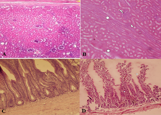
Histological structure of sheep kidney (A, B) and intestine (C, D). Control kidney (A), sheep kidney infected with C. perfringens type D (B) showing leukocyte infiltration, cast deposition, tubular epithelial cells and severe degeneration in tubular epithelium. Control intestine (C), intestine of infected sheep (D) showing sloughing of villi and leukocytic infiltration (D). **Stain: H&E (Hematoxcillin and Eosin); Magnification: A, B, 100x; C, D, 400X.
References
- Aschfalk, A., Valentin-Weig, P., Muller, W. and Goethe, R., 2002. Vet. Rec., 151: 210–213. https://doi.org/10.1136/vr.151.7.210[CrossRef]
- Bachhil, N. and Jaiswal, T.N., 1989. Indian Vet. Med. J., 13: 229–233. https://doi.org/10.17953/aicr.13.3-4.5613516316x12124[CrossRef]
- Bancroft, J.M. and Gamble, M., 2007. Theory and practice of histopathological techniques. Churchill Livingstone, London. pp. 125-138.
- Cheung, J.K., Awad, M.M., McGowan, S.J. and Rood. I., 2004. J. Vet. Microbiol., 105: 130-136.
- Fernandez Miyakawa, M.E. and Uzal, F.A., 2003. Vet. Res. Commun., 27: 231-241. https://doi.org/10.1023/A:1023348708599[CrossRef]
- Garmory, H.S., Chanter, N. and French, N.P., 2000. Epidemiol. Infect., 124: 61–67. https://doi.org/10.1017/S0950268899003295[CrossRef]
- Greco, G., Madio, A., Buonavoglia, D., Totaro, M., Corrente, M., Martella, V. and Buonavoglia, C., 2005. Vet. J., 170: 346-350. https://doi.org/10.1016/j.tvjl.2004.08.001[CrossRef]
- Kalender, H., Ertas, H.B., Cetinkaya, B., Muz, A., Arslan, N. and Kilic, A., 2005. Vet. Med. Czech., 50: 141-148.
- Kelender, H., Kili, A. and Atil, E., 2007. Turk. J. Vet. Anim. Sci., 31: 83-84.
- McClane, B.A., Uzal, F.A., Miyakawa, M.F., Lyerly, D. and Wilkins, T., 2006. The Enterotoxic Clostridia. In: The prokaryotes, Vol. 4 (eds. M. Dworkin, S. Falkow and E. Stackebrandt). Springer-Verlag, NY, pp. 698-752. https://doi.org/10.1007/0-387-30744-3_22[CrossRef]
- McClane, B.A., Robertson, S.L. and Chen, L.J., 2013. Clostridium perfringens. In: Food microbiology: Fundamentals and frontiers. (eds. R.L. Buchanan and M.P. Doyle). American Society for Mic, Washington, DC. pp. 465–489. https://doi.org/10.1128/9781555818463.ch18[CrossRef]
- Radad, K. and Khalil, S., 2011. Braz. J. Vet. Pathol., 4: 219-224.
- Sadique, U., Zafer, R., Younas, Z.U., Hassan, M., Idrees, M., Mushtaq, A., Sajid, M. and Sabtain, M., 2012. Pak. J. Anim. Pl. Sci., 22: 33-37.
- Wesonga, H.O., Bolske, G., Thiaucourt, F., Wanjohi, C. and Lindberg, R., 2004. Acta Vet. Scand., 45: 167-179. https://doi.org/10.1186/1751-0147-45-167[CrossRef]
Services
Article usage statistics
Loading…