
The current study was carried out in all administrative divisions of the province to explore the prevalence of Clostridium perfringens type D in small ruminants. A total of 204 fecal samples were collected from sheep and goats suffering from seasonal diarrhea. C. perfringens type D was identified through different biochemical tests and PCR. Antibiograms and lab animal trials were also carried out on local isolates of C. perfringens type D. Out of 204 analyzed samples, 89.18 % were found positive while 10.82 % were negative for C. perfringens type D. The division wise distribution showed that Quetta, Zhob, Sibi and Kalat were more effected zones of the Balochistan province. Sheep were more effected (54.78 %) than goats (34.4 %). C. perfringens was identified through different biochemical tests and PCR. Based on length of synthesized fragments by PCR, toxin gens of alpha and epsilon types were detected. C. perfringens type D was sensitive to tetracycline, ciprofloxacin, vancomycin, kanamycin, streptomycin, gentamicin, sulfamethoxazole and erythromycin, while showed resistance against carbenicillin, amoxicillin, penicillin g, colistin sulphate, bacitracin and metronidazole. In lab animal trial C. perfringens type D affected the fore stomach, intestine, liver, kidney, pancreas and gall bladder of mice.
Short Communication · Volume 50, Issue 6, 10 November 2018 · Open Access
Detection and Characterization of Clostridium perfringens Serotype D from Small Ruminants of Balochistan
· · · · · · · · ·
Affiliations & Notes ⌄
- 1Center for Advanced Studies in Vaccinology and Biotechnology, University of Balochistan, Quetta 2Livestock and Dairy Development Department, Balochistan, Pakistan 3Bolan Medical Complex Hospital, Quetta, Pakistan , * Corresponding author: taj_imran@yahoo.com
Article Info ⌄
- Journal:
- Pakistan Journal of Zoology
- Published:
- 10 November 2018

From the article
- The study result revealed that sheep (54.78 %) were more affected with C. perfringens type D as compared to goats (34.4 %).
- C. perfringens type D was confirmed through different biochemical tests (IMViC, Sugar fermentation, Catalase reaction, Oxidase tests, Gelatin hydrolysis, Litmus milk reaction and Lecithinase) and PCR.
- Different drugs result showed that C. perfringens type D was sensitive to tetracycline, ciprofloxacin, vancomycin, kanamycin, streptomycin, gentamycin, sulphamethoxazole and erythromycin, while carbenicillin, amoxicillin, penicillin g, colistin sulphate, bacitracin and metronidazole showed resistance.
- This study confirmed presence of Clostridium perfringens type D in small ruminants of the province.
Selected from the article's own text.
Abstract
Materials and methods
A total 204 fecal samples of enterotoxaemia-suspected sheep and goats suffering from seasonal diarrhea were collected from all divisions of Balochistan. The samples were collected aseptically in polyethylene sachets with UV treated contamination free with double volume of phosphate buffer solution (PBS) .
The samples were inoculated deep into reinforced clostridial medium RCM tubes (Oxoid-England). The tubes were placed in water bath for a period of 10-15 min at 80°C to eliminate the non-spore forming bacteria. Finally, the tubes were incubated in anaerobic jar at 37°C for 24-48 h. For pure colony isolation the culture was streaked on reinforced clostridial medium plate. The bacterial culture was identified with Gram staining. Different biochemical tests carried such as, fermentation reaction with different sugars, IMViC, H2S, Litmus milk reaction, Gelatin hydrolysis, Motility test, Egg yolk agar (Lecithinase and lipase) .
For detection of enterotoxin gene for C. perfringens type D, PCR was performed by using specific primers. Alpha toxin gene primers forward: 5’-GCTAATGTTACTGCCGTTGA-3’, reverse: 5’-CCTCTGATACATCGTGTAAG-3’ – Beta toxin gene primers forward: 5’-GCGAATATGCTGAATCATCTA-3’, revers: 5’-GCAGGAACATTAGTATATCTTC-3’ Epsilon toxin gene primers forward: 5’-GCGGTGATATCCATCTATTC-3’, reverse: 5’-CCACTTACTTGTCCTACTAAC- 3’ and Iota toxin gene primer forward: 5’-ACTACTCTCAGACAAGACAG-3’, reverse: 5’-CTTTCCTTCTATTACTATACG-3’ ; ; . Genomic DNA purificatation kit (HiMedia-India) was used for extraction of genomic DNA. The DNA template was (elute) stored at -20°C for further process. The amplifications alpha and epsilon gene of C. perfringens type D was done by using thermo cycler. The 30µl master mix contains 15µl 2X AmpMasterTM Tag (GeneAll-Korea), 2µl primers, 30ng template DNA and distilled water up to 30µl. All amplification reaction consisted of an initial denature 94°C for 5 min prior to 35 cycles of 94°C denaturation for 30 sec, 55°C annealing for 30 sec, and 72°C extension for 30 sec, followed by a final 72°C extension for 7 min. The PCR products were then analyzed by electrophoresis on 2.0% agarose gel.
Standardized antibiotic sensitivity test was performed on Mueller Hinton agar using disc diffusion bauer technique and McFarland turbidity standard method 0.5 following CLSI protocols. Isolates were considered as sensitive and resistant to a particular antimicrobial agent on the basis of inhibitory zone .
To test the pathogenicity of strain, lab animal (mice) has been selected and 0.5mL growth suspension having 1x10⁹cfu/mL of each sample was injected intra peritoneally. The post-mortem of the dead animal was performed . The data was analyzed through SPSS software version 6.
Results
C. perfringens type D was Gram-positive, non- motile, thick single or paired rods measuring 4-6×1µm. Total 204 fecal sample were collected from which 89.18 % were positive and 10.82 % were negative for C. perfringens type D. Figure 1 shows division wise distribution Zhob (16.17 %), Sibi (15.68 %), Quetta (15.19 %) and Kalat (14.7 %) divisions were highly infected with enterotoxaemia compared to other divisions of Balochistan.
The study result revealed that sheep (54.78 %) were more affected with C. perfringens type D as compared to goats (34.4 %).
Biochemical identification showed the C. perfringens biochemically was saccharolytic because it formed acid when it utilized glucose, sucrose, maltose and lactose but did not utilized mannitol and dulcitol. The gas of fermentation was hydrogen sulphide. Biochemical tests result revealed that C. perfringens was positive for litmus milk test, lecithinase test and gelatin liquefaction test while negative for catalase, indole, methyl red and voges-proskauer tests.
Molecular detection of alpha and epsilon toxin genes was carried from broth culture. After the extraction of DNA, alpha and epsilon toxin genes was amplified at annealing temperature 55˚C with amplicon size 324bp (alpha) and 655bp (epsilon) were observed as shown in Figure 2.
C. perfringens type D was sensitive to tetracycline (32 mm), ciprofloxacin (27 mm), sulfamethoxazole (25 mm), gentamicin (20 mm), erythromycin (20 mm), kanamycin (20 mm), and streptomycin (13 mm) while carbenicillin, amoxicillin, penicillin g, colistin sulphate, bacitracin and metronidazole showed resistance against C. perfringens type D.
Total 204 fecal sample were collected from which 89.18 % were positive and 10.82 % were negative for Clostridium perfringens type D. The division wise distribution showed that Quetta, Zhob, Sibi and Kalat were more effected zones of the Balochistan province. Species wise result showed that sheep (54.78 %) were more affected than goats (34.4 %). Our findings were similar with Haq et al. (2016). C. perfringens type D was confirmed through different biochemical tests (IMViC, Sugar fermentation, Catalase reaction, Oxidase tests, Gelatin hydrolysis, Litmus milk reaction and Lecithinase) and PCR. Both toxins alpha and epsilon of C. perfringens type D are considered to be the lethal toxins involved in the enterotoxaemia disease in sheep and goats. Alpha and epsilon toxins gens were amplified with amplicon size about 324bp and 665bp, respectively . Different drugs result showed that C. perfringens type D was sensitive to tetracycline, ciprofloxacin, vancomycin, kanamycin, streptomycin, gentamycin, sulphamethoxazole and erythromycin, while carbenicillin, amoxicillin, penicillin g, colistin sulphate, bacitracin and metronidazole showed resistance.
Conclusion
This study confirmed presence of Clostridium perfringens type D in small ruminants of the province. The polymerase chain reaction is good tool for identifying/confirming the clostridium perfringens type D from fecal samples. The local isolates will help researcher to build a good vaccine against enterotoxaemia.
Acknowledgements
The author wishes to thank Mr. Aurangzaib, Mr. Abdullah, and staff of CASVAB, who helped in the research. The author also acknowledges contribution of Dr. Hameed-u-allah Khethran, Executive District Officer, Livestock and Dairy Development Department Balochistan in this study.
Statement of conflict of interest
Authors have declared no conflict of interest.
Figures
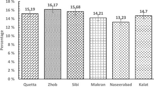
Divisional situation of C. perfringens type D in Balochistan at provincial level.
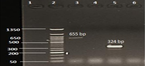
Agarose gel electrophoresis of PCR product: Lane 1, negative control; Lane 2, DNA ladder (50 bp); Lane 3, epsilon toxin encoding gene, (655 bp); Lane 4, beta toxin encoding as negative; Lane 5, alpha toxin encoding gene as positive (324 bp), Lane 6, Iota toxin encoding as negative control.
References
- Ahsani, M.R., Abadi, M.R. and Shamsaddini, M.B., 2010. J. Venom. Anim. Toxins incl. Trop. Dis., 16: 573-578. https://doi.org/10.1590/S1678-91992010000400006[CrossRef]
- Haq, A., Kamran, M.T., Imran, T., Sana, A., shfaq, A.A., Ghulam, M., Zaheer, A., Zahoor, A., Farhat, A. and Abdul, S., 2016. Int. J. biol. Sci., 4: 1-8.
- Carlos, A.D.O.J., Silva, R.O.S., Amanda, N.D., Pires, A.N., Salvarani, P.S.P., Assis, F.M. and Lobato, R.A.D., 2016. Semina: Ciênc. Agrár. Lond., 37: 257-262.
- Hadimli, H.H., Erganiș, O., Sayin, Z. and Aras, A., 2012. Turk. J. Vet. Sci., 36: 409-415.
- Jamshid, R., Gholam, T., Ali, R., Mehrnaz, R.M. and Ahmad, 2014. Iranian J. Microbiol., 6: 31-36.
- Jones, A.L., Dagleish, M.P. and Caldow, G.L., 2015. Vet. Rec., 177: 390.
- Kumar, V. and Sreenivasulu, N.D., 2016. Vet. World, 7: 1132-1136. https://doi.org/10.14202/vetworld.2014.1132-1136[CrossRef]
- Linden, J.R., Ma, Y., Zhao, B., Harris, J.M., Rumah, K.R., Schaeren-Wiemers, N. and Vartanian, T., 2015. mBio, 6: e02513. https://doi.org/10.1128/mBio.02513-14[CrossRef]
- Li, J., Miyamoto, K., Sayeed, S. and McClane, B.A., 2010. PLoS One, 5: e10932.
- Nagwa, A., Khairy, E.A., Dorgham, S.M. and Zaki, M.S., 2013. Life Sci. J., 10: 1599-1602.
- Nasir, A.A., Younus, M., Rehman, M.U., Lateef, M., Khaliq, S.A., Ahmad, I. and Abbas, M., 2006. J. Anim. Pl. Sci., 23: 1553-1558.
- Nasir, A.A., Younas, M., Rashid, M., Khaliq, A.K., Shah, S.E., Aslam, S.H., Ghumman, A. and Joiya, M.H., 2015. Iranian J. Vet. Res., 16: 94-99.
- Naz, S., Ghuman, M.A., Anjum, A.A., Manzoor, A.W., Rana, W. and Akhter, R., 2012. J. Vet. Anim. Sci., 2: 89-94.
- Piet, D. and Deperz, E.C.B.H., 2015. Vet Rec., 177: 388-389. https://doi.org/10.1136/vr.h5428[CrossRef]
- Radhika, B., Kumar, N.V. and Sreenivasulu, D., 2016. Vet. World, 9: 60-64. https://doi.org/10.14202/vetworld.2016.60-64[CrossRef]
- Rahaman, M.S., Akther, M.R., Adullah, M., Sayed-Khan, M.A., Jahan, M.S., Zia-ul-Haque, A.K.M. and Lutful-Kabir, S.M., 2013. J. Vet. Microbiol., 2: 83-88.
- Sinosh, S., Mahajanakatti, A.B., Biradar, U.B., Sharma, N. and Abhilash, M., 2010. Int. J. pharma. Sci. Rev. Res., 42: 164-172.
- Uzal, F.A., Giannitti, F., Finnie, J.W. and García, J.P., 2016. Diseases produced by Clostridium perfringens type D. In: Clostridial disease of animals. John Wiley & Sons, Inc., pp. 157-172. https://doi.org/10.1002/9781118728291.ch13[CrossRef]
- Yuan, T., Wen, M., Lian, B., Cheng, J., Wang, K. and Zhou, B., 2012. J. Vet. Anim. Sci., 36: 153-158.
- Zhenyu, C., Hui, Z., Khalid, M., Mujeeb, U.R., Xiadan, Y., Zhaoyang, L., Fazal, N., Xiaoxing, W., Xinxing, T., Xueting, L., Jingen, X. and Dongha,i Z., 2017. Pakistan. J. Zool., 49: 2249-2255.
Services
Article usage statistics
Loading…