Research Article · Volume 38, Issue 1, 4 July 2023 · Open Access
Lithium Induced Histological and Thyroid Hormone Alterations Observed in Channa punctatus and Oreochromis niloticus
· · · ·
Affiliations & Notes ⌄
1PG - Postgraduate and Research Department of Biochemistry, Mohamed Sathak College of arts and science, Sholinganallur- 600119, Chennai, India 2PG and Research Department of Biotechnology, Mohamed Sathak College of arts and science, Sholinganallur- 600119, Chennai, India 3Department of Zoology, Faculty of Life Sciences, University of Okara, Okara, Pakistan Corresponding author: V. Anuradha vanuradha2712@gmail.com
Aquatic pollution is a major threat and this environment pollution directly affected the aquatic animals, metals, minerals and trace elements have vital role in living organism but certain heavy metals do not have any biological role like lithium.
This research helps to prove that prolonged exposure of lithium damaged fish internals and affected hormone secretion.
Short-term exposure of lithium doesn’t show any significant changes.
Compared to short-term studies, long-term studies used to find out the metabolic stress and alteration occurs in the cell morphology of the fish species under study.
Selected from the article's own text.
Abstract
Abstract | Thyroid hormones secreted from thyroid gland has vital role for all living organisms such as mammals, vertebrates etc., for their regular growth and development. The secretion of thyroid hormones may get disrupted in marine organisms due to environmental pollution. Heavy metal (chromium, beryllium etc.,) or trace metal (lithium, titanium etc.,) pollution alters the thyroid hormone secretion not only in human beings; but also affects the aquatic species such as fish. Low or nil secretion of thyroid hormone will leads to metabolic stress that in turn breakdown the basic metabolic processes that eventually ends in disturbing the marine ecosystem. The current research work deals with observed alteration in the thyroid metabolism (causes hypothyroidism) and morphological (histology) changes occurred in fish species (selected organism– Channa punctatus and Oreochromis niloticus) exposed to sublethal concentration of Lithium [30%] for a chronic period of 60 days. The levels of FT3, FT4 and TSH tested were significantly decreased in treated groups when compared to the normal. Thus, it is evident that determination of thyroid hormone secretion and histological studies (FESEM – field emission scanning electron microscopy) were useful in determining the effect of trace metal pollution and its effect in freshwater fish species.
Novelty Statement | The study is based on the hypothesis that the deposition and exposure of heavy metal lithium is arising as environmental threat. Lithium exposed fresh water fishes can accumulate the ingested lithium in their vital organs. Further, it can cause acute toxic effects to both fresh water biota and humans upon feeding on these lithium exposed fishes. Thus, by eliminating the exposure of heavy metal into the water bodies, we can save the ecosystem.
Introduction
Environmental pollutants are substances that have detrimental effects on the environment either from natural sources or from human activity. A variety of organisms is affected by environmental pollution, such as humans, animals, plants, and even associated biota . Mankind is faced with several types of pollution such as (air, water, noise, soil, thermal, metal pollution etc.). Water is the most abundant natural resource on earth and is crucial for both the survival of biotic communities and the advancement of contemporary technologies. Now a day, the major threat in the world after air pollution is water pollution. The emergence of rapid industrialization, agricultural operations and urbanization and their interaction with the environment , causing pollution in the following order: Air pollution> water pollution> soil pollution. Second major pose is aquatic pollution, where aquatic environment gets polluted by natural reservoirs, anthropogenic activities, volcanic eruption and man-made activities. Heavy metals and trace elements gets accumulated into aquatic environment through reservoirs, pharmaceutical discharge and industrial wastes etc.,
Fishes are one of the most widely dispersed organisms accounting for approximately one-third of all organisms in the aquatic ecosystem and due to their susceptibility to metal contamination; they may provide insight into the magnitude of the biological impacts of metal pollution in the waters.
Fishes are among the species that are most widely dispersed in aquatic ecosystems , and being vulnerable to metal contamination, may reveal the amount of the biological impacts of metal pollution in waters . Fish species are typically considered as one of the crucial components for evaluating the quality of aquatic fields and all aquatic environments are populated by them, and resident fish constitute a vital part of the population that is exposed to toxins (Van der Oost et al., 2003). All this process revealed varying metal concentrations in the organ system at various levels . Heavy metals are difficult to eradicate from aquatic biota because they can easily bio-accumulate within aquatic biota through the food chain. Several negative effects, such as cardiovascular illnesses, hepatic cell damage, kidney disorders, and even death, can be brought on by heavy metals toxicity in human body through fish consumption. The following heavy metals such as arsenic (As), beryllium (Be), trivalent aluminium (Al III), antimony (Sb), hexavalent chromium (Cr VI), organic and inorganic mercury (Hg) etc., and trace elements; vanadium (V), molybdenum (Mo), lithium (Li), osmium (Os) etc., can disturb the structural morphology and metabolic alterations in aquatic species.
Lithium is fairly widely distributed in nature and is about twice as plentiful as lead. The important lithium bearing minerals are Lithia mica (1.5% of lithium), petalite (2.7–3.7% of lithium), spodumene (4–6% of lithium) and triphylite (4% of lithium). Lithium salts also occur in certain spring waters and in some plants such as tobacco, beet and sugar cane etc., . Lithium is a monovalent electropositive element, which is less reactive when compared among the alkali metals . Lithium was first offered to the medical profession in the mid-1800s as a cure-all for many common ailments. Especially, bipolar disorders (Mental illness) are commonly treated with lithium salts, particularly the carbonate (Li2CO3 - lithium carbonate) and acetate (LiCH3COO - lithium acetate) forms. Lithium therapy can helps to enhance leukocyte counts and lowers the thyroxine production by interfering with iodination of tyrosine .
Since lithium is a geochemically highly mobile element, the environmental and occupational health and safety issues connected with lithium in brine are substantial . Metallic lithium is somewhat hazardous as taken orally; nausea, vomiting, and mild diarrhoea occur primarily, but can be avoided by starting at minimal doses. Nevertheless, the physical tolerance of individuals varies from person to person, and the central nervous systems are the prime target organs for lithium poisoning . There are no known biological applications for lithium, and it doesn’t seem to be a necessary component of life ; Lenntech, 2007). A wide range of organisms, from cellular slime mould to humans, are affected by lithium in terms of cell growth, neural communication, and metabolism. Hence, the current study deals with the comparative toxicity studies of changes in histology and thyroid hormone secretion occurred due to lithium (Li) exposed to higher concentration in two fresh water fish species.
Thyroid hormone (TH)
History: The significance of the thyroid gland in terms of physiology was realized as a result of Graves and Basedow’s (1835, 1840) association of thyroid gland hypertrophy with the clinical signs of Graves disease and Gull (1874) found that myxedema to its atrophy and Yazbeck (2012) revealed that the thyroid system is a vital system for maintaining the homeostasis of the body and ensuring that the neurological, cardiovascular, reproductive, and body growth control systems are all functioning properly.The present research work deals with the effects of lithium toxicity on Channa punctatus (spotted snake head) and Oreochromis niloticus (Nile tilapia).
Materials and Methods
Selection of organism
The study focused mostly on fish species since the buildup of heavy metals in their bodies can enter the human system directly. Especially fish can able to survive in both fresh water and brackish water could be easily maintained in laboratory condition and available throughout the year. Hence in view of the above-mentioned advantages, Channa punctatus (spotted snake head) and Oreochromis niloticus (Nile tilapia) was selected for the study.
Fish acclimatization
The selected fish specimen (Channa punctatus and Oreochromis niloticus) of the weight (2±0.84 and 1.5±0.53 respectively) and length (16±3.14 and 16.2±2.01, respectively) was collected from Puzhal lake, Pothur, Chennai, Tamilnadu. Fishes were immediately transferred to laboratory (glass aquaria) and acclimatized for 30days before starting the treatment. Fishes were fed by commercially available fish feed (Taiyo grow contains crude protein, crude fat, crude fiber and moisture). Only healthy fishes were used for thyroid hormone analysis and histological studies.
Toxicity studies
Acute and chronic toxicity studies: 24, 48, 72 and 96 h exposure was set to study the acute toxicity. The studies were carried out by using Probit scale (LC₅₀ - lethal concentration). Based on acute studies, chronic studies was followed for (10, 20, 30 and 60 days of exposure for both species– channa punctatus, oreochromis niloticus). Fishes were made to 24hr fasting prior to the analysis.
Collection of blood and tissue sample
The blood samples were obtained through heart puncture and examined for thyroid hormone levels. After that, the fishes were sacrificed for histological studies.
Thyroid hormone analysis
This analysis was carried out by using kit method (VAST–3 analytes 1 kit ELISA microwells). The quantitative determination of free thyroxine (FT₄); free triiodothyronine (FT3); thyrotropin (TSH) concentration was analyzed colorimetrically by microplate enzyme immunoassay.
Field emission scanning electron microscopy (FESEM)
Tissue samples were examined using a FIELD EMISSION SCANNING ELECTRON MICROSCOPE CARL ZEISS, SUPRA 55VP for histological studies. The samples should be in dried condition and it can be thin film of 1cm² area and 5mm thickness in size. The procedure includes washing the surface, stabilizing, rinsing, dehydrating, and drying the specimen before mounting and coating it .
Results and Discussion
Endocrine parameter (Thyroid hormone)
The current study reveals the level of thyroid hormones such as tri-iodo thyronine (FT3), thyroxine (FT₄) and thyroid stimulating hormone (TSH) in Channa punctatus and Oreochromis niloticus. There is no alteration in the levels of thyroid hormones when exposed for an acute period (24, 48, 72 and 96) and so, the observations were made at the end of exposure of chronic periods (10, 20, 30 and 60 days), respectively for both the species under study.
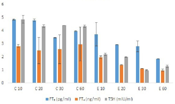
Table 1 and Figure 1 showed the level of FT3 (pg/ml) and FT₄ (ng/ml) and TSH in Channa punctatus. Which showed a significant decreasing level at 30% sublethal concentrations when compared to control. Decreased levels of FT3 (pg/ml) were recorded in 30% concentrations which shows as for (control: 4.85; 4.78; 3.45; 3.96 pg/ml and 3.73; 2.93; 2.8; 1.85 pg/ml for treated groups) studied at the end of 10, 20, 30 and 60days, respectively. Decreased values of FT₄ (ng/ml) were recorded in 30% concentrations which shows as for (control: 2.82; 2.48; 2.57; 2.97 and and 1.97; 1.38; 1.10; 0.97 ng/ml for treated groups) at 10, 20, 30 and 60 days, respectively. The levels of TSH (mIU/ml) were also decreased when recorded in 30% which shows as for (control: 4.85; 4.34; 4.39; 4.33 and 30% SLC: 2.18; 1.98; 0.97; 1.28 mIU/ml) at 10, 20, 30 and 60 days, respectively.
Table 1: Changes in FT₃ levels in blood of channa punctatus exposed to (10% Li) in different period of exposure (Days) (FT₃-free triiodothyroxine; FT₄- thyroxine; TSH-thyroid stimulating hormone).
Parameters
Samples
Control
Exposure days
10
20
30
60
10
20
30
60
FT₃ (pg/ml)
Blood
4.85±0.04
4.78± 0.1
3.45± 0.01
3.96± 0.04
3.73± 0.9
2.93± 0.01
2.8 ± 0.4
1.85± 0.01
FT₄ (ng/ml)
Blood
2.82± 0.12
2.48± 1.0
2.57± 1.1
2.97± 1.3
1.97± 0.10
1.38± 0.02
1.10± 0.01
0.97± 0.08
TSH (mIU/ml)
Blood
4.85± 0.3
4.34± 0.1
4.39± 0.01
4.33± 0.08
2.18± 0.08
1.98± 0.03
0.97± 0.04
1.28± 0.09
The values represented for two-way ANOVA: P-values were mentioned as: FT₃ - P>0.05 NS; FT₄ - P>0.05 NS; TSH – P>0.05 NS.
Table 2: Changes in FT₄ levels in blood of Oreochromis niloticus exposed to (10% of Li) in different period of exposure (Days) (FT₃-free triiodothyroxine; FT₄- thyroxine; TSH-thyroid stimulating hormone).
Parameters
Samples
Control
Exposure days
10
20
30
60
10
20
30
60
FT₃ (pg/ml)
Blood
4.45± 0.9
4.28± 0.40
4.96± 0.43
4.20± 0.96
4.42± 1.19
3.98± 0.10
2.78± 0.01
0.42± 0.02
FT₄ (ng/ml)
Blood
3.14± 1.2
2.97± 0.8
2.70± 1.1
2.68± 0.96
2.47± 1.2
2.68± 1.09
1.96± 0.07
0.98± 0.01
TSH (mIU/ml)
Blood
4.04± 0.28
4.78± 0.70
5.78± 0.96
4.85± 0.10
3.13± 0.9
3.01± 0.08
2.85± 0.01
1.42± 0.11
The values represented for two-way ANOVA: P-values were mentioned as: FT₃ - P>0.05 NS; FT₄ - P>0.05 NS; TSH – P>0.05 NS.
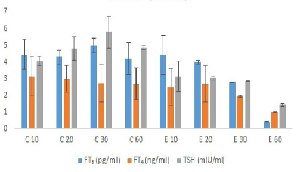
Table 2 and Figure 2 showed the level of FT₃ (pg/ml) and FT₄ (ng/ml) and TSH in oreochromis niloticus. In Oreochromis niloticus, the FT₃, showed a significant decreasing tendency at 30% sublethal concentration compared to control. Decreased values were recorded as for (control: 4.45; 4.28; 4.96; 4.20 pg/ml and 4.42; 3.98; 2.78; 0.42 pg/ml for treated groups) observed at 10, 20, 30 and 60 days respectively. The FT₄, showed a significant decreasing tendency at 30% sublethal concentration when compared to control. Decreased values were recorded as for (control: 3.14; 2.97; 2.70; 2.68 30% SLC: 2.47; 2.68; 1.96; 0.98 ng/ml) determined at 10, 20, 30 and 60 days respectively. The levels of TSH (mIU/ml) was also decreased when recorded in 30% which shows as for (control: 4.04; 4.78; 5.78; 4.85 and O. niloticus 30% SLC: 3.13; 3.01; 2.85; 1.42 mIU/ml) at 10, 20, 30 and 60 days, respectively.
FESEM
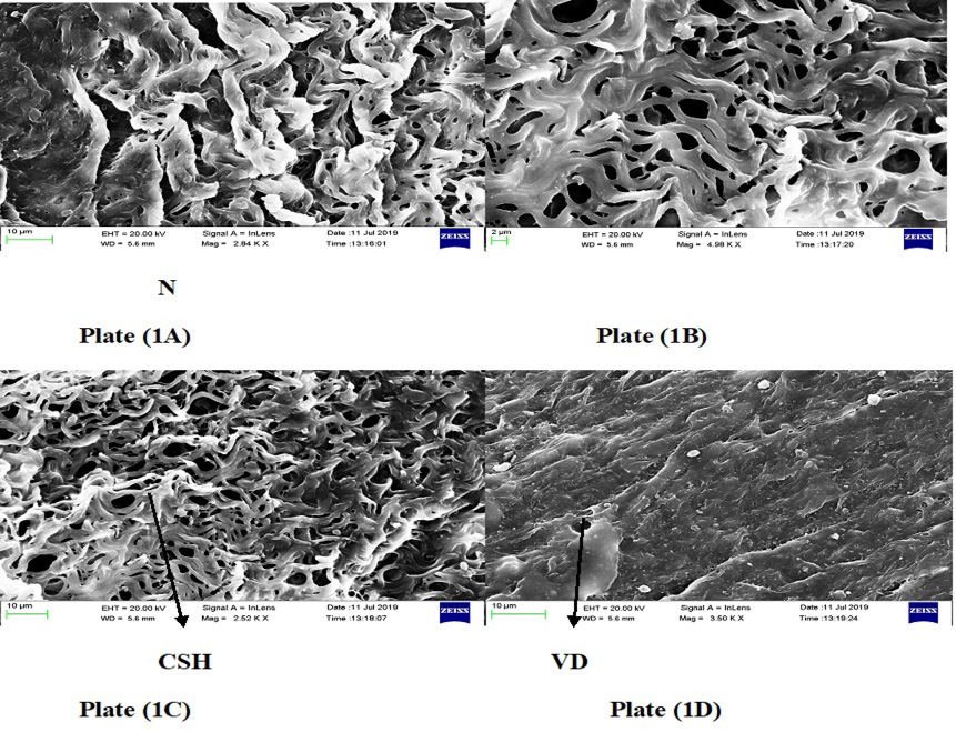
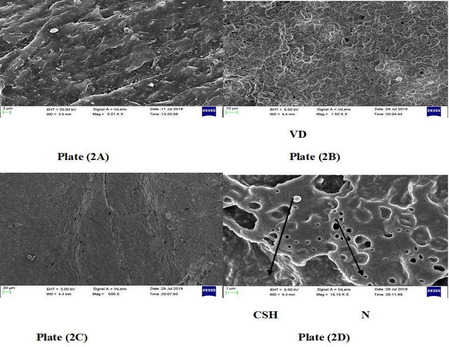
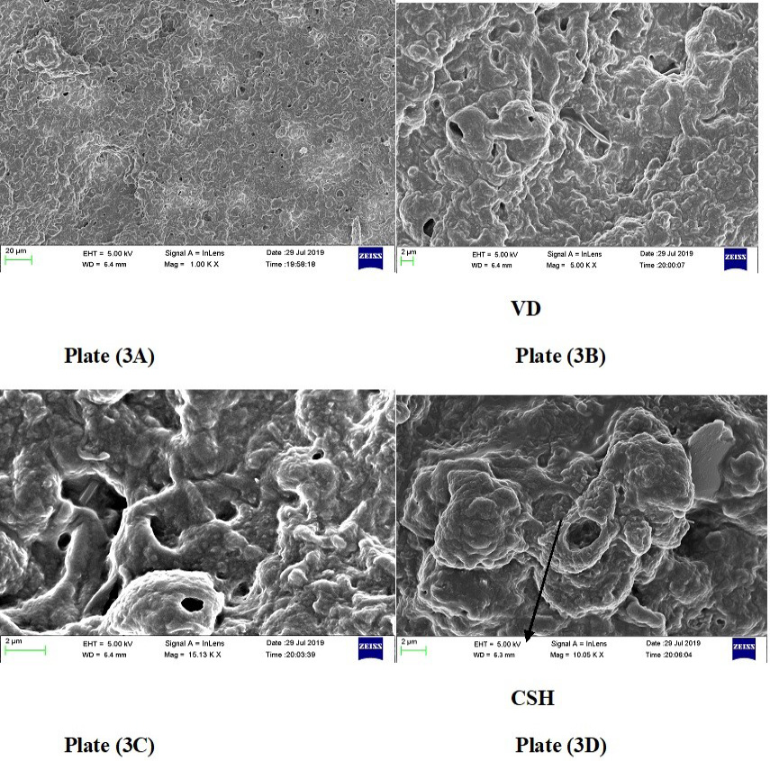
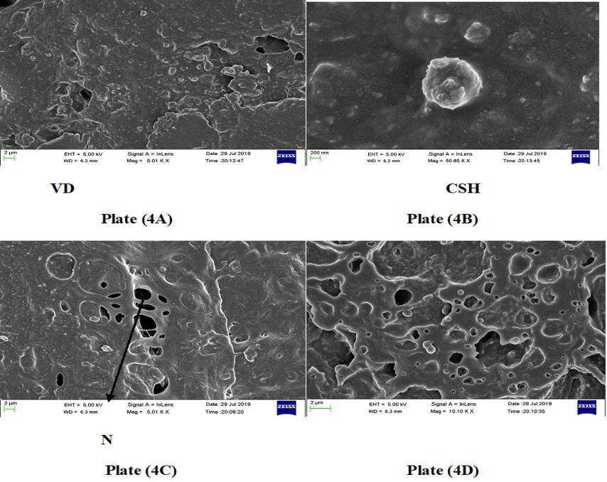
Figures 3 to 6 depicted FESEM images of histological changes identified in the hepatic tissue of both the species (C. punctatus and O. niloticus). After 30 and 60 days of exposure, liver tissue in the group of channa punctatus treated with Li showed the damaged CSH, cloudy swelling of hepatocytes; VD, vascular degeneration and N, necrosis at the surface when compared with the normal control [Plate 1A to 1D] and [Plate 2A to 2D] respectively. After 30 and 60 days of exposure in liver tissue of O. niloticus treated with Li also showed the CSH – cloudy swelling of hepatocytes and VD, vascular degeneration [Plate 3A to 3D] and [Plate 4A to 4D], respectively.
Thyroid hormone
Compared to control, lithium exposed species shows decreased value of thyroid hormone (FT3, FT4 and TSH). From the results, decreased level of thyroid hormone shows the hypothyroidism due to prolonged exposure of lithium (60 days) for both fish species. Due to metabolic stress, lithium interfere the thyroid hormone synthesis, short-term of lithium exposure does not alter hormone synthesis, When the duration of exposure time increases, lithium play a critical role in fish mechanism and it shows the decreased or nil secretion of thyroid hormone. Lithium not only affected thyroid hormone synthesis it also affected secretory pathway of other hormones. Lithium interferes with iodine absorption or involving in secretary mechanism while thyroid gland started to synthesis thyroxine and ultimately leading to decrease in thyroid hormone secretion. The same results were also observed in alterations of plasma thyroid of fish and reported by many authors like nitrite in Oreochromis mossambicus , cadmium in Peralichty dentatus . Cyprinus carpio to environmental acidic , environmental salinity in telesots, nitrite in sea beam and teleost Sparus saeba . Fish eggs contain maternally derived thyroid hormone. In Rainbow trout interaction between the endocrine showed a gradual acclimation improved non specific immunity in hormones. Due to interaction the Brown trout Salmontrutta also showed activation of lysozyme and phagocytosine , 1995). In Oreochromis mossambiocus showed an increased secretion due to effect of water salinity on immune function. During acclimation of Brown trout, there are improvements in immunological parameters and an increase in hormone levels has been observed . Several Teleost species have adapted to a hyperosmotic environment through hormone stimulation as well as body growth. In response to environmental salinity, endogenous hormone secretion is stimulated. T4 probably plays a more important role in the development of hypo-osmoregulatory ability than in hyper-osmoregulation. Hagfish , cat fish Ictaluruspunctuates ; , Atlanticsalmonsalar, channel cat fish and buffer fish Spheroidscephalous , Rainbow trout and Japanese flounder paralichthy olivaceus showed a decreased in T3 and T4 hormone due interaction between endocrine and immune system. Cyprimuscarpio grown in to acidic water resulted a decreased in hormones T3 and T4. The decrease in level of thyroid hormone was also noted when the fish species were exposed to metals, such as copper, aluminum and cadmium . From the thyroid hormone analysis, it is evident that metal stress in aquatic environment increases the metabolic burden to fish species.
Histology of liver
Liver is the principal organ, where the metabolism of carbohydrates, lipids and proteins take place . The given results reveals that, control fish liver cells show normal architecture for both the species (Channa punctatus and Oreochromis niloticus) when seen under FESEM. Lithium treated fish liver cells showed abnormal architecture with prolonged time of exposure gradually affecting the organ. In lithium exposed (30 and 60 days) Channa punctatus and Oreochromis niloticus, from the figure it clearly indicated organ damage due to metal exposure from 30 to 60 days, liver cell’s architecture deeply shows cloudy swelling of hepatocytes (CSH), vascular degeneration (VD) and necrosis (N). The similar results shown by Peebua et al. (2008) indicates hydropic swelling and vacuolation of hepatocytes in Nile tilapia exposed to acute toxicity with alachlor. In addition, the effect of pentachlorophenol was reported by Pandey and Dubey (2015) following exposure of Heteropneustes fossilis (Cat fish) to minimum and maximum doses of sublethal concentrations of pentachlorophenol for a period of 21 days. Based on the histological results, cell architecture studies helps to find out damage of the internal organs.
Conclusions and Recommendations
Aquatic pollution is a major threat and this environment pollution directly affected the aquatic animals, metals, minerals and trace elements have vital role in living organism but certain heavy metals do not have any biological role like lithium. Lithium has lots of industrial applications. Though Lithium is explored by pharmaceutical industries for psychiatric treatment like bipolar disorders, long- term consumption of lithium or absorption of lithium directly affected the metabolic mechanism especially hormone secretion and prolonged exposure of lithium damaged internal organs. Endocrine parameters and histology studies are sensitive tools to detect the consequences of prolonged exposure of heavy metals upon the metabolism of any organism. This research helps to prove that prolonged exposure of lithium damaged fish internals and affected hormone secretion. Short-term exposure of lithium doesn’t show any significant changes. Compared to short-term studies, long-term studies used to find out the metabolic stress and alteration occurs in the cell morphology of the fish species under study.
Acknowledgments
The authors would like to thank Mohamed Sathak Trust for providing facilities.
Availability of data and materials
The datasets analyzed during the current study are available from the corresponding author on reasonable request.
Consent for publication
All authors are agreed to publish the current version of the manuscript.
Ethics approval consent to participate
Not applicable.
Conflict of interest
The authors have declared no conflict of interest.
Figures
Figure 1:.
Changes in free thyroid hormones levels in blood of Channa punctatus exposed to sublethal concentration (30%) of Lithium in different exposure days. Values are expressed as mean ± SD for above observation. – or + indicate percent decrease or increase over control. For two-way ANOVA P-values mentioned as: P>0.05 NS; FT4- P> 0.05 NS; TSH- P> 0.05 NS.
Figure 2:.
Changes in free thyroid hormones levels in blood of Oreochromis niloticus exposed to sublethal concentration (30%) of Lithium in different exposure days. Values are expressed as mean ± SD for above observation. – or + indicate percent decrease or increase over control. For two-way ANOVA P-values mentioned as: P>0.05 NS; FT4- P> 0.05 NS; TSH- P> 0.05 NS.
Figure 3:.
FESEM (field emission scanning electron microscopy) of liver tissues in the group of Channa punctatus treated with lithium chloride. CSH, cloudy swelling of hepatocytes; VD, vascular degeneration and N, necrosis. (Upon 30 days exposure of lithium).
Figure 4:.
FESEM images of liver tissue in the group of Channa punctatus treated with lithium chloride. CSH, cloudy swelling of hepatocytes; VD, vascular degeneration and N, necrosis. (Upon 60 days exposure of lithium).
Figure 5:.
FESEM imaged of liver tissue in the group of Oreochromis niloticus treated with lithium chloride. CSH, cloudy swelling of hepatocytes; VD, vascular degeneration and N, necrosis. (Upon 30 days exposure of lithium).
Figure 6:.
FESEM of liver tissue in the group of Oreochromis niloticus treated with lithium chloride. CSH, cloudy swelling of hepatocytes; VD, vascular degeneration and N, necrosis. (Upon 60 days exposure of lithium).
References
Abdel Warith, A.A., Younis, E.M, Al-Asgah, N.A. and Wahbi, O.M., 2011. Effect of zinc toxicity on liver histology of Nile tilapia, Oreochromis niloticus. Sci. Res. Ess., 6: 3760-3769. https://doi.org/10.5897/SRE11.883[CrossRef]
Anderson, T.W., Nerio, L.C., Schremiber, G.B., Talbot, F.D.F. and Zdrojewski, A., 1975. Ischemic heart disease, water hardness and myocardial magnesium. Can. Med. Assoc. J., 113: 119 – 203.
Bervoets, L.R., Blust, and Verheyen, R., 2001. Accumulation of metals in the tissues of three spined stickleback Gasterosteus aculeatus from natural fresh waters. Ecotoxicol. Environ. Saf., 48: 117–127. https://doi.org/10.1006/eesa.2000.2010[CrossRef]
Carvalho, C.S. and Fernandes, M.N., 2008. Effect of copper on liver key enzymes of anaerobic glucose metabolism from freshwater tropical fish Prochilodus lineatus. Comp. Biochem. Physiol., 151A: 437–442. https://doi.org/10.1016/j.cbpa.2007.04.016[CrossRef]
Chatterjea, M.N. and Rana, S., 2008. Textbook of medical biochemistry, Jaypee publications, 7th edition, pp. 616. https://doi.org/10.5005/jp/books/10917_37[CrossRef]
De Menezes, C.C., Loro, V.L., Da Fonseca, M.B., Cattaneo, R., Pretto, A., Dos Santos, M.D. and Santi, A., 2011. Oxidative parameters of Rhamdia quelen in response to commercial herbicide containing clomazone and recovery pattern. Pestic. Biochem. Physiol., 100: 145–150. https://doi.org/10.1016/j.pestbp.2011.03.002[CrossRef]
Eder, M. and Gedigk, P., 1983. Lehrbuch uer Allgeminen Aathologie und der patholo-gischem Anatomie. Springer, Berlin. https://doi.org/10.1007/978-3-642-96760-3[CrossRef]
Hal, A. and Angelica, V.S., 2008. Toxicity of lithium to humans and the environment. A literature review. J. Ecotoxiol. Environ. Safe., 70: 349–356. https://doi.org/10.1016/j.ecoenv.2008.02.026[CrossRef]
Hawkes, J.W., 1980. The effect of xenobiotic on fish tissues: Morphological studies. Fed. Proc., 39: 3230.
Heath, A.G., 1987. Water pollution and fish physiology. CRC press,. Florida, USA. pp. 245.
Heath, A.G., 1995. Water pollution and fish physiology. CRC press. Florida, USA, pp. 245.
Hoole, D., Schuwerack, M.M. and Lewis, J.W., 2003. Cadmium-induced cellular and immunological responses in Cyprinus carpio infected with the blood parasite, Sanguinicola inermis. J. Helmin., 77: 341-350. https://doi.org/10.1079/JOH2003190[CrossRef]
Kjolholt, J., Stuer-Lauridsen, F., Skibsted M.A. and Havelund, S., 2003. The elements in the second rank-lithium. Miljoministeriet, Copenhagen, Denmark. (www2.mst.dk/commo/Udgivramme/Frame.asp?pg=http://www2.mst.dk/udgiv/publications/2003/87-7972-491-4/html/bill08_eng.htm).
Klaverkamp, J.F., Madonald, W.A., Duncan D.A. and Wagemann, R., 1984. Metallothionein and accumulation to heavy metals in fish: A review. In: Contaminant effects on fisheries (eds. V.W. Cairns, PV Hodson and J.O. Nriagu). Wiley, New York, pp. 99-113.
Lenntech, L., 2007. Lithium and water: Reaction mechanisms, environmental impact and health effects. www.lenntech.com/elements-and-water/lithium-and-water.htm.
Leonard, A., Hantson, P.H. and Gerber, G.B., 1995. Mutagenicity, carcinogenicity and teratogenicity of lithium compounds. Mutat. Res. Rev. Genet. Toxiol., 339: 131–137. https://doi.org/10.1016/0165-1110(95)90007-1[CrossRef]
Loganathan, K., Velmurugan, B., Hongray, H.J., Selvanayagam, M. and Patnaik, B.B., 2006. Zinc induced histological changes in brain and liver of Labeo rohita (Ham.) J. Environ. Biol., 27: 107-110.
Metelev, V.V., Kanaev, A.L. and Diasokhva, N.G., 1971. Water toxicity. Amerind Publishing Co. Pvt. Ltd, New Delhi.
Moustfa, M., Zeitoun and El-Sayat, E.M., 2014. Impact of water pollution with heavy metals on fish health: Overview and updates. IDOSI publications. Glob. Vet., 12: 219-231.
Nagae, M., Ogawa, K., Kawahara, A., Yamaguchi, M., Nishimura, T. and Ito, F., 2001. Effect of acidification stress on endocrine and immune functions in carp, Cyprinuscarpio. Water. Air. Soil. Poll., 130: 893-898. https://doi.org/10.1023/A:1013855501445[CrossRef]
Pandey, A.K. and Dubey, S., 2015. Histological changes in liver and kidney of cat fish, heteropneustes fossilis, exposed to pentachlorophenol (PCP). Pl. Arch., 1: 1117–1120.
Peebua, P., Kruatrachue, M., Pokethitiyook, P. and Singhakaew, S., 2008. Histopathological alterations of Nile tilapia oreochromis niloticus in acute and subchronic alachlor exposure. J. Environ. Biol., 29: 325–331.
Puri, B.R., Sharma, L.R. and Kalia, K.C., 2003. Principles of inorganic chemistry. Vallabh publications, 28th edition, pp. 1291, 432–435.
Sanjay, P.K., Ravindra, S., Shilpi, N.S., Nagpure, K., Satish, S., Srivastava, S. and Verma, M.S., 2006. Acute toxicity of chromium exposed Channa marulius. Indian J. Environ. Prot., 38: 118–121.
Soni, P.L., 1963. A textbook of inorganic chemistry. Sultan Chand and Sons, 11th edition, pp. 543–544.
Subash, P., Leji, J., Babitha, G., Rejitha, S. and Vijayasree, A.S., 2007. Thyroidal and osmoregulatory responses in tilapia (Oreochromis mossambicus) to the effluents of coconut husk retting mechanism of thyroid hormone action. Trends Endocrinol. Metab., 5: 65-72.
Tripathi, K.D., 2010. Essentials of medical pharmacology. Jaypee publications, 6th edition, pp. 434 – 436.
Van Der Oost, R., Beyer, J. and Vermeulen, N.P.E., 2003. Fish bioaccmulation and biomarkers in environmental risk assessment: A review. Environ. Toxicol. Pharmacol., 13: 57-149. https://doi.org/10.1016/S1382-6689(02)00126-6[CrossRef]
Yada, T., Azuma, T. and Takagi, Y., 2001. Stimulation of non-specific immune functions in sea water-adapted rainbow trout, Oncorhynchus mykiss, with reference to the role of growth hormone. Comp. Biochem. Physiol., 1298: 695-701. https://doi.org/10.1016/S1096-4959(01)00370-0[CrossRef]
Yazbeck, S., 2012. Thyroid disorders during pregnancy. Med. Clin. N. Am., 96: 235-256. https://doi.org/10.1016/j.mcna.2012.01.004[CrossRef]