Research Article · Volume 38, Issue 1, 4 July 2023 · Open Access
Isolation, Identification, and Screening of Keratinase Producing Bacteria from Soil and Production Optimization Using Feather Waste as Substrate
· ·
Affiliations & Notes ⌄
1Department of Biotechnology, University of Sargodha, Sargodha, Pakistan 2Food and Biotechnology Research Center, Pakistan Council of Scientific and Industrial Research Laboratories Complex Ferozpur Road, Lahore, Pakistan. Corresponding author: Muhammad Irfan irfan.ashraf@uos.edu.pk
In this investigation, seven distinct bacterial strains that can produce keratinase were isolated and identified from the abattoir region. A transparent, clear zone was evident in each isolate during the initial primary screening on skimmed milk agar plates. The secondary screening was then evaluated via submerged fermentation utilizing raw feathers as a substrate for keratinase production. On skimmed milk agar plates with streaked pure culture growth patterns, these seven strains gave a positive appearance of sizable dramatic zones around the growth patterns. These seven strains such as Bacillus velezensis FD1, Bacillus cereus FD2, Bacillus subtilis FD3, Bacillus altitudinis FD4, Bacillus licheniformis FD5, Bacillus flexus FD6 and Alcaligenes sp. FD7 were verified by the molecular identification investigation using 16S rRNA sequence technology respectively. Out of these seven isolates, the Bacillus cereus FD2 strain produced the most keratinase (298U/mL), and was selected for more research. The Bacillus cereus showed the highest keratinase activity at neutral pH 7.0, with 1% inoculum size, 1% substrate and after 72h of the incubation period. This current study revealed that these isolates have ability to be used in many biotechnologies prospective applications like in hydrolysis of keratin-containing substrate. Recently, it has been extremely beneficial and a major priority to use microorganisms for the enzymatic destruction of keratin waste material rather of various conventional procedures that are expensive and not ecologically friendly.
Novelty Statement | The study reports proteolytic/keratinolytic bacteria for the first time from abattoir region.
Introduction
The third most important polymer found in nature, after cellulose, an organic substance, and chitin, a fibrous polysaccharide, is keratin, an insoluble structural protein and a highly extraordinary stable macromolecule . In order to produce keratinase, keratinous materials such as chicken feathers, hooves, and hair are needed as substrate materials . Moreover, many types of non keratinous substrates can also act as keratinase inducers such as casein, skimmed milk, gelatin and soybean meal . Keratin is a major fundamental unit of skin and in addition to this; it is also a major constitutional and structural element of hooves, feathers, skin, cloves, nails, horns, and beaks (Mckittrick et al., 2012; ; . Due to vast increase of poultry industry business, an enormous compost waste is being generated every day amongst which feathers are the most important ingredient factor which could be utilized. According to Abdelmoteleb et al. (2023), 10% of the chicken’s body weight is thought to be made up of keratin protein, which makes up 90% of the feather structure. Based on the amount of sulphur in its structure, keratins are divided into two type’s hard and soft keratin . According to Parry and North (1998), the supercoiled polypeptide chain created by the close packing of alpha helix/beta sheets results in mechanical stability. Keratinase enzymes can also transform keratin into beneficial bioproducts.
The keratin maximum stability is due to presence of disulphide and hydrogen bonding that makes its resistance against proteolytic hydrolysis. For biodegradation of keratin, disulfide bonds an extracellular protease enzyme keratinase is used . In their experiments, Nnolim et al. (2021) demonstrated how some microorganisms produce the keratinase enzyme when there are substrates present that include keratin. Actinomycetes, fungi, and bacteria are only a few of the microbes that have been identified as producing keratinase (Brandelli, 2005). More recently, keratinase derived from other Bacillus strains, including Bacillus pumilus, Bacillus licheniformis, and Bacillus subtilis, also displayed maximal intensity of variations, which gained significance due to their capacity for keratin breakdown quickly. According to Zhang et al. (2002), variations in molecular configuration, features of protein sequencing and conserved residues in nature may be the cause of the exceptionality of keratinase enzymes from various bacterial origins.
In addition to keratinolytic bacteria, keratin and keratinase have impressive uses in a number of industries, such as waste management, biodegradation, and industrial biotechnology ; ; . Different cultural factors, such as temperature, pH, inoculum size, nitrogen sources, and carbon type, have an impact on the production of the keratinase enzyme by bacteria. Keratinase synthesis is also influenced by the medium’s substrate concentration and aeration level, and these characteristics can be optimized to increase keratinase production .
Environmental wastes generated by human actions have a huge amount of proteins and other carbon compounds. One such waste is feathers that are produced in massive quantities in places of poultry treating and processing plants as a surplus secondary product at the commercial level . Chicken is a delicacy demanded not only in Pakistan but around the world. Because of population increases, the demand for this food is also increasing worldwide. Consideration should be given to scientifically utilizing this waste. Moreover, the increase of such wastes can be a severe cause of pollution as well as health problems so we can overcome such tragic environmental issues by resourceful way of avoiding environmental pollution . Previous works have given attention to the depletion method of some polymeric wastes which was mostly based on feather waste products (Sweltana and Jain, 2010; . In this study, 16S rRNA sequencing techniques were used to extract, screen, and identify bacteria that produce keratinase from places where poultry manure is dumped.
Materials and Methods
Soil sampling
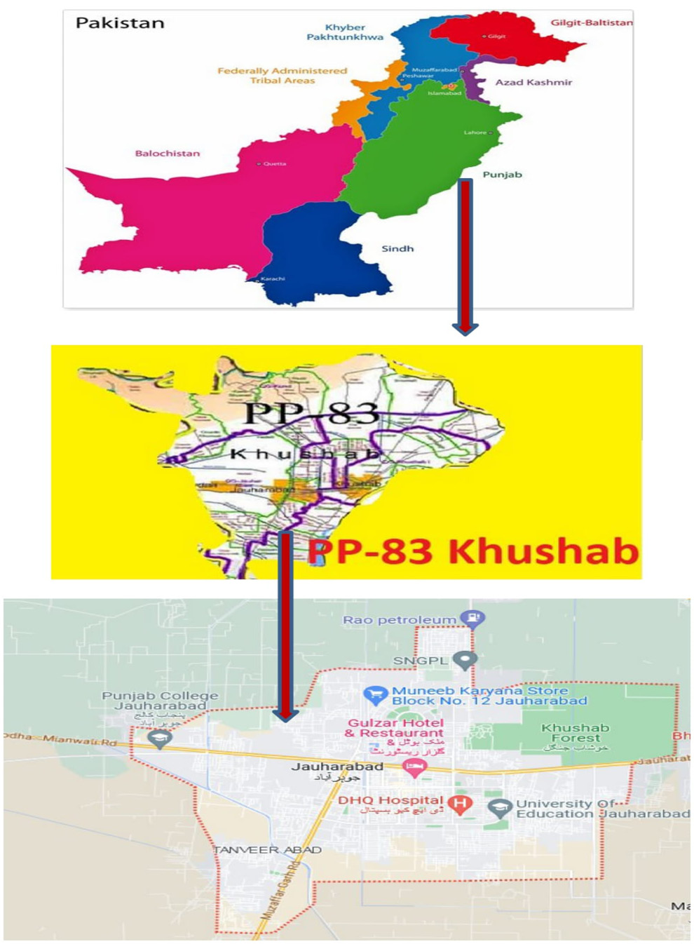
The three different kinds of soil samples were taken from the locations of poultry farms in the Jauharabad area of Khushab, Punjab Province, Pakistan, as indicated in Figure 1, in order to identify bacteria that produce keratinase. Samples were gathered and brought to be processed in polythene bags.
Substrate preparation
From poultry farms and slaughterhouses in market areas, chicken feathers were taken. The feathers were thoroughly cleaned and rinsed in warm distilled water to remove any dust, then dried in an oven at 60°C for two days. The feathers were kept at room temperature for use in additional experiments.
Isolation and initial screening of keratinolytic bacteria
Techniques like serial dilution and spread plate were used to isolate keratinolytic bacteria. A test tube containing 9 ml of sterilized distilled water and 0.1g of soil samples was filled, and then vigorously shaken. The following stage involved spreading each sample evenly over Keratinase positive strains were subsequently streaked on nutritional agar medium (nutritional agar 2.8% and skimmed milk powder 0.5%) plates and flooded with 10% Trichloroacetic acid to produce a pure culture of strains. The bacterial isolates on skimmed milk agar petri plates with the most transparent hydrolysis zone were subsequently chosen for the secondary screening step. To preserve and store the best keratinase-producing strains for use at 4oC in the future, glycerol was added to nutritional agar slants . The chosen strains colony characteristics were evaluated using measurements. The 100-microliter nutrient agar plates underwent a 24-h incubation period at 37°C.
Inoculum preparation
For inoculum preparation, the Erlenmeyer flask of 100mL containing 25 ml nutrient broth media sterilized at 121°C, 15 Ib/in for 15 min. After sterilization, the media was cooled and 24 h old loopful culture of Bacillus cereus FD2 strain was aseptically transferred then kept in a shaker and incubate at 37°C for 24 h.
Secondary screening and keratinase production in submerged fermentation
Secondary screening was applied to isolates that formed transparent zones on skimmed milk agar petri plates. For secondary screening, distilled water was mixed with the basal salt feather meal media, which included (g/L) feathers 10g, NH4Cl 1g, NaCl 1g, K2HPO4 0.6g, KH2PO4 0.8g, MgCl2.6H2O 0.48 g, yeast extract 0.2 g (Rajesh et al., 2016). After that, each flask containing 100 ml of feather meal media was inoculated with 1 ml of the inoculum from a selection of bacterial strains, and the flasks were rotated on a rotary shaker at 120 rpm for one week at 37°C. Centrifugation of the fermented media was place for 10 min at a speed of 5000 rpm. The culture supernatant served as the source of the enzymatic extract for the keratinase activity assay and protein estimate. For secondary screening, the visual feather hydrolysis procedure was also examined for a week.
Enzyme assay
The powder from the hooves was used as a substrate for a keratinase assay, which was observed. The ingredients for the reaction mixture are 0.005 g of powdered hooves, 0.5 ml of pH 9.0 Tris-HCl-buffer, and 0.5 ml of a crude enzyme solution. After 30 min of incubation at 50°C with the experimental mixture, the reaction was stopped by adding 0.5 ml of 10%-Trichloroacetic Acid (TCA) solution. The reaction mixture was then centrifuged against the control for 10 min at 5,000 rpm and 40°C. A UV-1100 spectrophotometer from Robes Technologies was used to measure the absorbance of the samples. The amount of enzyme that causes an increase in absorbance of 0.01 per ml/min at 280 nm under standard conditions is referred to as one unit (U) of keratinase activity.
Estimation of total proteins
According to Lowery et al. (1951) method, the content of all soluble proteins in the culture supernatants was measured using bovine serum albumin (BSA) as a protein standard. A spectrophotometer was used to take the values at 600 nm.
Molecular identification of bacterial isolates
Gene sequencing technology (16S rRNA) was used for the molecular identification. A DNA extraction kit was used to obtain the genomic DNA of the Bacillus cereus FD2 strain. The amplification of the 16S rRNA gene was accomplished using the primers 785F (5’GGATTAGATACCCTGGTA) and 907R (5’CCGTCAATTCMTTTRAGTTT). Sequencing of the isolated DNA was done. In order to receive accession numbers, the sequence was eventually submitted to GenBank. The nucleotide sequence was compared to the reference sequence using the basic local alignment search tool (BLAST) available on the NCBI website .
Phylogenetic analysis
A phylogenetic tree was created using MEGA6 software employing closely related sequences in order to determine the perspective of evolutionary history .
One factor at a time (OFAT) technique of keratinase production optimization
The different cultural parameters like pH, incubation period, concentration of substrate and inoculum size were optimized to obtain highest production of keratinase enzyme. The selected strains were optimized by using one factor at a time method. The entire fermentation experiments were conducted in duplicate in the Erlenmeyer pyrex flask of (250 mL) contained 50ml of the basal salt feather meal media which was inoculated with 24 h old loopful culture of Bacillus cereus FD2 strain. The effect of the different optimization parameters such as incubation period 24, 48, 72, 96, inoculum size 1, 3, 5, 7% a variety of pH values, including 5.0, 6.0, 7.0, 8.0, and 9.0 and substrate concentrations 1, 2, 3, 4 and 5g/100 ml were developed for the submerged fermentation method to produce the most keratinase possible from the Bacillus cereus FD2 strain. Daily samples of the crude enzyme supernatant were obtained to assess keratinase activity and estimate protein .
Statistical analysis
All experiments were run in duplicate and statistical analysis was performed using the analysis of variance (ANOVA).
Results and Discussion
Primary screening of keratinase producing isolates
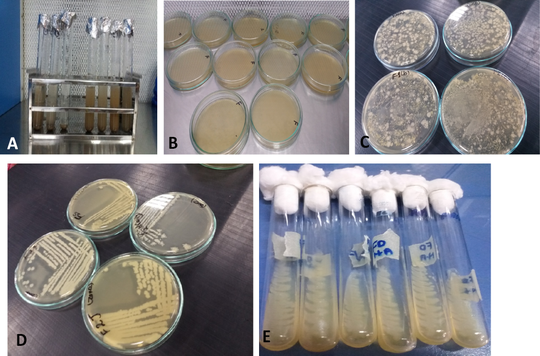
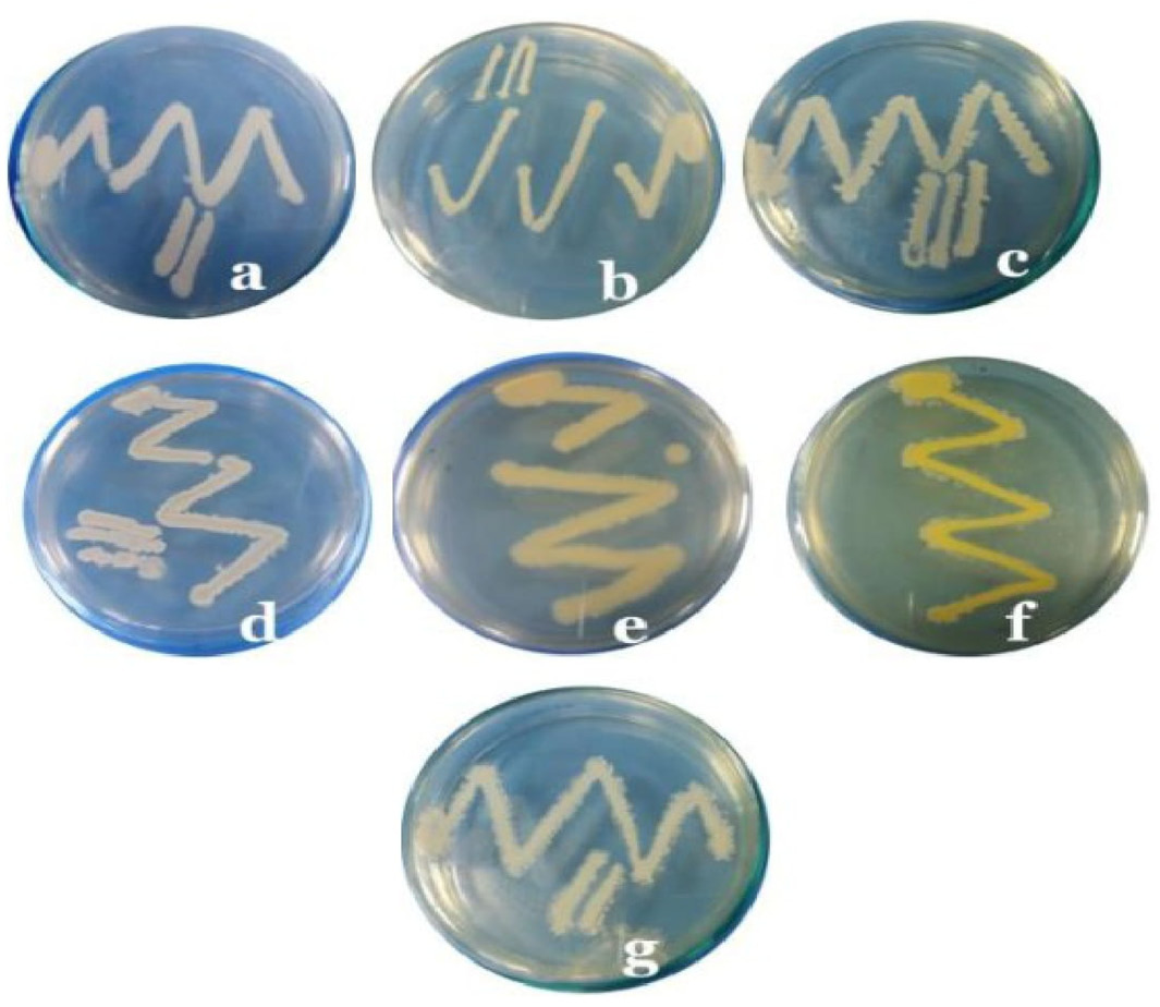
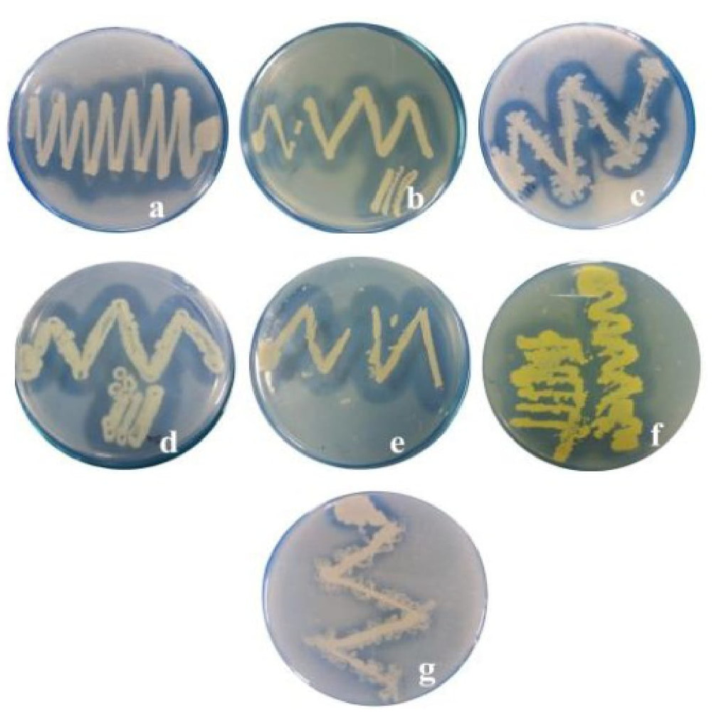
In the current investigation, seven bacterial strains with keratinolytic characteristics were identified from composted soil-samples collected from chicken farms. The prepared suspension was processed on nutritional agar medium, and well-formed single colonies were preferred to streak further on nutritional agar medium in various petri plates to generate pure cultures of strains. Figure 2, illustrates the many steps in the isolation process for distinct bacterial strains using various approaches, including serial dilution, the pour plate method, and the streak plate technique. The growth patterns of seven strains were shown in Figure 3. The morphological identification was performed and characteristics are shown in Table 1, respectively. Following the isolation stage, the main screening method was chosen based on the translucent, intense zones that developed on skimmed milk agar plates when flooded with 10% Trichloroacetic acid, as shown in Figure 4 for each. The best keratinase producers were determined to be seven bacterial strains based on the development of transparent zones on skimmed milk agar plates. Because due to transparent zone formation bacterial strains were considered as best keratinase producers. Among all seven isolates Bacillus cereus FD2 strain was selected for further studies due to maximum zone production on skimmed milk agar plates.
Table 1: Morphological identification of bacterial strains showing keratinase activity on skimmed milk agar plates and molecular identification by 16S RNA.
S
Isolate name
Acession number
Colony colour
Shape
Margin
Elevation
Consistency
1
Bacillus velezensis FD1
MG 952530
White,
Spherical
Irregular edges
Flat
Creamy
2
Bacillus cereus FD2
MG 952538
Grey yellow
Irregular
Opaque
Flat
Creamy
3
Bacillus subtilis FD3
MG 952539
Light yellowish
Filamentous
Opaque and smooth
Flat
Gummy
4
Bacillus altitudinis FD4
MG 952540
Whitish
Irregular
Rough and wrinkled
Flat
Mucoid
5
Bacillus licheniformis FD5
MG 952567
Fuzzy white
Wavy
Jagged edges
Flat
Brittle
6
Bacillus flexus FD6
MG 952576
Dark yellowish
Irregular
Erose
Convex
Gummy
7
Alcaligenes sp FD7
MG 952568
Off white
wrinkled
Regular margin
Convex
Creamy
According to Figure 3A, the Bacillus cereus FD2 had grey-white granular colonies with less wavy edges and opaque margins. Seven bacterial strains that produced the most keratinase was identified for the current study project from soil samples. In the presence of keratinolytic bacteria such as Bacillus paseudofirmus, Bacillus cereus, and Bacillus subtilis, which have been found by multiple researchers, the putrefaction of leftover poultry feathers persisted. ; ; Rajesh et al., 2016; Nanolim et al., 2020; Szabo et al., 2000). In this study, the creation of transparent zones on the surface of skim milk agar plates served as the primary indicator of screening procedures. Rajesh et al. (2016) isolated Chrysosporium keratinophilum and Bacillus subtilis and discovered that the development of the zone on skimmed milk agar medium exhibited keratinolytic capabilities. Mukhthar et al. (2019) and Nanolim et al. (2020) reported findings about Bacillus strains and Arthrobacter sp. that were similar to these findings.
Secondary screening of bacteria for keratinase production
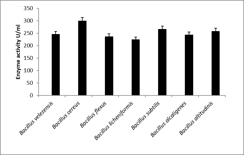
The only carbon source used in the submerged fermentation procedure for secondary screening was chicken feathers. The keratinase production values for all selected strains were shown in Figure 4, respectively. Bacillus licheniformis FD5 had the lowest production of keratinase (223 U/ml) while Bacillus cereus FD2 had the highest production of keratinase (298 U/ml) during 72 h of fermentation. The seven selected strains that exhibited prominent zones were measured as the best keratinase producer and out of seven isolates, Bacillus cereus FD2 was considered the best enzyme producer and selected for further study as depicted in Figure 5.
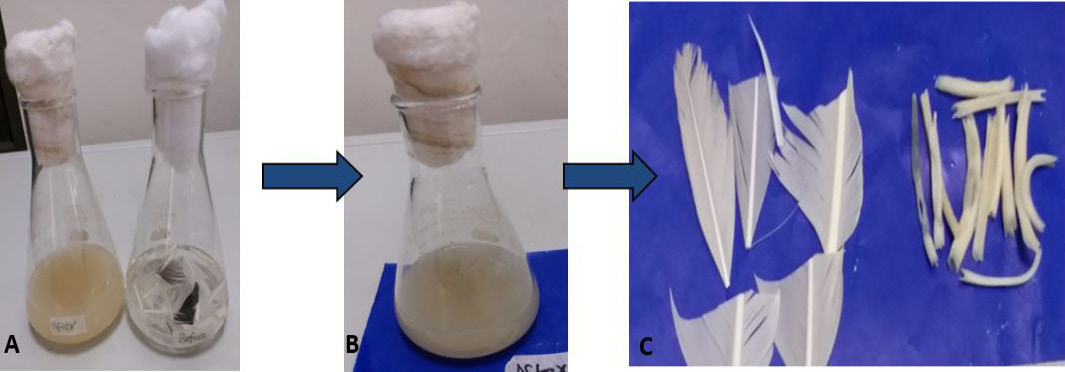
After primary screening of isolates by skim milk agar plate next step was to predict the most productive strain for keratinase production so these selected isolates were again processed for the secondary screening phase through the submerged fermentation technique. The maximum keratinase production was recorded for Bacillus cereus FD2 isolate, and most important point to be noted during submerged fermentation through visual observation was feathers complete degradation and on the fourth day of optimization experimental media appearance was quite milky as shown in Figure 6. Similar results from different bacterial strains were observed and reported by some studies ; Qui et al., 2022).
Molecular identification of keratinolytic bacterial isolates
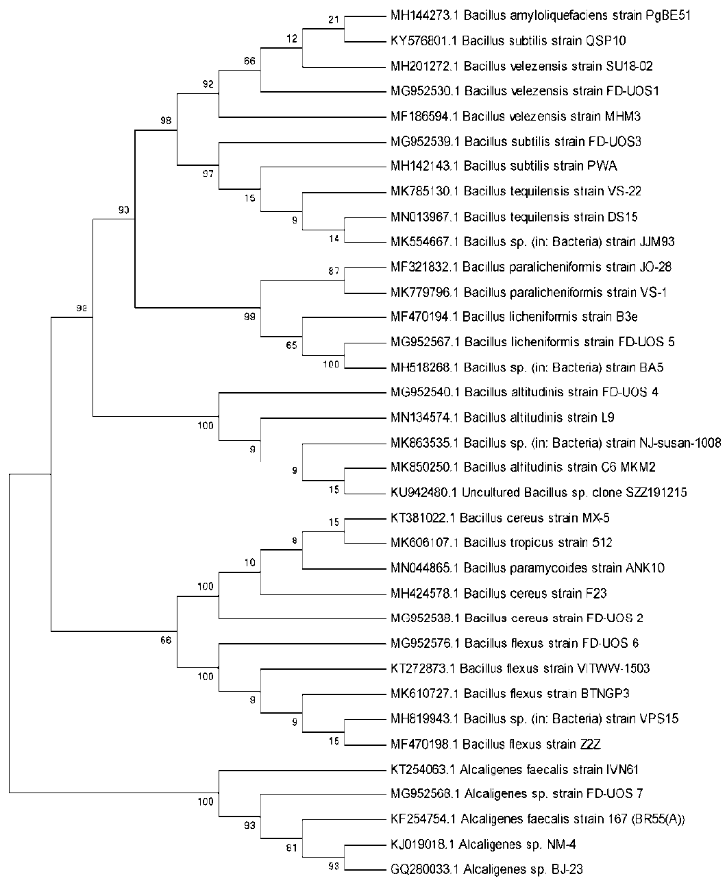
Seven bacterial strains were identified and tested in this investigation for their ability to break down keratin. Through the analysis of the 16S rRNA gene, these top keratinase-producing isolates were found. Results of all selected strains with their gene bank accession number are shown in constructed phylogenetic tree in Figure 7, respectively. The strain FD1 (MG952530.1) was recognized as Bacillus velezensis which has 100% similarity to previously reported strains of the genus Bacillus. The second isolate with accession number FD2 (MG 952538.1) was recognized as Bacillus cereus as revealed by blast analysis a study. The isolate FD3 (MG 952539.1) which was identified as Bacillus subtilis showed 100% similarity with previous related strains. The isolate FD4 with accession number (MG 952540.1) was identified as Bacillus altitudinis which shows resemblance with other species of Bacillus. The FD5 strain which has accession number (MG 952567.1) confirmed as Bacillus licheniformis and shows high sequence similarity to other species of this genus. Phylogenetic analysis of FD6 and FD7 with accession numbers MG 952576.1 and MG 952568.1 showed close lineage to Bacillus flexus and Alcaligenes sp., respectively. The all isolated seven strains evolutionary history with their closed genera in the form of phylogenetic tree depicted in Figure 7, respectively.
Optimization of culture conditions for production of the keratinase
Optimization of incubation time for keratinase production by seven isolated strains
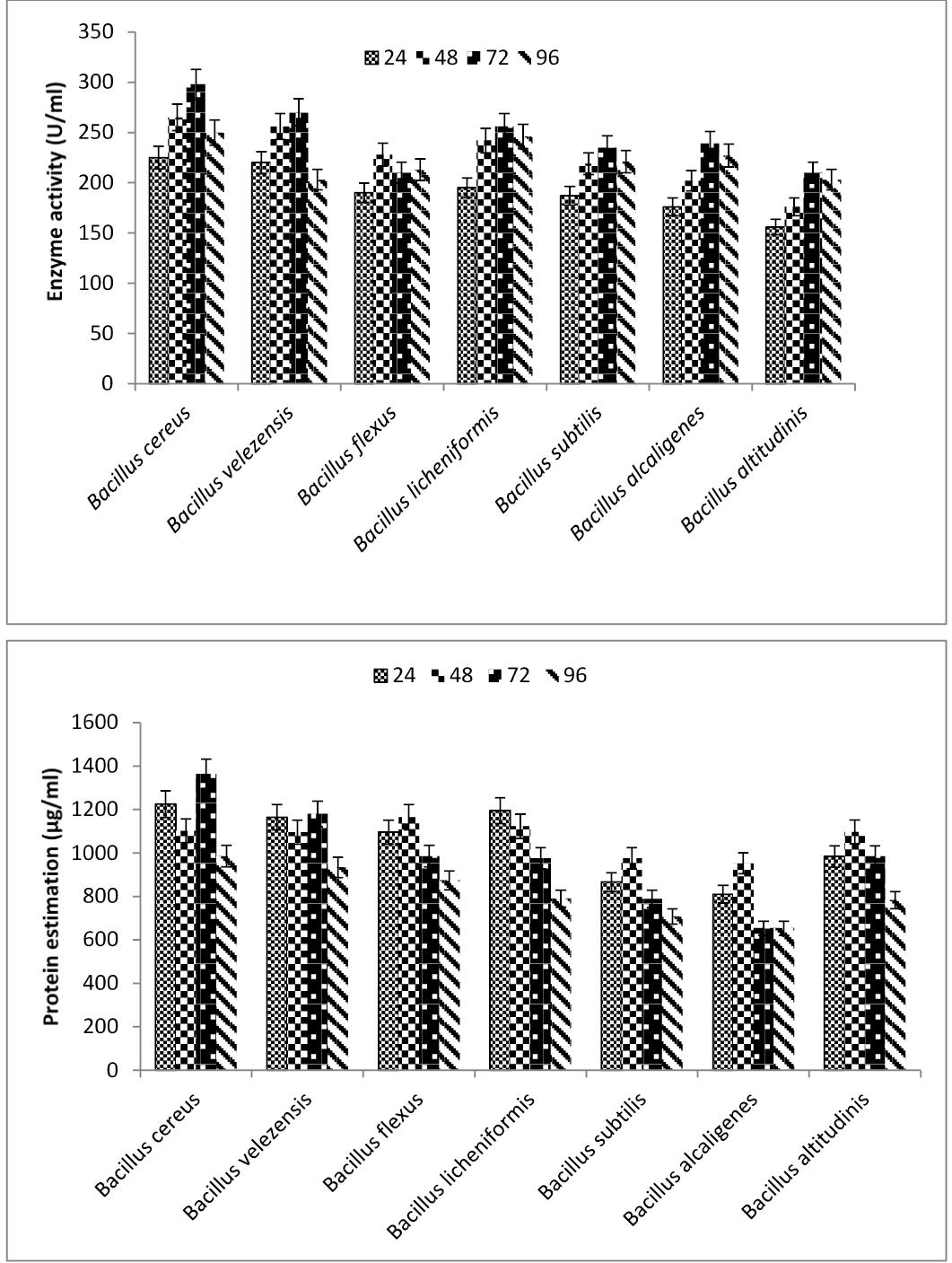
During the experiment, different incubation times like 24, 48, 72, and 96h on seven selected strains at 37°C were observed for keratinase production. The enzyme production was initiated after 24 h, but the maximum keratinase enzyme production value 298U/ml was observed after 72 h of incubation from Bacillus cereus FD2, respectively, among the seven strains depicted in Figure 8. During the current study, the maximum incubation period was recorded as 72 h for Bacillus cereus FD2. When further incubation period increased enzyme production rate decreased. Sivakumar et al. (2013) observed the same time period from Bacillus thuringiensis at 72 h, and Ire and Onyenama (2017) reported results with maximum keratinase enzyme production 72 h for Bacillus licheniformis. Similar findings were also reported by Akram and Jabbar (2020), who discovered that Bacillus sp. produced the most keratinase at pH 7.0, 40oC after 72 h of incubation.
Optimization of pH for keratinase production by Bacillus cereus FD2
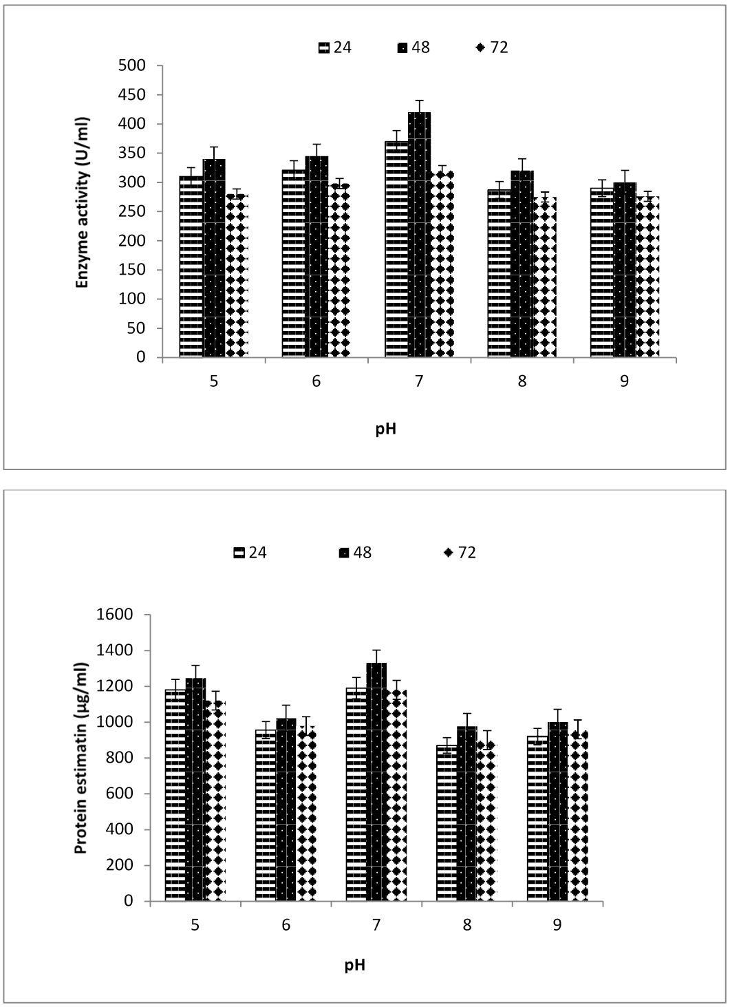
By modifying different pH values between 5.0 and 9.0, it was possible to observe how the pH factor affected the growth of the Bacillus cereus FD2 strain and the generation of keratinase. According to Figure 9, the isolate Bacillus cereus FD2 strain’s keratinase activity and soluble protein concentration peaked at 420 U/ml and 1330 g/ml, respectively, and fell to 235 U/ml and 840 g/ml at their lowest points. The Bacillus cereus FD2 best keratinase production was recorded at pH 7.0. Significant change in keratinase activity at pH 7 was observed at day 3 as compared to previous days and the shake flask feather media milky appearance and feathers digestibility shown neutral pH considered as best pH range on the other hand acidic and basic pH shown significant results as compared to neutral pH medium. So, it was determined that for Bacillus cereus FD2, pH 7.0 was ideal for achieving the highest level of keratinase activity. Additionally, Kainoor and Naik (2010) noted that pH 7.5 and 37°C were the ideal conditions for Bacillus berevis to produce the most keratinase. According to some other researchers , the pH that produced the most enzymes was neutral.
Optimization of inoculum size for keratinase production by Bacillus cereus FD2
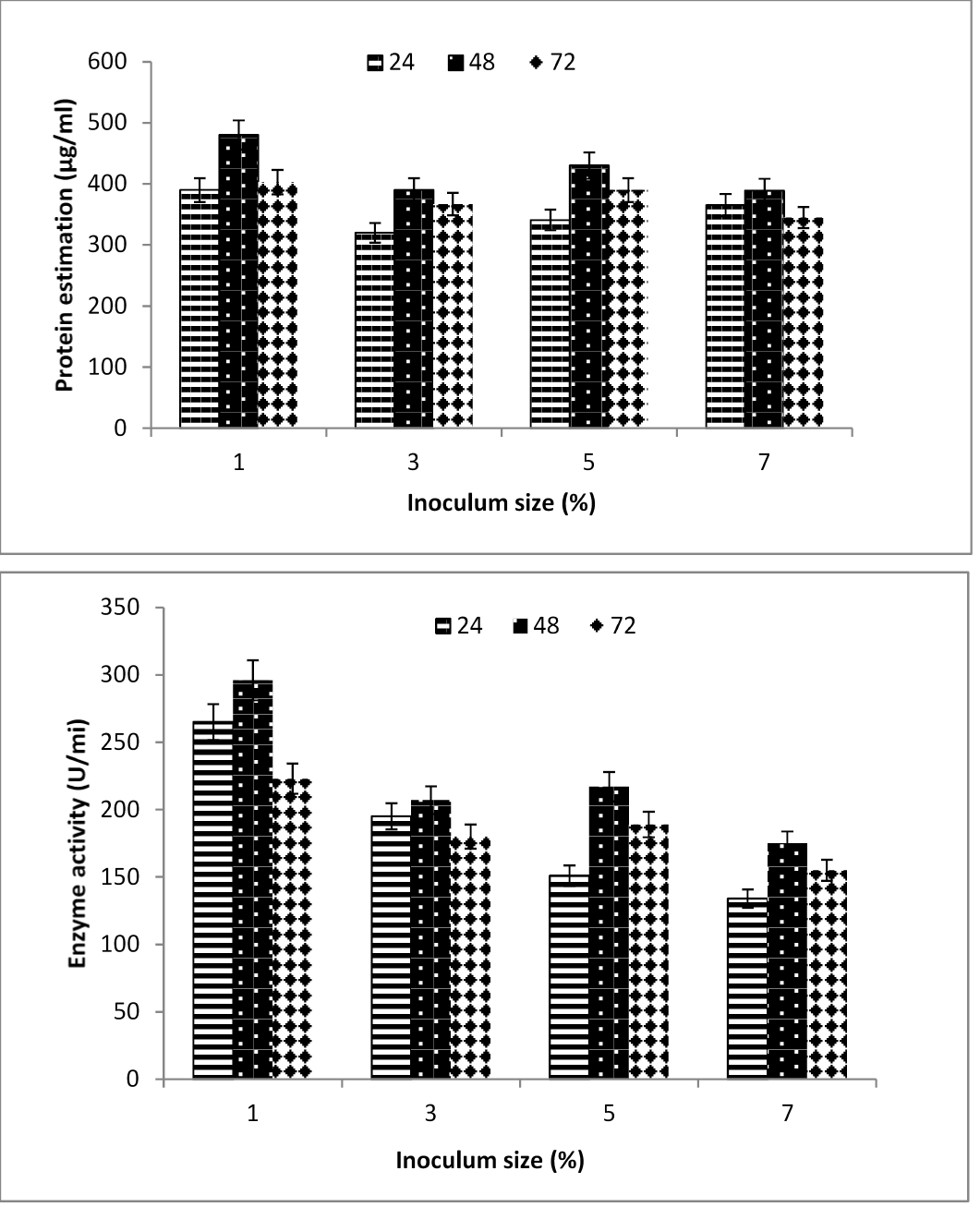
Bacillus cereus FD2 recorded the highest levels of keratinase production and soluble protein when media was inoculated with various inoculum sizes, such as 1, 3, 5, and 7%, as shown in Figure 10. Significant drops in keratinolytic activity and soluble protein concentrations were seen at 3, 5 and 7%, respectively, as inoculum size was increased. It was discovered that inoculum size had an impact on both cell growth and enzyme production.
Optimization of substrate concentration for keratinase production by Bacillus cereus FD2
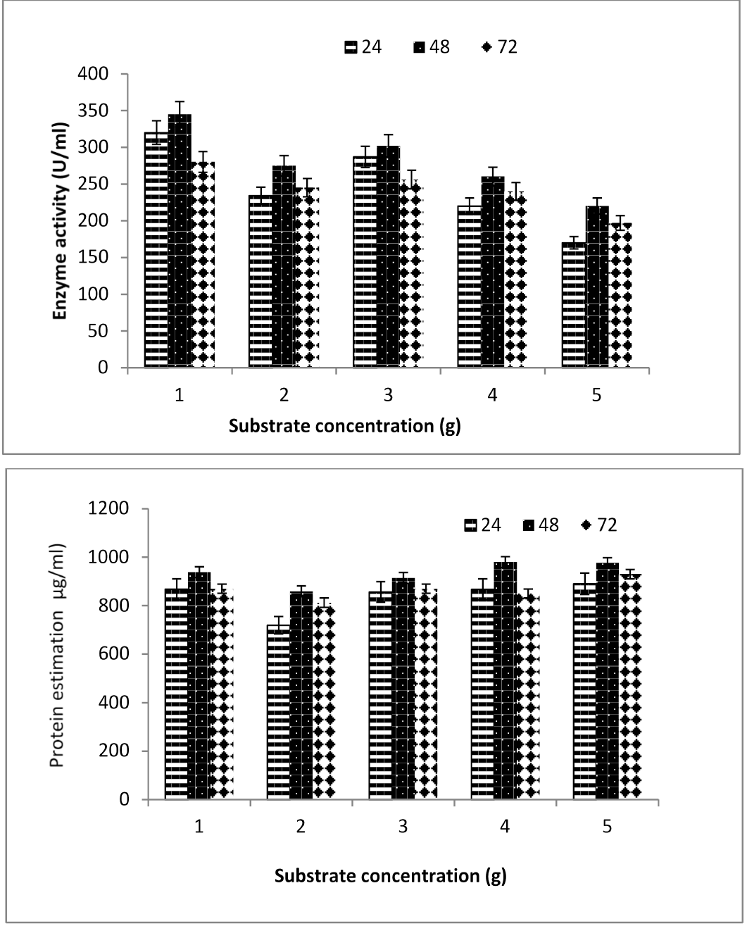
In this study, substrate feather meal was accordingly used at 1, 2, 3, 4, and 5% as the main carbon source for keratinase production. Figure 11 depicts the Bacillus cereus FD2 strain producing extremely high levels of keratinase and soluble protein concentrations 345U/ml and 980g/ml
respectively, when the substrate concentration was 1%. The highest level of keratinase production, according to Kate and Pethe (2014), was attained with just 1% of chicken feathers. With 1% substrate, the experimental isolate Bacillus cereus FD2 demonstrated maximal enzyme production. Less protein concentrations and keratinase activity were seen at substrate concentrations greater than 1%. According to Singh et al. (2017), keratinase activity is slowed down by a 1% or greater rise in substrate concentration.
Conclusions and Recommendations
The results of the current investigation show that all culture factors, including pH, inoculum size, incubation time, and substrate concentration, significantly influence how much keratinase Bacillus cereus FD2 can produce. Recycling garbage containing keratin would be advantageous from an economic and environmental standpoint. These isolates may eventually be the most effective options for boosting keratinase production on an industrial scale.
Acknowledgments
Authors are highly grateful to the Department of Biotechnology, University of Sargodha, Sargodha for supporting this research work.
Availability of data and materials
The sequence of the bacterial strains are accessible from the NCBI website as mentioned in the manuscript with accession number.
Consent for publication
All authors are agreed for publication of this manuscript.
Ethics approval consent to participate
Not applicable
Conflict of interest
The authors have declared no conflict of interest.
Figures
Figure 1:.
Map of District Khushab Jauharabad, Pakistan sampling sites.
Figure 2:.
Different isolation phases of keratinase-producing bacterial strains isolated from feather composted sites and soil samples (A) Serial dilution method; (B) Pouring method; (C) After spreading method, bacterial colonies are obtained on nutrient agar plates; (D) Streaking method; (E) Slants of pure culture are saved.
Figure 3:.
Growth pattern of Bacillus cereus (a), Bacillus altitudinis (b), Bacillus subtilis (c), Bacillus velezensis (d), Bacillus flexus (e), Bacillus alcaligenes (f) and Bacillus licheniformis (g) on nutrients agar plates isolated from soil.
Figure 4:.
Primary screening of keratinase producing strains on skimmed milk agar plate after flooded with 10% Trichloroacetic acid demonstrated transparent zone of hydrolysis and identified as (a) Bacillus cereus (b) Bacillus altitudinis (c) Bacillus subtilis (d) Bacillus velezensis (e) Bacillus flexus (f) Bacillus alcaligenes and (g) Bacillus licheniformis.
Figure 5:.
Screening of seven bacterial isolates for keratinase production under submerged fermentation (SMF) method.
Figure 6:.
Secondary screening and successive feather degradation s of Bacillus cereus FD2 for keratinase production under submerged fermentation (SMF) method.
Figure 7:.
Phylogenetic analysis of identified strains isolated from feather-compost soil samples.
Figure 8:.
Effect of incubation time on keratinase production and protein estimation by different bacterial strains.
Figure 9:.
Effect of pH and protein estimation on keratinase production of Bacillus cereus FD2.
Figure 10:.
Effect of inoculums size on keratinase production and protein estimation by Bacillus cereus FD2.
Figure 11:.
Effect of substrate concentration on keratinase production and protein estimation by Bacillus cereus FD2.
References
Akram, F. and Jabbar, Z., 2020. Keratinolytic enzyme-mediated biodegradation of recalcitrant poultry feathers waste by newly isolated Bacillus sp. NKSP-7 under submerged fermentation. Folia Microbiol., 65: 823-834. https://doi.org/10.1007/s12223-020-00793-6[CrossRef]
Adelere, I.A. and Lateef, A., 2022. Valorization of feather by Bacillus safensis and Aquamicrobium defluvii for growth promotion in leafy vegetables. Waste Biomass Valori., pp. 1-15. https://doi.org/10.1007/s12649-022-01904-9[CrossRef]
Abdelmoteleb, A., Gonzalez-Mendoza, D., Tzintzun-Camacho, O., Grimaldo-Juárez, O., Mendez-Trujillo, V., Moreno-Cruz, C. and Roumia, A.F., 2023. Keratinases from Streptomyces netropsis and Bacillus subtilis and their potential use in the chicken feather degrading. Fermentation, 9: 96. https://doi.org/10.3390/fermentation9020096[CrossRef]
Arokiyaraj, S., Varghese, R., Ahmed, B.A., Duraipandiyan, V. and Al-Dhabi, N.A., 2019. Optimizing the fermentation conditions and enhanced production of keratinase from Bacillus cereus isolated from halophilic environment. Saudi J. biol. Sci., 26: 378-381. https://doi.org/10.1016/j.sjbs.2018.10.011[CrossRef]
Bach, E., Cannavan, F.S., Duarte, F.R., Taffarel, J.A., Tsai, S.M. and Brandelli, A., 2011. Characterization of feather-degrading bacteria from Brazilian soils. Int. Biodeterior. Biodegrad., 65: 102-107. https://doi.org/10.1016/j.ibiod.2010.07.005[CrossRef]
Barman, N.C., Zohora, F.T., Das, K.C., Mowla, M., Banu, N.A., Salimullah, M. and Hashem, A., 2017. Production, partial optimization and characterization of keratinase enzyme by Arthrobacter sp. NFH5 isolated from soil samples. AMB Exp., 7: 1-8. https://doi.org/10.1186/s13568-017-0462-6[CrossRef]
Brandelli, A., 2005. Hydrolysis of native proteins by a keratinolytic protease of Chryseobacterium sp. Annls Microbiol.,
Casarin, F., Cladera-Olivera, F. and Brandelli, A., 2008. Use of poultry byproduct for production of keratinolytic enzymes. Fd. Bioprocess Technol., 1: 301-305. https://doi.org/10.1007/s11947-008-0091-9[CrossRef]
Cortezi, M., Cilli, E.M. and Contiero, J., 2008. Bacillus amyloliquefaciens: A new keratinolytic feather-degrading bacteria. Curr. Trends Biotechnol. Pharm., 2: 170-177.
Friedrich, J., Gradišar, H., Vrecl, M. and Pogačnik, A., 2005. In vitro degradation of porcine skin epidermis by a fungal keratinase of Doratomyces microsporus. Enzyme Microb. Technol., 36: 455-460. https://doi.org/10.1016/j.enzmictec.2004.09.015[CrossRef]
Gopinath, S.C., Anbu, P., Lakshmipriya, T., Tang, T.H., Chen, Y., Hashim, U. and Arshad, M.K., 2015. Biotechnological aspects and perspective of microbial keratinase production. BioMed. Res. Int., 8: 124-134. https://doi.org/10.1155/2015/140726[CrossRef]
Gupta, R. and Ramnani, P., 2006. Microbial keratinases and their prospective applications: An overview. Appl. Microbiol. Biotechnol., 70: 21-33. https://doi.org/10.1007/s00253-005-0239-8[CrossRef]
Gupta, R., Rajput, R., Sharma, R. and Gupta, N., 2013. Biotechnological applications and prospective market of microbial keratinases. Appl. Microbiol. Biotechnol., 97: 9931-9940. https://doi.org/10.1007/s00253-013-5292-0[CrossRef]
Ire, F.S. and Onyenama, A.C., 2017. Effects of some cultural conditions on keratinase production by Bacillus licheniformis Strain NBRC 14206. J. Adv. Biol. Biotechnol., 13: 1-13. https://doi.org/10.9734/JABB/2017/32726[CrossRef]
Kainoor, P.S. and Naik, G.R., 2010. Production and characterization of feather degrading keratinase from Bacillus sp. JB 99. Indian J. Biotechnol., 9: 384–390.
Kate, S. and Pethe, A., 2014. Study of efficiency of keratinase production by Arthrobacter creatinolyticus KP015744 isolated from leather sample. Int. J. Adv. Res., 2: 992-999.
Kiani, A.M., Doudi, M. and Ahadi, A.M., 2018. Isolation and molecular identification of keratinase-producing bacteria from the sludge of Qeshm Island. Med. Lab. J., 12: 34-40. https://doi.org/10.29252/mlj.12.3.34[CrossRef]
Kim, J.M., Lim, W.J. and Suh, H.J., 2001. Feather-degrading Bacillus species from poultry waste. Process Biochem., 37: 287-291. https://doi.org/10.1016/S0032-9592(01)00206-0[CrossRef]
Kokwe, L., Nnolim, N.E., Ezeogu, L.I., Sithole, B. and Nwodo, U.U., 2023. Thermoactive metallo-keratinase from Bacillus sp. NFH5: Characterization, structural elucidation, and potential application as detergent additive. Heliyon. https://doi.org/10.1016/j.heliyon.2023.e13635[CrossRef]
Lin, X., Shih, J. and Swaisgood, H.E., 1996. Hydrolysis of feather keratin by immobilized keratinase. Appl. Environ. Microbiol., 62: 4273-4275. https://doi.org/10.1128/aem.62.11.4273-4275.1996[CrossRef]
Lowry, O., Rosebrough, N., Farr, A.L. and Randall, R., 1951. Protein measurement with the Folin phenol reagent. J. biol. Chem., 193: 265-275. https://doi.org/10.1016/S0021-9258(19)52451-6[CrossRef]
Manczinger, L., Rozs, M., Vágvölgyi, C. and Kevei, F., 2003. Isolation and characterization of a new keratinolytic Bacillus licheniformis strain. World J. Microbiol. Biotechnol., 19: 35-39. https://doi.org/10.1023/A:1022576826372[CrossRef]
Matikeviciene, V., Grigiskis, S., Levisauskas, D., Sirvydytė, K., Dizavicienė, O., Masiliūnienė, D. and Ancenko, O., 2011. Optimization of keratinase production by Actinomyces fradiae 119 and its application in degradation of keratin containing wastes. Environ. Technol. Resour. Proc. Int. Sci. Pract. Conf. 1: 294-300. https://doi.org/10.17770/etr2011vol1.905[CrossRef]
McKittrick, J., Chen, P.Y., Tombolato, L., Novitskaya, E.E., Trim, M.W., Hirata, G.A. and Meyers, M.A., 2010. Energy absorbent natural materials and bioinspired design strategies: A review. Mater. Sci. Eng. C, 30: 331-342. https://doi.org/10.1016/j.msec.2010.01.011[CrossRef]
Mukhtar, H., Ahmad, M. and Arshad, Y., 2019. Isolation and screening of keratinase producing bacteria from soil. Biol. Pak.., 65: 1-6.
Najafi, M.F., Deobagkar, D.N., Mehrvarz, M. and Deobagkar, D.D., 2006. Enzymatic properties of a novel highly active and chelator resistant protease from a Pseudomonas aeruginosa PD100. Enzyme Microb. Technol., 39: 1433-1440. https://doi.org/10.1016/j.enzmictec.2006.03.031[CrossRef]
Nnolim, N.E., Okoh, A.I. and Nwodo, U.U., 2020. Proteolytic bacteria isolated from agro-waste dumpsites produced keratinolytic enzymes. Biotechnol. Rep.., 27: 1-8. https://doi.org/10.1016/j.btre.2020.e00483[CrossRef]
Nnolim, N.E. and Nwodo, U.U., 2021. Microbial keratinase and the bio-economy: A three-decade meta-analysis of research exploit. AMB Express, 11: 1-16. https://doi.org/10.1186/s13568-020-01155-8[CrossRef]
Parry, D.A. and North, A.C.T., 1998. Hard α-keratin intermediate filament chains: Substructure of the N-and C-terminal domains and the predicted structure and function of the C-terminal domains of type I and type II chains. J. Struct. Biol., 122: 67-75. https://doi.org/10.1006/jsbi.1998.3967[CrossRef]
Qiu, J., Barrett, K., Wilkens, C. and Meyer, A.S., 2022. Bioinformatics based discovery of new keratinases in protease family M36. New Biotechnol., 68: 19-27. https://doi.org/10.1016/j.nbt.2022.01.004[CrossRef]
Rajesh, T.P., Rajasekar, S., Mathan, R.K.H. and Anandaraj, B., 2016. Isolation and identification of feather degrading bacteria from feather-dumped soil. Int. J. Environ. Sustain. Dev., 15: 293-299. https://doi.org/10.1504/IJESD.2016.077393[CrossRef]
Rayudu, K. and Jayaraj, Y.M., 2013. Keratinolytic protease production from keratinaceous wastes. J. Recent Adv. Appl. Sci., 28: 69-77.
Saha, S. and Dhanasekaran, D., 2010. Isolation and screening of keratinolytic actinobacteria form keratin waste dumped soil in Tiruchirappalli and Nammakkal, Tamil Nadu, India. Curr. Res. J. Biol. Sci., 2: 124-131.
Saran, S., Isar, J. and Saxena, R.K., 2007. A modified method for the detection of microbial proteases on agar plates using tannic acid. J. Biochem. Biophys. Methods, 70: 697-699. https://doi.org/10.1016/j.jbbm.2007.03.005[CrossRef]
Singh, S., Masih, H., Jeyakumar, G.E., Lawrence, R. and Ramteke, P.W., 2016. Optimization of fermentative production of keratinase by Bacillus subtilis strain S1 in submerged state fermentation using feather waste. Int. J. Curr. Microbiol. Appl. Sci., 6:1499-1510
Singh, S., Masih, H., Jeyakumar, G.E., Lawrence, R. and Ramteke, P.W., 2017. Optimization of fermentative production of keratinase by Bacillus subtilis strain S1 in submerged state fermentation using feather waste. Int. J. Curr. Microbiol. App. Sci., 6: 1499-1510. https://doi.org/10.20546/ijcmas.2017.612.167[CrossRef]
Sivakumar, T., Balamurugan, P. and Ramasubramanian, V., 2013. Characterization and applications of keratinase enzyme by Bacillus thuringiensis TS2. Int. J. Future Biotechnol., 2: 1-8.
Snajder, M., Vilfan, T., Černilec, M., Rupreht, R., Popović, M., Juntes, P. and Ulrih, N.P., 2012. Enzymatic degradation of PrPSc by a protease secreted from Aeropyrum pernix K1. PLoS One, 7: e39548. https://doi.org/10.1371/journal.pone.0039548[CrossRef]
Subathra, C., Shankar, R., Kumar, S., Mohanasrinivasan, V. and Vaishnavi, B., 2018. Production of keratinase from a newly isolated feather degrading Bacillus cereus VITSDVM4 from poultry waste. Natl. Acad. Sci. Lett., 41: 307-311. https://doi.org/10.1007/s40009-018-0664-8[CrossRef]
Suzuki, Y., Tsujimoto, Y., Matsui, H. and Watanabe, K., 2006. Decomposition of extremely hard to degrade animal proteins by thermophilic bacteria. J. Biosci. Bioeng., 102: 73-81. https://doi.org/10.1263/jbb.102.73[CrossRef]
Swetlana, N. and Jain, P.C., 2010. Feather degradation by strains of Bacillus isolated from decomposing feathers. Braz. J. Microbiol., 41: 196-200. https://doi.org/10.1590/S1517-83822010000100028[CrossRef]
Szabo, I., Benedek, A., Szabó, I.M. and Barabas, G.Y., 2000. Feather degradation with a thermotolerant Streptomyces graminofaciens strain. World J. Microbiol. Biotechnol., 16: 253-255. https://doi.org/10.1023/A:1008950032017[CrossRef]
Tamura, K., Stecher, G., Peterson, D., Filipski, A. and Kumar, S., 2013. MEGA6: Molecular evolutionary genetics analysis version 6.0. Mol. Biol. Evolut., 30: 2725-2729. https://doi.org/10.1093/molbev/mst197[CrossRef]
Zerdani, I., Faid, M. and Malki, A., 2004. Feather wastes digestion by new isolated strains Bacillus sp. in Morocco. Afr. J. Biotechnol., 3: 67-70. https://doi.org/10.5897/AJB2004.000-2012[CrossRef]
Zhang, B., Jiang, D., Zhou, W., Hao, H. and Niu, T., 2009. Isolation and characterization of a new Bacillus sp. 50-3 with highly alkaline keratinase activity from Calotes versicolor faeces. World J. Microbiol. Biotechnol., 25: 583-590. https://doi.org/10.1007/s11274-008-9926-9[CrossRef]
Zhang, R.X., Wu, Z.W., Cui, H.Y., Chai, Y.N., Hua, C.W., Wang, P. and Yang, T.Y., 2022. Production of surfactant-stable keratinase from Bacillus cereus YQ15 and its application as detergent additive. BMC Biotechnol., 22: 1-13. https://doi.org/10.1186/s12896-022-00757-3[CrossRef]
Isolation, Identification, and Screening of Keratinase Producing Bacteria from Soil and Production Optimization Using Feather Waste as Substrate · ResearchersLinks