
The present study was designed to find the association of two stress enzymes i.e., monooxygenases and glucathione S-transferases with breast cancer. For enzyme estimation plasma samples from the patients suffering from breast cancer and age matched healthy individuals (control) were collected. The results revealed decline in the level of these enzymes in breast cancer patients as compared to healthy individuals because of this decline the overall level of protein was also found to be decline. It is concluded from the study that these enzymes can be used as biomarker in the diagnosis of breast cancer.
Research Article · Volume 33, Issue 2, 12 December 2018 · Open Access
Levels of Monooxygenases and Glutathione S-Transferases in Breast Cancer Patients
· · · · · · · ·
Affiliations & Notes ⌄
- 1Department of Zoology, University of Sargodha, Sargodha, Pakistan 2Department of Zoology, GC University Lahore, Pakistan 3UCMD, University of Lahore, Pakistan 4Lahore General Hospital, Lahore 5University of Education, Lower Mall Campus, Lahore, Pakistan 6Punjab Agriculture Research Board, Gulberg III, Lahore, Pakistan , *Corresponding Author: Hafiz Muhammad Tahir, hafiztahirpk1@yahoo.com
Article Info ⌄
- Journal:
- Punjab University Journal of Zoology
- Published:
- 12 December 2018

From the article
- In this study we compared the level of monooxygenases and glutathione S-transferases (GSTs) in the plasma of breast cancer patients.
- We observed that there was decrease in the level of these enzymes in the plasma of breast cancer patients as compared with healthy individuals.
- So this study and few previous studies demonstrate and confirm that the correlation obtained in this manner show a powerful approach that these enzymes can be used as biomarkers for the diagnosis of breast cancer.
- The lower level of these enzymes is seen in breast cancer patients with increase in oxidative stress support that this enzyme level is inversely related to oxidative stress in breast cancer patients.
Selected from the article's own text.
Abstract
Introduction
Breast cancer is responsible for the death of 3.3 million women and 14 % of total cancer related deaths worldwide ; ; . Approximately, one in every nine Pakistani women is expected to suffer from breast cancer . In Pakistan, breast cancer is a huge public health problem, because its frequency is frighteningly high and death rate is the highest in any Asian population ; ; .
Although, a lot of information is available on breast cancer . However, there is a need for more information to understand the biochemistry of breast cancer initiation. Oxidative stress is a major risk factor for breast cancer . Many reports indicate that cancer cells are under a constant oxidative stress ; ; . These cells under oxidative stress produce (ROS) Reactive oxygen species. These (ROS) cause oxidative stress in cancerous cells . These species lead to extensive damage to DNA, proteins and lipids. This damage produces the mutations that initiate the tumors ; ; .
Some stress enzymes that are involved in the detoxification system also participate in the removal of reactive oxygen species (ROS). The main enzymes of this system are glutathione S-transferases (GSTs) and monoxygenases (Li et al., 2007). These are important oxidative enzymes that cause oxidative metabolism of a wide variety of endogenous and exogenous compounds andbl detoxification of many xenobiotics in mammals ; ; ; ; . These are widely distributed in nature and are found in essentially all eukaryotic species ; ; . These enzymes also catalyze detoxification of alkylating agents used in chemotherapy and causing the detoxification of products of reactive oxidation .
As these enzymes are regulated by diverse range of xenobiotics i.e ROS. These enzymes also control tumor development, tumor response to therapy and are recognized as tumor biomarkers, that’s why these are used in diagnosis, monitoring, classification, staging, and localization of tumor action of antibodies and effectiveness of therapy . Aim of present study was to find out the relationship of monooxygenases and Glutathione S- transferases with breast cancer patients. Based on the results of this study we will be able to decide whether we can use these enzymes as biomarker for diagnosis of breast cancer or not?
Materials and Methods
Sample collection
The subjects (n=200) were categorized into two groups. One group comprised of diseased individuals (n=100) and the other consisted of age matched healthy individuals (n=100). The blood samples of cancerous patients were collected from different localities i.e., Niazi laboratory, Sahara laboratory, Rehman laboratory of Sargodha city, Punjab, Pakistan. The samples were collected from June, 2014 to March, 2015 considering ethics and norms. All blood samples (5cc each) were taken by cubital vein puncture using sterilized syringes (BD, USA). Blood samples were collected in EDTA coated vials (BD, USA) for enzyme estimation. All breast cancer patients included in this study were subjected to chemotherapy. The blood samples were collected from the patients before the treatment.
Fresh blood samples were centrifuged at 1300rpm for 5 minutes to separate the plasma. The plasma was collected in Eppendorf tubes (1.5ml) by using micropipette. Plasma samples were kept at -20°C until further study. This plasma was used as enzyme source for biochemical estimation of glutathione S-transferases (GSTs), monooxygenases and total proteins.
Biochemical estimation of monooxygenases
The activity of monooxygenases was determined by following the method described by Vulule et al. (1999). Reaction mixture comprised of 20μl of plasma, 200μl of 3,3, 5’,5’- Tetramethyl benzidine (TMBZ) solution [TMBZ solution was made by dissolving 0.01g TMBZ in 5ml ethanol and 15ml of 0.25M sodium acetate buffer (pH 5.0)], 100μl of 0.625M potassium phosphate buffer (PPB) at pH 7.0 and 30μl of 3% hydrogen peroxide. Reference solution contained 120µl TMBZ, 600μl of 0.625M potassium phosphate buffer (PPB) at pH 7.0 and 30μl 3% hydrogen peroxide. After ten minutes of reagent mixing readings were recorded at the wavelength of 620nm by using ELISA reader. The cytochrome C was used for preparing standard curve. The quantity of monooxygenases was calculated from a standard curve of cytochrome C.
Biochemical estimation of glutathione s-transferases
The activity of glutathione S-trasferases towards 1-chloro-2, 4-dinitrobenezene (CDNB) was estimated according to the method of Habig et al. (1974). The total volume of the mixture was comprised of 40µl of 1.0mM reduced Glutathione, 20µl 1.0mM 1-chloro-2, 4-dinitrobenzene (CDNB) and 200µl phosphate buffer (100mM, pH 7.0) and 20µl of supernatant. Reference solution for reaction mixture contained 20µl 1.0mM CDNB, 220µl phosphate buffers (100mM, pH 7.0) and 40µl 1.0mM reduced Glutathione. The absorbance was measured at 340 nm after five minutes of the reaction. Absorbance values were converted to units of concentration using a molar extinction coefficient (ε) of 9.6mM cm-1 for CDNB-GSH conjugate. Then the enzyme activity was calculated as:

Total protein assay
To measure the total protein contents dye binding method of Bradford (1976) was used. The reaction mixture contained 10µl of plasma, 90µl phosphate buffer (100mM, pH 7.0) and 100µl of Bradford dye reagent. It was mixed by shaking. Reference solution only contained 100µl buffer and 100µl Bradford dye reagent. The ELISA plates were covered and incubated at 30°C approximately for 15 minutes. Then the absorbance was recorded at the wavelength of 595nm on ELISA plate’s reader (Thermo scientific multiskan FC ELISA plate photometer). By using the standard curve of Bovine Serum Albumin (BSA) the amount of protein in the sample was measured. To make standard curve, BSA was dissolved in distilled water and diluted to make different concentration (µg/ml). Reaction mixture for standard curve contained solution from different dilutions and Bradford dye reagent.
Statistical analysis
Normality of data was assessed before analyzing the data Mann-Whitny U-test (a nonparametric test) was used to compare the level of protein and enzymes between breast cancer patients and healthy individuals. Difference was considered significance, if the p-value was ˂ 0.05.
Results
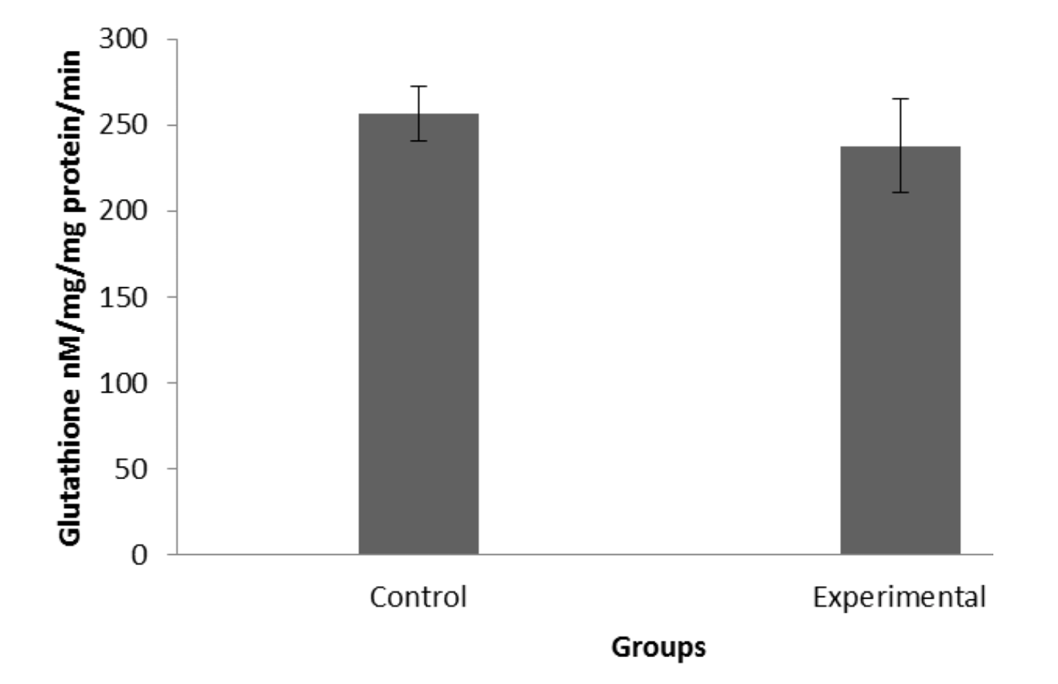
The level of total protein was 105.63µg/ml and 91.40µg/ml in healthy individuals (control group) and breast cancer patients, respectively. The amount of total protein in diseased individuals was significantly lower as compared with the control group (Mann-Whitny U = 1073.5; P =0.004; Figure 1). The levels of Glutathione S-transferases in the control group was higher (256.5nM /mg protein /min) than the patients (237.8nM /mg protein /min). Statistically the level of glutathione S-transferases differed significantly between two groups (Mann-Whitny U =1523.0; P =0.0148; Figure 2). The level of monooxygenases was high (3 1.26µg/min/mg) in the control group as compared to diseased individuals (29.87µg/min/mg), However, Statistically the difference was not significant (Mann-Whitny U =1367.5; P=0.5704; Figure 3).
Discussion
In this study we compared the level of monooxygenases and glutathione S-transferases (GSTs) in the plasma of breast cancer patients. We observed that there was decrease in the level of these enzymes in the plasma of breast cancer patients as compared with healthy individuals. Kadam et al. (2013) recorded significant decline in the level of GST in the breast cancer patients due to increase in oxidative stress. Oxidative stress in the body increases due the increase in the system toxicity. Different carcinogens in the body increase the toxicity of the system. Monooxygenases and GSTs are involved in the metabolism of human breast carcinogens (Ambrasone et al., 1995), so their level decreases in the breast cancer patients. These are also known as antioxidant or metabolizing enzymes and their level varies in the plasma of breast cancer patients due to change in oxidative stress.
The cytochrome P450 (CYP) is also an important oxidative enzyme that metabolizes many new plasma carcinogens and anticancer drugs. As these enzymes influence tumor development and they are recognized tumor biomarkers (Murray et al., 2010). The results of present study showed that the level of cytochrome p450 decreased non-significantly in breast cancer patients as compared with healthy individuals. El-Rayes et al. (2003) and Helzlsouer et al. (1998) also reported down regulation of CYP in the tumor tissues of cancer patients. As there is change in the level of P450 in tumor tissues so these enzymes could be used as biomarkers in the diagnosis of the disease. The balance between oxidative stress and antioxidant defence mechanism may be impared by the decline in the level of these enzymes level in the plasma. We perceive that failure of antioxidant defence mechanism against oxidative stress may be an important feature in the cause of breast cancer. The results of present study also demonstrate that the level of protein in the plasma of breast cancer patients was decreased. This decline of protein level in the body is due to overall decline in the level of these enzymes in the plasma of breast cancer patients.
In this study it is found that there is a strong association of these enzymes and breast cancer patients. To our knowledge, there have been only few previous studies evaluating the association between these enzymes and the risk of breast cancer. So this study and few previous studies demonstrate and confirm that the correlation obtained in this manner show a powerful approach that these enzymes can be used as biomarkers for the diagnosis of breast cancer. The lower level of these enzymes is seen in breast cancer patients with increase in oxidative stress support that this enzyme level is inversely related to oxidative stress in breast cancer patients. So these can be used as good biomarkers for the diagnosis of breast cancer.
Conflict of interest statement
The authors have no conflict of interest to declare.
Figures
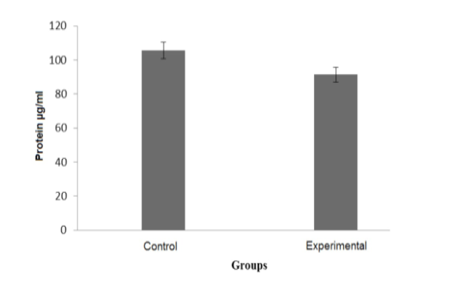
Total proteins (µg/ml) in control group and breast cancer patients
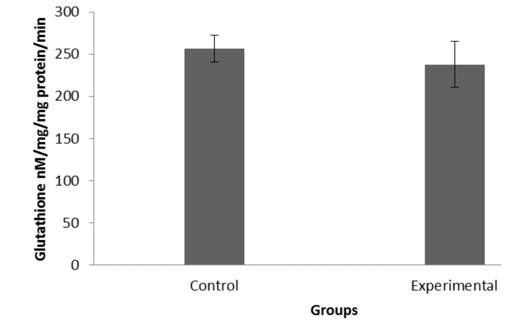
Activity of glutathione-S-transferases nM /mg protein /min in control and breast cancer pa-tients.
Activity of monooxygenase in control group and breast cancer patients
References
- Ambrosone, C. B., Freudenheim, J. L., Graham, S., Marshall, J. R., Vena, J. E., Brasure, J. R. and Ford, T. D., 1995. Cytochrome P4501A1 and glutathione S-transferase (M1) genetic polymorphisms and postmenopausal breast cancer risk. Cancer Res., 55(16): 3483-3485.
- Bayoumi, M. M. M., Elbasuny, M. M. M., Nasser, A. M. A. and Abdullah, K. M., 2012. Saudi young females’ level of knowledge regarding cervical and breast cancer. Int. J. Res. Nurs., 2: 47-52.
- Bieche, I., Narjoz, C., A, T., Vacher, S., Marcellin, P., Lidereau, R. AND DE Waziers, I., 2007. Reverse transcriptase-PCR quantification of mRNA levels from cytochrome (CYP) 1, CYP2 and CYP3 families in 22 different human tissues. Pharmacog. Genom., 17: 731-742. https://doi.org/10.1097/FPC.0b013e32810f2e58[CrossRef]
- Brown, N. S. AND BicknelL, R., 2001. Hypoxia and oxidative stress in breast cancer. Oxidative stress: its effects on the growth, metastatic potential and response to therapy of breast cancer. Breast Cancer Res., 3: 323-327. https://doi.org/10.1186/bcr315[CrossRef]
- Bhurgri, Y., Bhurgri, A., Hassan, S. H., ZaidI, S., Rahim, A., Sankaranarayanan, R. and Parkin, D. M., 2000. Cancer incidence in Karachi, Pakistan: first results from Karachi cancer registry. Int. J. Cancer. Oncol., 85: 325-329.
- Bhurgri, Y., Kayani, N., Faridi, N., Pervez, S., Usman, A., Bhurgri, H. And Hasan, S. H., 2007. Patho-epidemiology of Breast Cancer in Karachi 1995-1997’. APJCP., 8: 215.
- Devi, G. S., Prasad, M. H., Saraswathi, I., Raghu, D., RAO, D. N. and Reddy, P. P., 2000. Free radicals antioxidant enzymes and lipid per oxidation in different types of leukemias. Clinica. Chimica. Acta., 293: 53-62. https://doi.org/10.1016/S0009-8981(99)00222-3[CrossRef]
- Devi, P. U., 2004. Basics of carcinogenesis. Health Adm., 17: 16-24.
- El-Rayes, B. F., Ali, S., Heilbrun, L. K., Lababidi, S., Bouwman, D., Visscher. and Philip, P. A., 2003. Cytochrome p450 and glutathione transferase expression in human breast cancer. Clin. Cancer Res., 9(5): 1705-1709.
- Forrester, L. M., Hayes, J. D., Millis, R., Barnes, D., Harris, A. L., Schlager, J. J. and Wolf, C. R., 1990. Expression of glutathione S-transferases and cytochrome P450 in normal and tumor breast tissue. J. Carcinog., 11: 2163-2170. https://doi.org/10.1093/carcin/11.12.2163[CrossRef]
- Gonzalez, F. J. and Gelboin, H. V., 1994. Role of human cytochromes P450 in the metabolic activation of chemical carcinogens and toxins. Drug. Metab. Rev., 26: 165-183. https://doi.org/10.3109/03602539409029789[CrossRef]
- Grover, P. L. and Martin, F. L., 2002. The initiation of breast and prostate cancer. J. Carcinog., 23: 1095–102. https://doi.org/10.1093/carcin/23.7.1095[CrossRef]
- Gut, I., Danielova, V., Holubova, J., Souček, P. and Klučková, H., 2000. Cytotoxicity of cyclophosphamide, paclitaxel, and docetaxel for tumor cell lines in vitro: effects of concentration, time and cytochrome P450-catalyzed metabolism. Arch. Toxicol. Suppl., 74: 437-446. https://doi.org/10.1007/s002040000141[CrossRef]
- Habig, W. H., Pabst, M. J. and Jakoby, W. B., 1974.Glutathione S-transferase, the first enzymetic step in mercapturic acid formation. J. Biol. Chem. 249: 7130-7139.
- Hayes, J. D. and Pulford, D. J., 1995. The Glut athione S-Transferase Supergene Family: Regulation of GST and the Contribution of the lsoenzymes to Cancer Chemoprotection and Drug Resistance Part II. Crit. Rev. Biochem. Mol. Biol., 30: 521-600. https://doi.org/10.3109/10409239509083492[CrossRef]
- Helzlsouer, K. J., Huang, H. Y., Hoffman, S., Alberg, A. J., Comstock, G. W., Strickland, P. T. and BelL, D., 1998. Association between glutathione S-transferase M1, P1, and T1 genetic polymorphisms and development of breast cancer. J. Natl. Cancer. Inst., 90: 512-518. https://doi.org/10.1093/jnci/90.7.512[CrossRef]
- Huang, Z., Roy, P. and Waxman, D. J., 2000. Role of human liver microsomal CYP3A4 and CYP2B6 in catalyzing N-dechloroethylation of cyclophosphamide and ifosfamide. Biochem. Pharmacol., 59: 961-972. https://doi.org/10.1016/S0006-2952(99)00410-4[CrossRef]
- Kadam, C. Y. and Abhang, S. A., 2013. Evaluation of serum levels of reduced glutathione, glutathione-s-transferase and nitric oxide in breast cancer patients undergoing adjuvant chemotherapy. Ijcrar., 5: 51-57.
- Kelley, M. K., EngqvisT-Goldstein, A., Montali, J. A., Wheatley, J. B., Schmidt, D. and Kauvar, L. M., 1994. Variability of glutathione S-transferase isoenzyme patterns in matched normal and cancer human breast tissue. Biochem. J., 304: 843-848. https://doi.org/10.1042/bj3040843[CrossRef]
- Ketul, R. Chaudhary., Sri Nagarjunbatchu. and John, M. Seubert., 2009. Cytochrome P450 Enzymes and the Heart. Iubmbiubmb Life., 61: 954–960. https://doi.org/10.1002/iub.241[CrossRef]
- Kryston, T. B., Georgiev, A. B., Pissis, P. and Georgakilas, A. G., 2011. Role of oxidative stress and DNA damage in human carcinogenesis. Mutat. Res. /Fundamen. Mole. Mechan. Mutagen., 711: 193-201. https://doi.org/10.1016/j.mrfmmm.2010.12.016[CrossRef]
- Leong, S. P., Shen, Z.-Z., Liu, T.-J., Agarwal, G., Tajima, T., Paik, N.-S. and Foulkes, W. D., 2010. Is breast cancer the same disease in Asian and Western countries? World. J. Surg., 34: 2308-2324. https://doi.org/10.1007/s00268-010-0683-1[CrossRef]
- LI, X., Schuler, M. A. and Berenbaum, M. R., 2007. Molecular mechanisms of metabolic resistance to synthetic and natural xenobiotics. Annu. Rev. Entomol., 52: 231-253. https://doi.org/10.1146/annurev.ento.51.110104.151104[CrossRef]
- Murray, G. I., Paterson, P. J., Weaver, R. J., Ewen, S. W., Melvin, W. T. and Burke, M. D., 1993. The expression of cytochrome P-450, epoxide hydrolase, and glutathione s-transferase in hepatocellular carcinoma. Cancer., 71: 36-43. https://doi.org/10.1002/1097-0142(19930101)71:1%3C36::AID-CNCR2820710107%3E3.0.CO;2-J[CrossRef]
- Naeem, M., Khan, N., Aman, Z., Nasir, A., Samad, A. and Khattak, A., 2008. Pattern of breast cancer: experience at lady reading hospital, peshawar. J. Ayub. Med. Coll. Abbottabad., 20: 22-25.
- Pervaiz, S. and Clement, M.-V., 2004. Tumor intracellular redox status and drug resistance-serendipity or a causal relationship? Curr. Pharma. Design., 10(16): 1969-1977. https://doi.org/10.2174/1381612043384411[CrossRef]
- Raijmakers, M., Steegers, E. and Peters, W., 2001. Glutathione S-transferases and thiol concentrations in embryonic and early fetal tissues. Human Reprod., 16: 2445-2450. https://doi.org/10.1093/humrep/16.11.2445[CrossRef]
- Salinas, A. E. and Wong, M. G., 1999. Glutathione S-transferases-a review. Curr. Med. Chem., 6: 279-310.
- Schumacker, P. T., 2006. Reactive oxygen species in cancer cells: live by the sword, die by the sword. Cancer Cell., 10: 175-176. https://doi.org/10.1016/j.ccr.2006.08.015[CrossRef]
- Sohail, S. and Alam, S. N., 2007. Breast cancer in Pakistan awareness and early detection. J. Coll. Physicians. Surg. Pak., 17: 711–2
- Vulule, J. M., Beach, R. F., Atieli, F. K., Mcallister, J. C., Brogdon, W. G., Robert, J. M., Mwangi, R. W. and Hawley, W. A., 1999. Elevated oxidase and esterases levels associated with permenthrin tolerance in Anopheles gambiae from Kenyan villages usung permethrin impregnated nets. Med. Veterin. Entomol., 13: 239-244. https://doi.org/10.1046/j.1365-2915.1999.00177.x[CrossRef]
- Wiseman, H. and Halliwell, B., 1996. Damage to DNA by reactive oxygen and nitrogen species: role in inflammatory disease and progression to cancer. Biochem. J., 313: 17-29. https://doi.org/10.1042/bj3130017[CrossRef]
- Wu, L. L., Chiou, C.-C., Chang, P.-Y. and WU, J. T., 2004. Urinary 8-OHdG: a marker of oxidative stress to DNA and a risk factor for cancer, atherosclerosis and diabetics. Clinica. Chimica. Acta., 339: 1-9. https://doi.org/10.1016/j.cccn.2003.09.010[CrossRef]
Services
Article usage statistics
Loading…