
The global spread of Carbapenem-Resistant Klebsiella pneumoniae (CRKP), particularly the high-risk sequence type 147 (ST147) known for its epidemic potential and carbapenem resistance, represents a critical threat to public health. Bacteriophages offer a promising alternative for combating such multidrug-resistant (MDR) clones. This study aimed to isolate and characterize a novel bacteriophage specific to CRKP ST147 strains. A novel bacteriophage, RMN1, was isolated from hospital wastewater using a CRKP ST 147 strain from a clinical sample as a host. Phage purification was performed via double-agar overlay assay, followed by morphological characterization using transmission electron microscopy (TEM). Genomic DNA was extracted and analyzed for whole genome sequencing (WGS). Phage RMN1 effectively inhibited CRKP ST 147 clinical isolate and possessed a 44,226 bp linear dsDNA genome containing 58 open reading frames (ORFs), classifying it within the Drulisvirus genus. Genomic analysis confirmed the absence of lysogenic or virulence genes, suggesting its suitability for therapeutic use. Phage RMN1 exhibited a podovirus-like morphology with an icosahedral head (~56 nm) and a short tail. Novel phage RMN1 showed strong lytic activity against CRKP ST 147, making it a promising candidate for phage therapy. Notably, phage RMN1 represents the first K. pneumoniae phage isolated from Indonesian hospital environments, with whole-genome analysis confirming its unique genomic features. Further in vivo studies are warranted to evaluate its efficacy and safety in clinical settings.
Research Article · Volume 10, Issue 1, 22 January 2026 · Open Access
Isolation of Novel Carbapenem-Resistant Klebsiella pneumoniae (CRKP) ST 147 Phage RMN1
· · · · · ·
Affiliations & Notes ⌄
- 1Master’s Programme in Biomedical Sciences, Faculty of Medicine, Universitas Indonesia, Jakarta, Indonesia
- 2Eijkman Research Center for Molecular Biology, National Research and Innovation Agency (BRIN), Cibinong, West Java, Indonesia
- 3Department of Civil Engineering, Environmental Engineering Study Program, Faculty of Engineering, Universitas Indonesia, Depok, Indonesia. *Correspondence | Rosantia Sarassari, Eijkman Research Center for Molecular Biology, National Research and Innovation Agency (BRIN), Cibinong, West Java, Indonesia
- 4Email: rosa001@brin.go.id
- 5Department of Microbiology, Faculty of Medicine, Universitas Indonesia, Dr. Cipto Mangunkusumo Hospital, Jakarta, Indonesia
Article Info ⌄
- Journal:
- Novel Research in Microbiology Journal
- Published:
- 22 January 2026
From the article
- Our findings demonstrated that phage RMN1 formed clear plaques, indicating strong lytic activity against its CRKP host.
- The identified genomic variations in phage RMN1, particularly in non-contractile tail fiber proteins and hypothetical proteins of unknown function, suggested evolutionary adaptations to specific K. pneumoniae hosts.
- This close phylogenetic relationship suggested conserved mechanisms of host recognition, possibly mediated by capsule-specific interactions, as phage RMN1 targeted ST 147 KL128-capsulated K. pneumoniae.
- Combining phage RMN1 with other phages or antibiotics could further mitigate resistance development; a strategy gaining traction in precision medicine.
Selected from the article's own text.
Abstract
Introduction
The rapid global spread of carbapenem-resistant Klebsiella pneumoniae (CRKP) has become a critical public health concern, particularly in hospital-acquired infections (HAIs). CRKP has emerged as a severe challenge for clinical management, considering increased mortality rates and elevated healthcare costs . World Health Organization (WHO) has classified CRKP, as “critical“ priority pathogens requiring urgent research into alternative therapies .
The global dissemination of CRKP sequence type 147 (ST147); a high-risk clone associated with multidrug resistance (MDR) and hospital outbreaks, poses a critical threat to public health systems worldwide . ST147 strains exhibit enhanced virulence and plasmid-mediated resistance to last-resort antibiotics, limiting treatment options and increasing mortality rates in clinical settings . The difficulties in treatment options, necessitating alternative approaches such as phage therapy, which utilizes bacteriophages to specifically target and lyse bacterial pathogens .
Bacteriophages offer several advantages over antibiotics, including high specificity, self-replication at infection sites, and minimal disruption to the host microbiota . Despite their therapeutic potential, the isolation and characterization of phages against CRKP ST 147 remain underexplored, particularly in clinical settings. While numerous phages with lytic activity against MDR K. pneumoniae have been reported ; ; nevertheless, phage efficacy against CRKP remains limited due to restricted host range . Commonly isolated CRKP-specific phages often display narrow tropism, typically targeting only specific sequence types (e.g., ST258, ST11) or capsule variants (e.g., KL64, KL107) . Furthermore, environmental CRKP strains frequently harbor temperate phages, which may contribute to lysogenic conversion and potential enhancement of bacterial virulence or antibiotic resistance . Therefore, discovering novel, strictly lytic phages with broad-spectrum activity is crucial for advancing phage therapy.
Several recent studies on bacteriophages targeting CRKP have primarily focused on non-ST147 lineages ; ; , leaving a significant gap in our understanding of phage therapeutics for this high-risk clone. While several phages such as vB_KpS_GP-1 and vB_KpP_GP-2 have demonstrated efficacy against CRKP, their activity against ST147 (KL128) strains remains largely unexplored . Notably, ST147-specific phages are exceptionally rare in literatures, despite this clone’s global dissemination and association with carbapenem resistance . The few reported phages active against ST147, such as vB_KpP_GP-4 showed limited host range, highlighting the need for broader host spectrum . The ST147 clone includes multiple K-types, defined by variations in the cps loci, which influence phage susceptibility ; . The objective of this study was to to isolate and characterize a novel bacteriophage, RMN1, capable of lysing CRKP clinical isolates. We assessed its morphological features and genomic stability to evaluate its therapeutic potential. The obtained findings contribute to the growing body of researches on using phage-based alternatives to combat antibiotic-resistant infections, offering a potential solution to the escalating crisis of CRKP.
Materials and Methods
A bacterial strain, media, and growth conditions
The clinical bacterial strain used in this study was isolated from a urine specimen obtained from a hospitalized patient at Dr. Soetomo Regional General Hospital, Surabaya, Indonesia. A bacterial stock was maintained on Tryptic Soy Agar (TSA; BD Difco™, USA) supplemented with 1.5% (w/v) agar with overnight incubation at 37°C. Mugo et al. (2025) protocol was followed for phage screening with slight modifications. The protocol started with the double agar overlay technique that employed TSA containing soft agar (0.7% agar in TSB) for both spot testing and phage titration through plaque assays. For broth cultures, individual colonies were inoculated into 3 mL of Tryptic Soy Broth (TSB; Oxoid, USA) and incubated at 37°C with continuous shaking at 150 rpm.
Phage isolation and purification protocols
A bacteriophage was isolated from wastewater treatment plant of a general hospital in Jakarta, Indonesia, following established protocols with slight modifications . The isolation procedure involved initial sample processing by centrifugation (6.000 x g, 10 min), followed by filtration using 0.22 µm pore-size membranes (Millipore filter) to remove particulate matter. For phage enrichment, the filtered supernatant was combined with 1 mL of exponential-phase bacterial culture and 15 mL of 10× concentrated TSB, followed by an overnight incubation at 37°C with agitation (150 rpm). Following enrichment, the lysate was clarified by centrifugation (6.000 x g, 10 min) and sterile filtration (0.22 µm). Initial phage detection was performed using the spot assay technique reported by Mugo et al. (2025) with slight modifications, where 5 µL aliquots of filtrate were applied to bacterial lawns prepared with the soft agar overlay method (0.7% agar in TSB). High-titter phage lysates (~1010 PFU/mL) were concentrated by centrifugation ((18.000 x g, 2 h, at 4°C) and the pellets were suspended in 1 mL Saline Magnesium (SM) buffer (0.05 M Tris-HCl, pH 7.5 containing 0.1 M NaCl, 0.008 M MgSO4•7H2O, and 0.01% gelatin).
Phage quantification was conducted using the standard double-layer agar technique with modifications . Briefly, serial dilutions of phage lysate were prepared in SM buffer and mixed with log phase bacterial culture in molten soft agar (0.7%), which was subsequently overlaid onto TSA plates. Plates were incubated at 37°C for 18–24 h to allow plaque development for titer determination.
Transmission electron microscopy (TEM) analysis
Phage morphology was characterized using high-resolution transmission electron microscopy (HR-TEM) following established protocols with minor modifications . For sample preparation, 10 μL aliquots of high-titter phage lysate (≥1010 PFU/mL) were adsorbed onto 200-mesh copper grids coated with Formvar film. The grids were subsequently negatively stained with 2% (w/v) uranyl acetate and air-dried at room temperature. Imaging was performed using a Talos F200C G2 transmission electron microscope (TEM, Thermo Fisher Scientific, USA). Viral capsid diameters were quantitatively analyzed using ImageJ software (version 1.54g; NIH) following the standardized protocols for nanoparticle characterization . Capsid diameters were measured using ImageJ software (version 1.54g; NIH) following the standardized protocols for nanoparticle characterization , with triplicate measurement performed for each particle to ensure measurement precision. This analytical approach enabled comprehensive morphological characterization of phage RMN1, including determination of its mean capsid diameter with associated standard deviation (±SD).
Bacterial genome sequencing and analysis
Genomic DNA extraction from the selected bacterial host was performed using Qiagen DNeasy blood and tissue kit (Qiagen, Carlsbad, CA, USA) following the manufacturer’s protocol. Whole-genome sequencing (WGS) was conducted on the Illumina NovaSeq 6000 PE150 coverage 100X platform (Illumina, USA) . Quality-controlled reads were assembled de novo using the automatic bacterial isolate assembly, annotation, and analyses (ASA3P) (https://github.com/oschwengers/asap) pipeline (v1.3.0) with Klebsiella pneumoniae HS11286 (NC_016845.1) as the reference genome . Subsequent genomic characterization was carried out using the PathogenWatch (https://pathogen.watch/) platform for comprehensive antimicrobial resistance such as genome length, GC content, sequence type (ST), K locus, capsular type determination using wzi gene sequencing, O locus, predicted O type, and antimicrobial resistance genes. wzi gene is one of six conserved genes (i.e., galF, orf2, wzi, wza, wzb, and wzc) located in cps locus for K typing .
Phage genome sequencing and analysis
High-purity phage genomic DNA was extracted from purified phage particles (≥1010 PFU/mL) using the Phage DNA isolation Kit (Norgen BioTek Corp., Thorold, ON, Canada). Phage WGS was conducted using Illumina NovaSeq 6000 PE150 coverage 100X (Illumina, USA). Bioinformatic analysis was performed using Geneious Prime 2025.0.2 build 2024-10-30 and its plugins . Bioinformatics processing included quality trimming using BBDuk adapter and de novo assembly using SPAdes v4.0.0 . Genome annotation was carried out using RASTtk (https://rast.nmpdr.org/) pipeline . Comparative genomics analysis was performed using BLASTn (https://blast.ncbi.nlm.nih.gov/Blast.cgi) against the National Center for Biotechnology Information (NCBI) non-redundant nucleotide database (nr/nt) for phylogenetic analysis and taxonomic classification, with matched genome database evaluated based on percent identity and query coverage thresholds (i.e., identity >90%, coverage >80%). Presence of integrase, tRNAs, transmembrane proteins, virulent factor, and antimicrobial resistant genes in the genome were detected with Phagescope ( . Phage genome packaging mechanisms and genome termini were determined through PhageTerm . Phage genome map was visualized via SnapGene Viewer 8.0 (http://www.snapgene.com/). Genome comparison with its highly similar phages (top five of highest identity and coverage threshold reference genome) was performed using MUMMER protein-based comparison, visualized by pyGenomeViz (https://pygenomeviz.streamlit.app/) to highlight regions of synteny and divergence. Phylogenetic analysis and Viral Proteomic Tree (VIPTree) 4.0. (https://www.genome.jp/viptree/) were used to generate a proteomic tree of phage genome based on genome-wide sequence similarity computed through tBLASTx with Drulisvirus-specific filters .
Results
Bacterial genome sequencing and analysis
Two bacterial isolates namely 032U and 118U were collected. Phenotypic characterization using the automated VITEK® 2 COMPACT system (bioMérieux, USA) confirmed both isolates as extended-spectrum β-lactamase (ESBL)-KP, which showed sensitivity to meropenem and imipenem. The bacterial genome analysis revealed 032U belonged to ST 147 and 118U belonged to ST 469. Therefore, 032U was selected as the host strain in this study. Genomic analysis using Pathogen Watch revealed that this isolate belonged to ST 147 with an unknown (KL128) capsule type and possessed an ompK35 mutation associated with carbapenem resistance. Based on this genotype, the isolate was classified as CRKP. Using VITEK® 2 COMPACT system (bioMérieux, USA), this isolate displayed resistance (100%) to several antibiotics; mainly third-generation cephalosporins, fluoroquinolones, penicillin, sulfonamides, tetracycline, and trimethoprim (Supplementary Table S1). The genome length was 5,452,315 bp with 57.3% GC content. This isolate expressed the presence of the blaCTX-M-15 resistance gene, confirming its identity as an extended-spectrum β-lactamase (ESBL)-KP isolate. Carbapenemase gene was not detected in this isolate.
Phage isolation and morphological characterization
The novel phage RMN1 was successfully isolated from wastewater treatment plant of General Hospital in Jakarta, Indonesia. Initial plaque assays demonstrated that phage RMN1 (1010 PFU/mL) produced distinct lytic zones (i.e., small and clear plaques) with a mean diameter of 0.25 ± 0.01 mm (Figure 1a). TEM analysis revealed that the viral particle possessed an icosahedral capsid architecture with an average diameter of 56 nm (Figure 1b), consistent with morphological characteristics of the Podovirus. This structural confirmation established phage RMN1 as a tailed phage with a podoviral morphology.
Phage RMN1 matched to the Drulisvirus genus
Whole-genome sequencing revealed that phage RMN1 possessed a 44,226 bp double-stranded DNA genome with a GC content of 53.9%, characteristic of the Drulisvirus genus. Genomic termini analysis identified 278-bp direct terminal repeats (DTRs) with redundant ends (Supplementary Figure S1). BLASTn analysis showed that phage RMN1 shared between 93.73% and 90% nucleotide similarity with Klebsiella phage vB_KpnP_Dlv622, P252, vB_KpP_AttikonH1, Pone, and VLC5 (Supplementary Table S2).
The phage RMN1 genome involved 58 putative open reading frames (ORFs), including 26 annotated and 32 hypothetical proteins. Functional annotation of the 58 predicted ORFs revealed five functional modules: (i) replication (phage DNA helicase, phage DNA-directed DNA polymerase, phage DNA-directed RNA polymerase, phage peptidase, and phage primase), (ii) packaging (endonuclease, exonuclease, phage phosphoesterase, HNH endonuclease, and phage terminase small and large subunits), (iii) structural assembly (capsid, major capsid protein, and non-contractile tail fiber protein), (iv) infection (internal virion protein A and phage non-contractile tail tubular protein), and (v) lysis (endolysin, holin, and spanin). Some ORFs exhibited combined functions, such as assembly and infection (collar and head to-tail connector protein) or lysis and infection (DNA ejectosome component and phage internal virion B) (Figure 2 and Supplementary Table S3), which were shared by several T7-like tail components.
The absence of virulence factors (e.g., toxin genes, immune modulators, or secretion system effectors) and antimicrobial resistance genes (particularly β-lactamases or qnr-type quinolone resistance) in phage RMN1’s genome aligns with World Health Organization (WHO) safety guidelines for therapeutic phages. Notably, the lack of tRNA genes further reinforced the phage’s obligatory lytic nature, minimizing risks of lysogenic conversion or host fitness enhancement.
Comparative analysis showed high similarity and gene synteny between the tested genomes, with some differences in hypothetical proteins, HNH endonuclease, and non-contractile tail fiber protein, suggesting divergent host recognition mechanisms and reinforcing phage RMN1’s host specificity (Figure 3a). A proteomic tree generated by VIPtree, included phage RMN1 and all available Klebsiella phages in database clustered phage RMN1 within the Drulisvirus genus (Figure 3b).
Genome availability
The complete genome sequence of phage RMN1 was deposited in the NCBI GenBank database under accession number PV804878, providing full open access to the genomic data.
Discussion
Genomic analysis of isolate 032U revealed a chromosome of 5,452,315 bp with 57.3% GC content, dimensions that aligned closely with the typical genomic architecture of Klebsiella pneumoniae, which generally ranged from 5.0 to 5.7 Mbp with a GC content of 57-58%, confirming a high-quality genome assembly suitable for accurate genotyping . The isolate’s predicted resistance profile spaned multiple antibiotic classes, including carbapenems, β-lactams (via blaCTX-M-15), and fluoroquinolones, characteristic of multidrug-resistant ST147 strains . Although no carbapenemase gene was detected, an ompK35 porin mutation was identified, supporting a carbapenem-resistant phenotype mediated by non-carbapenemase mechanisms . Collectively, all these attributes established 032U as a well-characterized, resistant ST147-KL128 variant, offering a stringent and clinically relevant host for evaluating the novel phage RMN1.
Successful isolation of bacteriophage RMN1 from hospital wastewater highlighted the potential of environmental sources as reservoirs for novel phages with therapeutic applications. Our findings demonstrated that phage RMN1 formed clear plaques, indicating strong lytic activity against its CRKP host. This aligns with recent studies showing that wastewater from healthcare facilities harbors diverse phages capable of infecting clinically relevant pathogens .
The successful isolation of podovirus RMN1 from hospital wastewater represented a substantial addition to the limited repertoire of phages active against CRKP ST147. The observed plaque morphology (0.25 ± 0.01 mm clear plaques) suggested robust lytic activity, comparable to other therapeutic podoviruses such as TC6 (0.5 mm plaques) , and it had an icosahedral head diameter of about 54 nm. The icosahedral capsid morphology (56 nm diameter) placed phage RMN1 within the typical size ranged for podoviruses infecting Enterobacteriaceae (55–60 nm) . Notably, the observed clear plaque phenotype indicated strictly lytic behavior; a crucial characteristic for therapeutic applications, as it minimizes the risks of lysogenic conversion and horizontal gene transfer .
The isolation of phage RMN1 from an Indonesian hospital wastewater expanded the geographical diversity of characterized K. pneumoniae phages, which had been predominantly isolated from European and American sources . This finding supported the hypothesis that environments may harbor unique phage diversity due to distinct microbial ecosystems, as suggested by recent metagenomic study . The podoviral morphology of phage RMN1 is particularly promising, as members of this family have demonstrated enhanced stability under physiological conditions compared to myoviruses, considered as an important consideration for pharmaceutical formulation . These characteristics positioned phage RMN1 as an excellent candidate for further development against the globally emerging ST147 clone.
The CRKP clinical isolate 032U characterized in this study represented a particularly concerning ST147 strain with an unusual KL128 capsule type and extensive MDR profile, including resistance to carbapenems and third-generation cephalosporins. This isolate carried the blaCTX-M-15 gene and an ompK35 porin loss mutation. This genetic profile is a high-risk due to its potential to developing carbapenem resistance. This finding aligns with recent global reports of ST147 as a high-risk clone demonstrating remarkable rapid dissemination of resistance determinants . The presence of CTX-M-15 gene detected in our isolate had been strongly associated with hospital outbreaks and treatment failures worldwide . Notably, the KL128 capsule type identified in our strain differed from the more commonly reported KL64 and KL10 variants in ST147 . This correlation between capsule type and sequence type observed in this study may be attributed to the impact of local clonal expansions of this pathogen . Klebsiella capsule type is the main determinant for phage tropism . Our genomic analysis identified capsule-type diversity within ST147, specifically KL128 variant. Variation in capsule type directly influences phage receptor recognition, thereby impacting therapeutic efficacy .
The identified genomic variations in phage RMN1, particularly in non-contractile tail fiber proteins and hypothetical proteins of unknown function, suggested evolutionary adaptations to specific K. pneumoniae hosts. This is consistent with the “arms race” co-evolutionary model between phages and bacterial capsular polysaccharides . Phage-host coevolution drives selective pressure for minor genomic changes that confer host specificity, even within highly conserved genomes . A structural study confirmed that even single-amino-acid changes in tail fibers can alter binding affinity to specific capsule types . Thus, while core genomic synteny was preserved, the critical tail fiber variations in phage RMN1 represented functional microevolution; –a hallmark of phage adaptation to bacterial surface diversity .
The combination of CTX-M-15 with resistance to last-resort antibiotics such as carbapenems creates particularly challenging clinical scenarios, as noted in intensive care unit (ICU) outbreaks where mortality rates exceed 40% . Our isolate’s resistance profile and ST147/KL128 characteristics provided a stringent model for evaluating phage therapeutics, as successful lysis demonstrated the phage potential against one of the most treatment-refractory CRKP variants currently emerging worldwide.
The genome shared < 94% identity with Drulisvirus Klebsiella phage vB_KpnP_Dlv622, P252, vB_KpP_AttikonH1, Pone, and VLC5 (below the 95% species threshold). Its distinct genetic signature and proteomic profile justified its classification as a novel phage with potentially unique therapeutic applications, confirming phage RMN1 as a novel member of the Drulisvirus genus . Meanwhile, the high degree of synteny conservation in core genomic regions supported the phage RMN1 functional similarity with these reference phages , and the observed variations in tail fiber proteins and HNH endonuclease domains likely accounted for its unique host range specificity. These findings align with a recent study demonstrating that even minor genomic differences in tail fiber genes can dramatically alter host recognition patterns, particularly against diverse K. pneumoniae capsule types .
The presence of 278-bp DTRs is particularly noteworthy as this genomic architecture, also observed in the closely related vB_KpnP_Dlv622 phage , enhanced DNA packaging efficiency and genomic stability . Of the 58 predicted ORFs, the 26 annotated genes included essential structural and lytic components, while the 32 hypothetical proteins presented intriguing targets for future functional studies. Crucially, the absence of integrase, virulence factors, antimicrobial resistance genes, and tRNA molecules satisfied key safety criteria for therapeutic phages .
The phylogenetic positioning of phage RMN1 among closely related Klebsiella phages underscored its evolutionary proximity and supported its taxonomic assignment. Phage RMN1 clusters were tight with other phages such as Klebsiella phage vB_KpnP_Dlv622 and VLC5, forming a coherent clade characterized by short branch lengths and high bootstrap support–evidence of a shared recent ancestor . The congruent genome sizes (~43–45 kb) and GC content (~54%) across this clade reinforced a conserved genome backbone and affirmed phage RMN1’s classification within this phylogenetic framework of Klebsiella podoviruses, similar to the KP-Rio/2015 bacteriophage . This close phylogenetic relationship suggested conserved mechanisms of host recognition, possibly mediated by capsule-specific interactions, as phage RMN1 targeted ST 147 KL128-capsulated K. pneumoniae.
The genomic features of phage RMN1 positioned it advantageously within the current phage therapeutic landscape. Its 53.9% GC content showed remarkable adaptation to its K. pneumoniae ST147 host (57–58% GC), potentially facilitating more efficient gene expression during infection . Compared to other Drulisvirus members, phage RMN1 demonstrated conserved synteny in structural gene clusters, including capsid, major capsid protein, and non-contractile tail fiber protein while maintaining unique variations in tail fiber proteins, likely responsible for its specific host recognition . These molecular characteristics combined with its strictly lytic nature, made phage RMN1 an exceptionally promising candidate for clinical development against CRKP, particularly as it targeted the high-risk ST147 lineage.
The genomic architecture of phage RMN1 revealed a sophisticated lytic toolkit, particularly noteworthy for its dual-component cell lysis system comprising holin and endolysin proteins. The holin protein formed pores in the bacterial cytoplasmic membrane at precisely programmed times, allowing endolysin to access and degrade the peptidoglycan layer; a mechanism shown to be highly effective against MDR K. pneumoniae . Endolysin protein displayed its function as an amidase, an enzyme that cleavesed the essential peptide cross-links within the peptidoglycan matrix . This precise targeting of the bacterial cell wall’s integrity presented a remarkable advantage over conventional antibiotics, as it avoided the common resistance mechanisms while maintaining high specificity for its CRKP ST147 host.
Equally intriguing was phage RMN1’s complement of structural proteins governing host recognition and DNA delivery. The non-contractile tail tubular proteins and DNA ejectosome component worked synergistically to recognize the KL128 capsule polysaccharide and created a channel for genome injection; a process recently visualized in near-atomic detail for related podoviruses . The presence of putative spanin proteins suggested that phage RMN1 employed a two-step lysis mechanism, while holin-endolysin disrupted the cell wall from within (holin forms a hole in the inner membrane, allowing endolysin to escape and destroy the peptidoglycan, resulting directly in bacterial lysis), spanins mediated an outer membrane disruption, ensuring complete bacterial host cell lysis . This comprehensive lytic system, combining precise host targeting with redundant lysis mechanisms, explained phage RMN1’s exceptional efficacy against CRKP ST147 and positioned it as a promising candidate for engineered phage therapeutics.
Recent studies emphasized the importance of capsule depolymerases in phage tropism ; , which may explain phage RMN1’s specificity. However, we were unable to detect a depolymerase gene in phage RMN1. The strictly lytic nature of phage RMN1 enhanced its therapeutic utility . The discovery of phage RMN1 expands the arsenal against CRKP ST 147, which increasingly evade last-resort antibiotics like carbapenems.
We recognized the limitations of this study. While phage RMN1 was able to lyse a single ST147-KL128 K. pneumoniae strain, its broader host range was not assessed. Furthermore, a depolymerase gene was not yet been detected in the phage RMN1 genome, suggesting an avenue for further investigations into its receptor-binding and host specificity mechanisms.
Conclusions and Recommendations
This study presented the first report of the isolation and characterization of a novel lytic Drulisvirus phage, which was selectively active against KL128 capsular CRKP. Its genomic safety, lytic efficiency, and capsule-specific targeting aligned with the urgent need for effective alternatives to antibiotics. We recommend that future studies should evaluate the structural mechanisms underlying phage attachment to the host, which determine its specificity for the capsule type rather than the sequence type. This specificity may be related to a receptor-binding domain. Combining phage RMN1 with other phages or antibiotics could further mitigate resistance development; a strategy gaining traction in precision medicine. Additionally, further in vivo studies and clinical trials are warranted to translate the current findings into therapeutic applications.
Acknowledgements
Thank you to wastewater plant treatment unit of Dr. Cipto Mangunkusumo General Hospital, Jakarta, Indonesia Team for helping the team during water sample collection and to all the technician at Genomic and CryoEM laboratory, the National Research and Innovation Agency of Indonesia.
Novelty Statement
This study isolated and characterized phage RMN1 as the first reported Drulisvirus isolated from wastewater treatment plant of General Hospital in Jakarta, Indonesia. This novel phage specifically targeted the Carbapenem-Resistant K. pneumoniae ST147 with the rare KL128 capsular type; a critical-priority pathogen. It highlights the phage RMN1’s specificity for the rare KL128 capsular type, its safety profile confirmed by genomic analysis, and its potential for phage therapy in clinical settings where antibiotic options are limited. The discovery of this phage RMN provides a crucial and locally sourced therapeutic candidate to combat globally circulating, high-risk bacterial pathogens in a region heavily affected by antimicrobial resistance.
Author’s Contribution
NAS: Conceptualization, data curation, formal analysis, investigation, project administration, methodology, visualization, writing–original draft, writing–review and editing.
MM: Data curation, formal analysis, investigation, methodology, software, validation, writing–original draft.
YM: Formal analysis, software.
IR: Investigation, resources.
DS: Resources, validation.
YRS: Conceptualization, funding acquisition, resources, supervision, validation.
RS: Conceptualization, formal analysis, funding acquisition, methodology, resources, supervision, validation, visualization, writing–original draft, writing–review and editing.
All authors have read and agreed to the published version of the manuscript.
Ethical approval
Funding source
This study was funded by BIMA DRTPM (Directorate of Research and Technology in Education and Learning) supported by the Ministry of Higher Education, Science, and Technology (Decree Letter No. 0459/E5/PG.02.00/2024 and Contract Agreement No. 051/E5/PG.02.00.PL/2024); Rumah Program, Organisasi Riset Kesehatan 2024 supported by National Research and Innovation Agency of Indonesia (No. B-11155/III.9/TK.02.02/12/2023).
Generative AI and AI-assisted technology statement
No generative AI or AI-assisted technology was employed in this study.
Supplementary material
There is supplementary material associated with this article. Access the material online at https://dx.doi.org/10.17582/journal.nrmj/2026/10.1.37.51
Conflict of interests
The authors have declared no conflicts of interest.
Figures
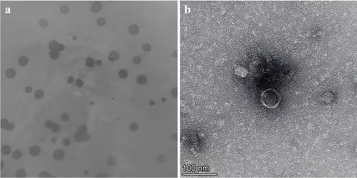
Morphological analysis of bacteriophage RMN1 using TEM. (a) Lytic plaques formed by phage RMN1 on a lawn of CRKP strain 032U, demonstrating clear zones of bacterial clearance with an average diameter of 0.25 ± 0.01 mm; (b) A micrograph of negatively-stained phage RMN1 showing characteristic podovirus morphology, including an icosahedral capsid (approximately 56 nm) measured using ImageJ software from 3 measurements.
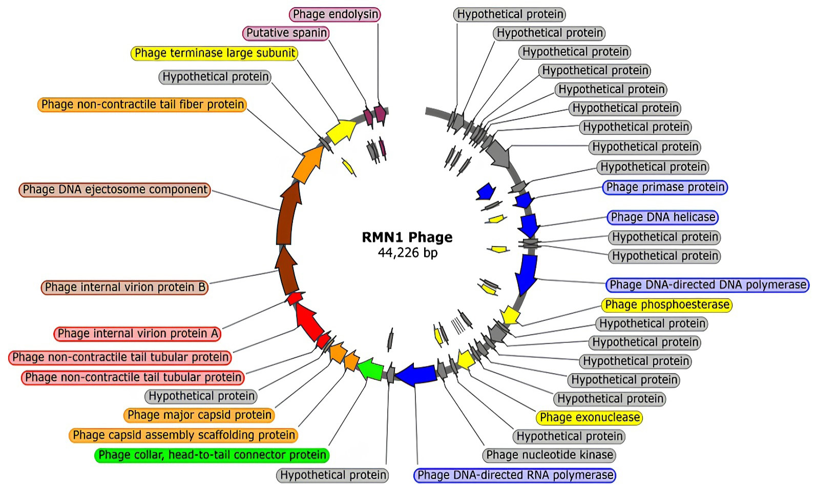
Circular genome map illustrating functional annotation of phage RMN1 ORFs, color-coded by protein function: replication (blue), packaging (yellow), structural assembly (orange), combined assembly and infection (green), infection (red), combined lysis and infection (brown), lysis (purple), and hypothetical proteins (gray).
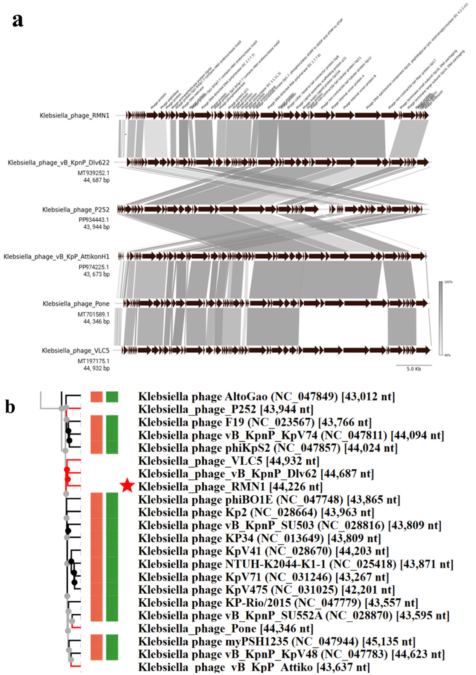
Genome comparison and phylogenetic tree of phage RMN1. (a) Genomic comparison of phage RMN1 with its highly similar phages using MUMMER protein-based, compared to highlight regions of synteny and divergence; (b) Viral proteomic tree of phage RMN1 against all Klebsiella phages existing in (NCBI) database.
References
- Altamirano, F.L.G. and Barr, J.J., 2021. Screening for lysogen activity in therapeutically relevant bacteriophages. Bio-Protoc. 11: e3997. https://doi.org/10.21769/BioProtoc.3997[CrossRef]
- Anjay, A., Kaushik, P., Prajapati, A., Jaiswal, R.K., Bhoomika, B., Roy, S.S., Kumar, A., Kumari, S., Archana, A., Dixit, B., 2025. Diverse lytic bacteriophages from India reveal genomic signatures and therapeutic potential against MDR Escherichia coli. https://doi.org/10.21203/rs.3.rs-6564877/v1[CrossRef]
- Argimón, S., David, S., Underwood, A., Abrudan, M., Wheeler, N.E., Kekre, M., Abudahab, K., Yeats, C.A., Goater, R., Taylor, B., Harste, H., Muddyman, D., Feil, E.J., Brisse, S., Holt, K., Donado-Godoy, P., Ravikumar, K.L., Okeke, I.N., Carlos, C., and Aanensen, D.M., 2021. Global Health Research Unit on Genomic Surveillance of Antimicrobial Resistance. Rapid genomic characterization and global surveillance of Klebsiella using Pathogenwatch. Clin. Infect. Dis. Off. Publ. Infect. Dis. Soc. Am., 73: S325–S335. https://doi.org/10.1093/cid/ciab784[CrossRef]
- Bankevich, A., Nurk, S., Antipov, D., Gurevich, A.A., Dvorkin, M., Kulikov, A.S., Lesin, V.M., Nikolenko, S.I., Pham, S., Prjibelski, A.D., Pyshkin, A.V., Sirotkin, A.V., Vyahhi, N., Tesler, G., Alekseyev, M.A. and Pevzner, P.A., 2012. SPAdes: A new genome assembly algorithm and its applications to single-cell sequencing. J. Comput. Biol. J. Comput. Mol. Cell Biol. 19: 455–477. https://doi.org/10.1089/cmb.2012.0021[CrossRef]
- Beamud, B., García-González, N., Gómez-Ortega, M., González-Candelas, F., Domingo-Calap, P. and Sanjuan, R., 2023. Genetic determinants of host tropism in Klebsiella phages. Cell Rep., 42. https://doi.org/10.1016/j.celrep.2023.112048[CrossRef]
- Bleriot, I., Trastoy, R., Blasco, L., Fernández-Cuenca, F., Ambroa, A., Fernández-García, L., Pacios, O., Perez-Nadales, E., Torre-Cisneros, J., Oteo-Iglesias, J., Navarro, F., Miró, E., Pascual, A., Bou, G., Martínez-Martínez, L. and Tomas, M., 2020. Genomic analysis of 40 prophages located in the genomes of 16 carbapenemase-producing clinical strains of Klebsiella pneumoniae. Microb. Genom., 6: e000369. https://doi.org/10.1099/mgen.0.000369[CrossRef]
- Burmeister, A.R., Tzintzun-Tapia, E., Roush, C., Mangal, I., Barahman, R., Bjornson, R.D. and Turner, P.E., 2023. Experimental evolution of the TolC-receptor phage U136B functionally identifies a tail fiber protein involved in adsorption through strong parallel adaptation. Appl. Environ. Microbiol., 89: e0007923. https://doi.org/10.1128/aem.00079-23[CrossRef]
- Cahill, J. and Young, R., 2019. Chapter two - phage lysis: Multiple genes for multiple barriers. In: Kielian, M., Mettenleiter, T.C., Roossinck, M.J. (Eds.), Advances in Virus Research. Academic Press, pp. 33–70. https://doi.org/10.1016/bs.aivir.2018.09.003[CrossRef]
- Casjens, S.R. and Gilcrease, E.B., 2009. Determining DNA packaging strategy by analysis of the termini of the chromosomes in tailed-bacteriophage virions. In: Clokie, M.R.J., Kropinski, A.M. (Eds.), Bacteriophages: Methods and Protocols, Volume 2 Molecular and Applied Aspects. Humana Press, Totowa, NJ, pp. 91–111. https://doi.org/10.1007/978-1-60327-565-1_7[CrossRef]
- Cassini, A., Högberg, L.D., Plachouras, D., Quattrocchi, A., Hoxha, A., Simonsen, G.S., Colomb-Cotinat, M., Kretzschmar, M.E., Devleesschauwer, B., Cecchini, M., Ouakrim, D.A., Oliveira, T.C., Struelens, M.J., Suetens, C., Monnet, D.L., Strauss, R., Mertens, K., Struyf, T., Catry, B., Latour, K., Ivanov, I.N., Dobreva, E.G., Andraševic, A.T., Soprek, S., Budimir, A., Paphitou, N., Žemlicková, H., Olsen, S.S., Sönksen, U.W., Märtin, P., Ivanova, M., Lyytikäinen, O., Jalava, J., Coignard, B., Eckmanns, T., Sin, M.A., Haller, S., Daikos, G.L., Gikas, A., Tsiodras, S., Kontopidou, F., Tóth, Á., Hajdu, Á., Guólaugsson, Ó., Kristinsson, K.G., Murchan, S., Burns, K., Pezzotti, P., Gagliotti, C., Dumpis, U., Liuimiene, A., Perrin, M., Borg, M.A., Greeff, S.C. de, Monen, J.C., Koek, M.B., Elstrøm, P., Zabicka, D., Deptula, A., Hryniewicz, W., Caniça, M., Nogueira, P.J., Fernandes, P.A., Manageiro, V., Popescu, G.A., Serban, R.I., Schréterová, E., Litvová, S., Štefkovicová, M., Kolman, J., Klavs, I., Korošec, A., Aracil, B., Asensio, A., Pérez-Vázquez, M., Billström, H., Larsson, S., Reilly, J.S., Johnson, A. and Hopkins, S., 2019. Attributable deaths and disability-adjusted life-years caused by infections with antibiotic-resistant bacteria in the EU and the European Economic Area in 2015: a population-level modelling analysis. Lancet Infect. Dis., 19: 56–66. https://doi.org/10.1016/S1473-3099(18)30605-4[CrossRef]
- Castillo, D., Christiansen, R.H., Espejo, R. and Middelboe, M., 2014. Diversity and geographical distribution of Flavobacterium psychrophilum isolates and their phages: patterns of susceptibility to phage infection and phage host range. Microb. Ecol., 67: 748–757. https://doi.org/10.1007/s00248-014-0375-8[CrossRef]
- Cheetham, M.J., Huo, Y., Stroyakovski, M., Cheng, L., Wan, D., Dell, A. and Santini, J.M., 2024. Specificity and diversity of Klebsiella pneumoniae phage-encoded capsule depolymerases. Essays Biochem., 68: 661–677. https://doi.org/10.1042/EBC20240015[CrossRef]
- Cox, P.B., Teo, J.Q.-M., Fouts, D.E., Clarke, T.H., Ruffin, F., Fowler, V.G., Thaden, J.T. and Kwa, A.L.-H., 2025. Molecular epidemiology and clinical characteristics of carbapenem-resistant Klebsiella pneumoniae bloodstream and pneumonia isolates. Microbiol. Spectr. 13: e00631-25. https://doi.org/10.1128/spectrum.00631-25[CrossRef]
- Dey, S., Gaur, M., Sykes, E.M.E., Prusty, M., Elangovan, S., Dixit, S., Pati, S., Kumar, A. and Subudhi, E., 2023. Unravelling the evolutionary dynamics of high-risk Klebsiella pneumoniae ST147 clones: Insights from comparative pangenome analysis. Genes, 14: 1037. https://doi.org/10.3390/genes14051037[CrossRef]
- Dhungana, G., Nepal, R., Houtak, G., Bouras, G., Vreugde, S. and Malla, R., 2024. Characterization and preclinical in silico safety assessment of three virulent bacteriophages targeting carbapenem-resistant uropathogenic Escherichia coli. https://doi.org/10.21203/rs.3.rs-3825307/v1[CrossRef]
- Fausti, E., Bonacorsi, A., Cesta, N., Giordano, C., Barnini, S., Marchetti, M., Campobasso, C., Lavigne, R., Altieri, A., D’Agostini, C., Iannetta, M., Malagnino, V., Tavanti, A., Sarmati, L. and Di Luca, M., 2025. Characterisation of four novel bacteriophages targeting carbapenem-resistant Klebsiella pneumoniae and their lytic activity alone and in combination. Curr. Res. Microb. Sci. 9: 100509. https://doi.org/10.1016/j.crmicr.2025.100509[CrossRef]
- Garneau, J.R., Depardieu, F., Fortier, L.-C., Bikard, D. and Monot, M., 2017. PhageTerm: A tool for fast and accurate determination of phage termini and packaging mechanism using next-generation sequencing data. Sci. Rep., 7: 8292. https://doi.org/10.1038/s41598-017-07910-5[CrossRef]
- Gorodnichev, R.B., Volozhantsev, N.V., Krasilnikova, V.M., Bodoev, I.N., Kornienko, M.A., Kuptsov, N.S., Popova, A.V., Makarenko, G.I., Manolov, A.I., Slukin, P.V., Bespiatykh, D.A., Verevkin, V.V., Denisenko, E.A., Kulikov, E.E., Veselovsky, V.A., Malakhova, M.V., Dyatlov, I.A., Ilina, E.N. and Shitikov, E.A., 2021. Novel Klebsiella pneumoniae K23-specific bacteriophages from different families: similarity of depolymerases and their therapeutic potential. Front. Microbiol., 12. https://doi.org/10.3389/fmicb.2021.669618[CrossRef]
- Hardy, J.M., Dunstan, R.A., Lithgow, T. and Coulibaly, F., 2022. Tall tails: Cryo-electron microscopy of phage tail DNA ejection conduits. Biochem. Soc. Trans. 50: 459–22W. https://doi.org/10.1042/BST20210799[CrossRef]
- Haudiquet, M., Le Bris, J., Nucci, A., Bonnin, R.A., Domingo-Calap, P., Rocha, E.P.C., Rendueles, O., 2024. Capsules and their traits shape phage susceptibility and plasmid conjugation efficiency. Nat. Commun., 15: 2032. https://doi.org/10.1038/s41467-024-46147-5[CrossRef]
- Holtappels, D., Alfenas-Zerbini, P. and Koskella, B., 2023. Drivers and consequences of bacteriophage host range. FEMS Microbiol. Rev. 47: fuad038. https://doi.org/10.1093/femsre/fuad038[CrossRef]
- Hsieh, P.-F., Lin, H.-H., Lin, T.-L., Chen, Y.-Y. and Wang, J.-T., 2017. Two T7-like bacteriophages, K5-2 and K5-4, each encodes two capsule depolymerases: Isolation and functional characterization. Sci. Rep., 7: 4624. https://doi.org/10.1038/s41598-017-04644-2[CrossRef]
- Huang, Ye, Huang, Yuan, Wu, Z., Fan, Z., Zheng, F., Liu, Y. and Xu, X., 2025. Characterization and genomic insights into bacteriophages Kpph1 and Kpph9 against hypervirulent carbapenem-resistant Klebsiella pneumoniae. Virulence, 16: 2450462. https://doi.org/10.1080/21505594.2025.2450462[CrossRef]
- Ismail, A., Saini, T., Al Qaffas, A., Erill, I., Caruso, S.M., Temple, L., Johnson, A.A., 2023. Evidence of a set of core-function genes in 16 bacillus podoviral genomes with considerable genomic diversity. Viruses, 15: 276. https://doi.org/10.3390/v15020276[CrossRef]
- Jamal, M., Andleeb, S., Jalil, F., Imran, M., Nawaz, M.A., Hussain, T., Ali, M., Ur Rahman, S. and Das, C.R., 2019. Isolation, characterization and efficacy of phage MJ2 against biofilm forming multi-drug resistant Enterobacter cloacae. Folia Microbiol. (Praha) 64: 101–111. https://doi.org/10.1007/s12223-018-0636-x[CrossRef]
- Kearse, M., Moir, R., Wilson, A., Stones-Havas, S., Cheung, M., Sturrock, S., Buxton, S., Cooper, A., Markowitz, S., Duran, C., Thierer, T., Ashton, B., Meintjes, P. and Drummond, A., 2012. Geneious basic: An integrated and extendable desktop software platform for the organization and analysis of sequence data. Bioinformatics, 28: 1647–1649. https://doi.org/10.1093/bioinformatics/bts199[CrossRef]
- Kim, B., Kim, S., Choi, Y.J., Shin, M. and Kim, J., 2024. Characterization of Newly Isolated Bacteriophages Targeting Carbapenem-Resistant Klebsiella pneumoniae. J. Microbiol., 62: 1133–1153. https://doi.org/10.1007/s12275-024-00180-7[CrossRef]
- Lemoine, F. and Gascuel, O., 2024. The Bayesian phylogenetic bootstrap and its application to short trees and branches. Mol. Biol. Evol., 41: msae238. https://doi.org/10.1093/molbev/msae238[CrossRef]
- Li, J., Feng, Y., Luo, H., Fang, Q., Yang, Y., Zong, Z., 2024. Ackermannviridae bacteriophage against carbapenem-resistant Klebsiella pneumoniae of capsular type 64. Front. Microbiol. 15. https://doi.org/10.3389/fmicb.2024.1462459[CrossRef]
- Lin, D.M., Koskella, B. and Lin, H.C., 2017. Phage therapy: An alternative to antibiotics in the age of multi-drug resistance. World J. Gastrointest. Pharmacol. Ther., 8: 162. https://doi.org/10.4292/wjgpt.v8.i3.162[CrossRef]
- Loh, B., Gondil, V.S., Manohar, P., Khan, F.M., Yang, H. and Leptihn, S., 2021. Encapsulation and delivery of therapeutic phages. Appl. Environ. Microbiol., 87: e01979-20. https://doi.org/10.1128/AEM.01979-20[CrossRef]
- Lu, J.Y., Pan, H.W., Sun, E.H., Li, W., Wang, H.X., Zhao, X.L., Wang, H.C., 2024. System analysis of the ecological distribution of bacteriophages in hospital wastewater. Zhonghua Yu Fang Yi Xue Za Zhi 58: 241–247.
- Mardiana, M., Teh, S.-H., Lin, L.-C. and Lin, N.-T., 2022. Isolation and characterization of a novel siphoviridae phage, vB_AbaS_TCUP2199, infecting multidrug-resistant Acinetobacter baumannii. Viruses, 14: 1240. https://doi.org/10.3390/v14061240[CrossRef]
- Mardiana, M., Teh, S.-H., Tsai, Y.-C., Yang, H.-H., Lin, L.-C. and Lin, N.-T., 2023. Characterization of a novel and active temperate phage vB_AbaM_ABMM1 with antibacterial activity against Acinetobacter baumannii infection. Sci. Rep., 13: 11347. https://doi.org/10.1038/s41598-023-38453-7[CrossRef]
- McNair, K., Aziz, R.K., Pusch, G.D., Overbeek, R., Dutilh, B.E. and Edwards, R., 2018. Phage Genome Annotation Using the RAST Pipeline. Methods Mol. Biol. Clifton N. J., 1681: 231–238. https://doi.org/10.1007/978-1-4939-7343-9_17[CrossRef]
- Meira, G.L.S., Campos, F.S., Albuquerque, J.P., Cabral, M.C., Fracalanzza, S.E.L., Campos, R.M., Vermelho, A.B. and Ferreira, D.F., 2016. Genome sequence of KP-Rio/2015, a novel Klebsiella pneumoniae (Podoviridae) Phage. Genome Announc., 4: 10.1128/genomea.01298-16. https://doi.org/10.1128/genomeA.01298-16[CrossRef]
- Modi, A., Vai, S., Caramelli, D., Lari, M., 2021. The Illumina Sequencing Protocol and the NovaSeq 6000 System, in: Mengoni, A., Bacci, G., Fondi, M. (Eds.), Bacterial Pangenomics: Methods and Protocols. Springer US, New York, NY, pp. 15–42. https://doi.org/10.1007/978-1-0716-1099-2_2[CrossRef]
- Mshana, S.E., Fritzenwanker, M., Falgenhauer, L., Domann, E., Hain, T., Chakraborty, T. and Imirzalioglu, C., 2015. Molecular epidemiology and characterization of an outbreak causing Klebsiella pneumoniae clone carrying chromosomally located blaCTX-M-15 at a German University-Hospital. BMC Microbiol. 15: 122. https://doi.org/10.1186/s12866-015-0460-2[CrossRef]
- Mugo, M., Musyoki, A., Makumi, A., Mutai, I., Kering, K., Muturi, P., Kebenei, C., Weber, K., Pietsch, M., Pilz, T., Drechsel, O., Hoffmann, T., Wieler, L., Mbae, C., Flieger, A., Kariuki, S., 2025. Isolation and characterization of bacteriophages with lytic activity against multidrug-resistant nontyphoidal Salmonella from Nairobi City county, Kenya. BMC Infect. Dis., 25: 940. https://doi.org/10.1186/s12879-025-11325-3[CrossRef]
- Natarajan, S.P., Teh, S.-H., Lin, L.-C., Lin, N.-T., 2024. In vitro and in vivo assessments of newly isolated N4-like bacteriophage against ST45 K62 capsular-type carbapenem- resistant Klebsiella pneumoniae: vB_kpnP_KPYAP-1. Int. J. Mol. Sci., 25: 9595. https://doi.org/10.3390/ijms25179595[CrossRef]
- Nishimura, Y., Yoshida, T., Kuronishi, M., Uehara, H., Ogata, H. and Goto, S., 2017. ViPTree: The viral proteomic tree server. Bioinformatics, 33: 2379–2380. https://doi.org/10.1093/bioinformatics/btx157[CrossRef]
- O’Connell, L.M., Coffey, A. and O’Mahony, J.M., 2024. Genomic analysis of seven mycobacteriophages identifies three novel species with differing phenotypic stabilities. Heliyon, 10: e27932. https://doi.org/10.1016/j.heliyon.2024.e27932[CrossRef]
- Pan, Y.-J., Lin, T.-L., Chen, Y.-Y., Lai, P.-H., Tsai, Y.-T., Hsu, C.-R., Hsieh, P.-F., Lin, Y.-T. and Wang, J.-T., 2019. Identification of three podoviruses infecting Klebsiella encoding capsule depolymerases that digest specific capsular types. Microb. Biotechnol., 12: 472–486. https://doi.org/10.1111/1751-7915.13370[CrossRef]
- Park, J., Shin, E., Kim, M.-K., Joo, S., Jeong, H.J., Kim, J.S., Yoo, J. and Kim, J., 2023. Dissemination of the high-risk clone ST147 carbapenem-resistant Klebsiella pneumoniae from a local tertiary care hospital in the Republic of Korea. https://doi.org/10.21203/rs.3.rs-2457972/v1[CrossRef]
- Parra, B., Matus-Köhler, M., Cerda-Leal, F., Suárez-Villota, E.Y., Hepp, M.I., Opazo- Capurro, A., González-Rocha, G., 2025. Isolation and characterization of a novel bacteriophage KpCCP1, targeting multidrug- resistant (MDR) Klebsiella strains. Science, 7: 157. https://doi.org/10.3390/sci7040157[CrossRef]
- Peirano, G., Chen, L., Kreiswirth, B.N. and Pitout, J.D.D., 2020. Emerging Antimicrobial-Resistant High-Risk Klebsiella pneumoniae Clones ST307 and ST147. Antimicrob. Agents Chemother., 64, 10.1128/aac.01148-20. https://doi.org/10.1128/AAC.01148-20[CrossRef]
- Pennone, V., Sanz-Gaitero, M., O’Connor, P., Coffey, A., Jordan, K., van Raaij, M.J. and McAuliffe, O., 2019. Inhibition of L. monocytogenes Biofilm formation by the amidase domain of the phage vB_LmoS_293 Endolysin. Viruses, 11: 722. https://doi.org/10.3390/v11080722[CrossRef]
- Ponsecchi, G., Olimpieri, T., Poerio, N., Antonelli, A., Coppi, M., Di Lallo, G., Gentile, M., Paccagnini, E., Lupetti, P., Lubello, C., Rossolini, G.M., Fraziano, M. and D’Andrea, M.M., 2024. Characterization of four novel bacteriophages targeting multi-drug resistant Klebsiella pneumoniae strains of sequence type 147 and 307. Front. Cell. Infect. Microbiol., 14. https://doi.org/10.3389/fcimb.2024.1473668[CrossRef]
- Rodrigues, C., Desai, S., Passet, V., Gajjar, D. and Brisse, S., 2021. Genomic evolution of the globally disseminated multidrug-resistant Klebsiella pneumoniae clonal group 147. https://doi.org/10.1101/2021.07.03.450759[CrossRef]
- Schneider, C.A., Rasband, W.S. and Eliceiri, K.W., 2012. NIH Image to ImageJ: 25 years of image analysis. Nat. Methods, 9: 671–675. https://doi.org/10.1038/nmeth.2089[CrossRef]
- Schwengers, O., Hoek, A., Fritzenwanker, M., Falgenhauer, L., Hain, T., Chakraborty, T., Goesmann, A., 2020. ASA3P: An automatic and scalable pipeline for the assembly, annotation and higher-level analysis of closely related bacterial isolates. PLOS Comput. Biol. 16, e1007134. https://doi.org/10.1371/journal.pcbi.1007134[CrossRef]
- Smolarska, A., Rabalski, L., Narajczyk, M. and Czajkowski, R., 2018. Isolation and phenotypic and morphological characterization of the first Podoviridae lytic bacteriophages ϕA38 and ϕA41 infecting Pectobacterium parmentieri (former Pectobacterium wasabiae). Eur. J. Plant Pathol., 150: 413–425. https://doi.org/10.1007/s10658-017-1289-3[CrossRef]
- Souli, M., Galani, I., Antoniadou, A., Papadomichelakis, E., Poulakou, G., Panagea, T., Vourli, S., Zerva, L., Armaganidis, A., Kanellakopoulou, K. and Giamarellou, H., 2010. An Outbreak of Infection due to β-Lactamase Klebsiella pneumoniae Carbapenemase 2–Producing K. pneumoniae in a Greek University Hospital: Molecular characterization, epidemiology, and outcomes. Clin. Infect. Dis., 50: 364–373. https://doi.org/10.1086/649865[CrossRef]
- Strathdee, S.A., Hatfull, G.F., Mutalik, V.K. and Schooley, R.T., 2023. Phage therapy: From biological mechanisms to future directions. Cell, 186: 17–31. https://doi.org/10.1016/j.cell.2022.11.017[CrossRef]
- Tacconelli, E., Carrara, E., Savoldi, A., Harbarth, S., Mendelson, M., Monnet, D.L., Pulcini, C., Kahlmeter, G., Kluytmans, J., Carmeli, Y., Ouellette, M., Outterson, K., Patel, J., Cavaleri, M., Cox, E.M., Houchens, C.R., Grayson, M.L., Hansen, P., Singh, N., Theuretzbacher, U., Magrini, N., Aboderin, A.O., Al-Abri, S.S., Jalil, N.A., Benzonana, N., Bhattacharya, S., Brink, A.J., Burkert, F.R., Cars, O., Cornaglia, G., Dyar, O.J., Friedrich, A.W., Gales, A.C., Gandra, S., Giske, C.G., Goff, D.A., Goossens, H., Gottlieb, T., Blanco, M.G., Hryniewicz, W., Kattula, D., Jinks, T., Kanj, S.S., Kerr, L., Kieny, M.-P., Kim, Y.S., Kozlov, R.S., Labarca, J., Laxminarayan, R., Leder, K., Leibovici, L., Levy-Hara, G., Littman, J., Malhotra-Kumar, S., Manchanda, V., Moja, L., Ndoye, B., Pan, A., Paterson, D.L., Paul, M., Qiu, H., Ramon-Pardo, P., Rodríguez-Baño, J., Sanguinetti, M., Sengupta, S., Sharland, M., Si-Mehand, M., Silver, L.L., Song, W., Steinbakk, M., Thomsen, J., Thwaites, G.E., Meer, J.W. van der, Kinh, N.V., Vega, S., Villegas, M.V., Wechsler-Fördös, A., Wertheim, H.F.L., Wesangula, E., Woodford, N., Yilmaz, F.O. and Zorzet, A., 2018. Discovery, research, and development of new antibiotics: The WHO priority list of antibiotic-resistant bacteria and tuberculosis. Lancet Infect. Dis., 18: 318–327. https://doi.org/10.1016/S1473-3099(17)30753-3[CrossRef]
- Tang, C., Deng, C., Zhang, Y., Xiao, C., Wang, J., Rao, X., Hu, F., Lu, S., 2018. Characterization and Genomic Analyses of Pseudomonas aeruginosa Podovirus TC6: Establishment of Genus Pa11virus. Front. Microbiol. 9. https://doi.org/10.3389/fmicb.2018.02561[CrossRef]
- Taslem Mourosi, J., Awe, A., Guo, W., Batra, H., Ganesh, H., Wu, X. and Zhu, J., 2022. Understanding bacteriophage tail fiber interaction with host surface receptor: The Key “Blueprint” for Reprogramming Phage Host Range. Int. J. Mol. Sci., 23: 12146. https://doi.org/10.3390/ijms232012146[CrossRef]
- Wagemans, J., Tsonos, J., Holtappels, D., Fortuna, K., Hernalsteens, J.-P., De Greve, H., Estrozi, L.F., Bacia-Verloop, M., Moriscot, C., Noben, J.-P., Schoehn, G. and Lavigne, R., 2020. structural analysis of jumbo coliphage phAPEC6. Int. J. Mol. Sci., 21: 3119. https://doi.org/10.3390/ijms21093119[CrossRef]
- Wakinaka, T., Matsutani, M., Watanabe, J., Mogi, Y., Tokuoka, M. and Ohnishi, A., 2023. Identification of capsular polysaccharide synthesis loci determining bacteriophage susceptibility in Tetragenococcus halophilus. Microbiol. Spectr., 11: e00385-23. https://doi.org/10.1128/spectrum.00385-23[CrossRef]
- Wang, C., Wang, S., Jing, S., Zeng, Y., Yang, L., Mu, Y., Ding, Z., Song, Y., Sun, Y., Zhang, G., Wei, D., Li, M., Ma, Y., Zhou, H., Wu, L. and Feng, J., 2024. Data-driven engineering of phages with tunable capsule tropism for Klebsiella pneumoniae. Adv. Sci., 11: 2309972. https://doi.org/10.1002/advs.202309972[CrossRef]
- Wang, R.H., Yang, S., Liu, Z., Zhang, Y., Wang, X., Xu, Z., Wang, J. and Li, S.C., 2024. PhageScope: A well-annotated bacteriophage database with automatic analyses and visualizations. Nucl. Acids Res., 52: D756–D761. https://doi.org/10.1093/nar/gkad979[CrossRef]
- Weinheimer, A.R., Aylward, F.O., Leray, M. and Scott, J.J., 2023. Contrasting drivers of abundant phage and prokaryotic communities revealed in diverse coastal ecosystems. ISME Commun., 3: 127. https://doi.org/10.1038/s43705-023-00333-6[CrossRef]
- Wyres, K.L., Nguyen, T.N.T., Lam, M.M.C., Judd, L.M., van Vinh Chau, N., Dance, D.A.B., Ip, M., Karkey, A., Ling, C.L., Miliya, T., Newton, P.N., Lan, N.P.H., Sengduangphachanh, A., Turner, P., Veeraraghavan, B., Vinh, P.V., Vongsouvath, M., Thomson, N.R., Baker, S. and Holt, K.E., 2020. Genomic surveillance for hypervirulence and multi-drug resistance in invasive Klebsiella pneumoniae from South and Southeast Asia. Genome Med., 12: 11. https://doi.org/10.1186/s13073-019-0706-y[CrossRef]
- Yao, Z., Feng, Y., Lin, J. and Zong, Z., 2017. Draft genome sequence of a colistin-resistant Klebsiella pneumoniae clinical strain carrying the blaNDM-1 Carbapenemase Gene. Genome Announc,. 5: 10.1128/genomea.01654-16. https://doi.org/10.1128/genomeA.01654-16[CrossRef]
- Zhang, J., Sun, X., Zeng, P., Ye, L., Dong, N., Gao, Z., Jiang, M., Chen, S.-Y., Huang, C., Chen, W., Lu, P., Leung, S.S.Y., Chen, S. and Cheng, Q., 2025. Two novel lytic bacteriophages with antibiofilm activity against carbapenem-resistant Klebsiella pneumoniae infections. Infection 53, 2587– 2600. https://doi.org/10.1007/s15010-025-02599-5[CrossRef]
Supplementary material
Services
Article usage statistics
Loading…