Research Article · Volume 12, Issue 3, 5 February 2024 · Open Access
Infertility in Female Buffaloes Due to Some Uterine Disorders
· · · ·
Affiliations & Notes ⌄
1Department of Pathology, College of Veterinary Medicine, King Faisal University, Saudi Arabia
2Animal Pathology Department, Animal Reproductive Research Institute, Egypt
3Department of Microbiology and Parasitology, College of Veterinary Medicine, King Faisal University, Saudi Arabia. *Correspondence | Mahmoud Hamouda, Department of Pathology, College of Veterinary Medicine, Faisal University, Saudi Arabia
The purpose of this study is to establish that certain uterine disorders may play a significant role in female buffalo infertility. A total of 340 reproductive tracts of female buffaloes have a previous history of infertility was collected from different farms in Egypt through the period of two years. Uteri have been visually and manually inspected as well as examined bacteriologically and histologically. Several uterine lesions either external or internal were noted in 78/340 animals (22.94%) as follows: internal lesions as endometritis in 33 animals (9.71%), metritis in 11 animals (3.24%), endometrial hyperplasia in 7 animals (2.06%), adenomyosis in 5 animals (1.47%) and fibroma in 3 animals (0.88%). External uteri lesions as perimetrial nodules in 10 animals (2.94 %). Perimetrial cysts in 9 animals (2.65%). Furthermore, many bacteria were isolated and the most common was Klebsiella oxytoca, Proteus vulgaris, Proteus vulgaris and Enterobacter aerogenes. The conclusion is that uterine disorders play a crucial role in female buffaloes’ infertility. It is highly recommended to use specific clinical parameters and biopsy techniques for early diagnosis in order to proceed with the appropriate treatment.
Keywords | Buffaloes, Uterus, Infertility
INTRODUCTION
The current world buffalo population is about 200 million . More than 97 % of the world’s population of water buffalo is kept in Asia. The uterine infertility in dairy animals is various and may be attributed to a number of conditions like hormonal disturbances, infectious diseases, nutritional causes, congenital and other pathological conditions. Among the most possible causes, uterine abnormalities were considered as one of the major causes like perimetritis, adenomyosis, hydrometera, mucometra, endometritis, pyometra, perimetritis and serositis. Out of the total affected buffaloes, the incidence of perimetritis was the highest (10.7%) followed by endometritis (7.14%), perimetrial cyst (5.35%), adenomyosis (3.57%) and pyometra (3.57%), hydrometra (1.78%), mucometra (1.78%) and serositis (1.78%) ; ; ; . The fertility of buffaloes still low due to bad nutrition, management and other environmental factors . The rate of uterine infection in buffaloes is more than in cows . Endometritis have been reported previously in buffaloes ; ; , . Postpartum metritis is a major disorders in buffaloes leading to a fetid vulvar discharge and uterine infections can be life-threatening, leading to prolonged inter calving intervals and involuntary culling . Bacterial species are involved in most cases of endometritis and in some cases fungal infection was recorded due to the irrational use of antibiotics ; Rashid et al., 2020). Subclinical endometritis in buffaloes is a main cause of infertility and represents a major role of repeat breeding syndrome in dairy animals ; . A repeat breeder is animal fails to conceive after three services associated with true oestrus. There are a number of factors impicated in this condition. In general, uterine infections together with anoestrus and environmental factors are the main causes (Saraswat and Purohit, 2016). In buffloes, failure of fertilization and early embryonic death has also been considered . There are different diagnostic techniques for the diagnosis of subclinical endometritis including biopsy, endometrial cytology, ultrasonography and vaginoscopy ; .Uterine tumours are very few and rare among dairy animals. However, leiomyomas are the most common tumour among cattle and buffaloes ; . In a conclusion, the present study clarified that uterine disorders play a major role in female buffaloes infertility.
MATERIALS AND METHODS
Study period and location
Uteri of 340 of buffaloes (3-6 years) with a history of infertility have been collected from different farms in Egypt for a period of two years (2021-2022).
Pathological examination
A gross inspection of the body of the uterus and two horns was performed for any external abnormalities. Endometrium was examined for colour, the nature of the fluid, and any internal lesions. Specimens from affected parts were collected and fixed in neutral buffered formalin (10%).The specimens were processed, embedded in paraffin, sectioned at 3-5 um, and stained with Hematoxylin and Eosin, as well as Masson’s trichome stains .
Microbiological study
The uteri were thoroughly inspected immediately after exsanguination and Swabs were collected from the lesions and put in buffered peptone water transport media. Samples were kept on ice bags and sent to the Microbiology laboratory of a College of Veterinary Medicine, King Faisal University. Identification of the bacteria was carried out as described previously by .
Statistical analysis
The collected data from female buffaloes was recorded and analysed via SPSS 16.0 program (SPSS 16.0 for Windows Evaluation Version Release 16.0; 06 Septemper 2007). The chi-square test was in the evaluation of different variables. P < 0.001 was considered statistically for seasonal variations.
RESULTS AND DISCUSSION
Uterine lesions were observed in 78 animals out of 340 (22.94%). Buffaloes were more susceptible to infection in summer comparable with spring, winter, autumn (Chi-square 70.44, P <0.001) (Table 1). In summer time, there are the suppression of ovarian activity and increasing embryonic deaths ; .
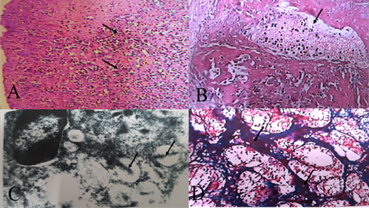
The acute endometritis was noticed in 6/33 animals (18.18%) and five of them were positive for bacteriological examination. Grossly, the uterine mucosa was severely congested and oedematous. Histologically, the lining epithelium of the endometrium was eroded and the endometrial stroma was highly infiltrated by neutrophils (Figure 1A). The stromal blood vessels were greatly congested and few of them showed fibrin thrombi (Figure 1B). Ultrastructural examination revealed several vacuoles inside neutrophils containing electron-dense rod-like structures (Figure 1C). The presence of bacteriaindicates recent infection and early stage of inflammation (Table 2).
Table 1: Pathological lesions in the uteri of buffaloes during different seasons.
Lesions
Total animals (340)
Seasons
Spring
Summer
Autumn
Winter
χ2 test
P
Endometritis
33
(9.71%)
2
28
3
0
70.44
<0.001
Metritis
11
(3.24%)
1
6
3
1
6.29
0.09
Endometrial hyperplasia
7
(2. 06%)
0
6
0
1
14.44
<0.01
Adenomyosis
5
(1.47%)
2
3
0
7
8.98
<0.05
Fibroma
3
(0.88%)
1
2
0
0
3.69
0.296
Perimetrial cysts
9
(2. 65%)
6
1
1
1
8.56
<0.05
Perimetrial nodules
10
(2.94%)
7
1
1
I
11.13
<0.05
Total
78
(22.94%)
19/78
47/78
8/78
11/78
χ2, Chi-square at (P<0.05) considered significant.
Table 2: Isolates of bacteria and its frequency in various types of inflammation of uteri.
Bacerial isolates
Endometritis
Metritis
Acute
Chronic
Acute
Chronic
Klebsiella oxytoca
2
4
-
1
Proteus vulgaris
-
2
-
-
Esherichia coli
1
-
-
-
Enterobacter aerogenes
1
1
-
-
Pseudomonas aeruginosa
1
-
-
-
Staphylococcus aureus
-
1
-
-
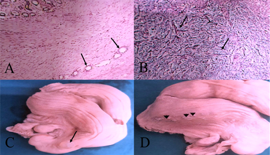
This finding was coincided with Bhadaniya et al. (2019). Chronic endometritis was noticed in 27/33 animals (81.82%) and one case was positive for bacteriological examination. Grossly, the uterine mucosa was corrugated and appeared dirty yellow in colour. Histologically, the endometrial surface was completely necrosed and the endometrial stroma was highly infiltrated by lymphoplasmacytic infiltration. Periglandular fibrosis (2-5 layers) was noticed in most cases (Figure 1D). The major chronic inflammatory cells were Lymphocytic type and/or plasma cells. Gonzalez et al. (1985) explained that the presence of these cells indicates the local production of sperm antibodies which interfere with conception. The presence of these types of cells may cause infertility and failure of implantation . Periglandular fibrosis was also seen in most cases of chronic endometritis and this finding was agreed with Salemi et al. (2020) in water buffaloes in Iran. Messier et al. (1984) clarified that perigandular fibrosis plays an important role in the reduction of uterine milk, early embryonic death, and consequently repeat breeder. Metritis was observed in 5 animals (1.47%) and all cases seemed to be chronic. Similar findings were observed in buffaloes and could be attributed to retained placenta or dystocia ; . Endometrial hyperplasia was recorded in 5 animals (1.47%). Similar finding was recorded by several authors ; . Cystic graafian follicles, ovarian neoplasms (granulosa cell tumors especially), and estrogens from plants are causes of endometrial hyperplasia in the cow . Adenomyosis was observed in 2 animals (0.58%). There were clusters of endometrial glands growing throughout the muscular layer of the uterus (Figure 2A). Similar percentage was recorded in buffloes . However, Alfaris and Fahid (2009) recorded a higher percentage (20%). Adenomyosis may arise as a malformation or due to hyperplastic overgrowth of the endometrium . Adenomyosis could be the cause of anoestrum and repeated breeding in buffaloes . Fibroma was observed in 3/340 animals (0.71%). There were bundles of collagen fibers running in different directions and mostly seen around blood vessels (Figure 2B). Perimetrial cysts were observed in 11 animals (3.24%). The cysts have thin walls containing a clear fluid and were firmly adhered to the outer uterine wall (Figure 2C). Similar cysts were recorded in buffaloes . McEntee (1990) explained that cysts developed in postpartum uterine involution in which the myometrium contracted leading to the infolding of perimetrium. Perimetrial nodules were observed in 10 animals (2.38%). The nodules appeared as single or multiple have red or greyish colour, and are of different sizes and shapes (0.4 in diameter) (Figure 2D).These nodules could be indicative of chronic reaction due to continuous irritation during an examination of animals with reproductive problems.
CONCLUSIONS AND RCOMMENDATIONS
The conclusion is that uterine disorders play a crucial role in female buffaloes infertility. It is highly recommended to use specific clinical parameters and biopsy techniques for early diagnosis in order to proceed with the appropriate treatment.
ACKNOWLEDGMENTS
This work was supported through the Annual Funding Track by the Deanship of Scientific Research, Vice Presidency for Graduate Studies and Scientific Research, King FaisalUniversity, Saudi Arabia (Grant number 5301).
NOVELTY STATEMENT
The present study tries to clarify that certain uterine disorders may play a major role in female buffalo infertility.
AUTHOR’S CONTRIBUION
MH designed the study. AJ examined histopathology. FH interpreted the data. AA prepared the manuscript. MH critically revised the manuscript. All the authors approved the final version of manuscript.
Ethical approval
The methods of the study were approved by the Research Ethics Committee at King Faisal University, Saudi Arabiba with Approval number KFU-REC-2022-NOV-ETHICS285.
Conflict of interest
The authors have declared no conflict of interest.
Figures
Figure 1:.
(A) Acute endometritis. Heavy infiltration with neutrophils in endometrium (arrows) HE X100. (B) Acute endometritis. Fibrin thrombi in endometrial blood vessels (arrow) HEX100. (C) Acute endometritis. Rod shape bacilli in phagosome of macrophage (arrow) (Uranyl acetate and lead citrate) X28000. (D) Chronic endometritis. Periglandular fibrosis of endometrial glands (3-5 layers of fibroblasts) (arrow) Masson’s trichrome stain X100.
Figure 2:.
(A) Uterus. Adenomyosis. Endometrial glands migrating throughout myometrium (arrows) HEX40. (B) Uterus, Fibroma. Fibroblastic proliferation and collagen fibers around blood vessels and in different directions (arrows) HEX40. (C) Uterus (right horn). The outer surface (perimetrium) has transparent cyst (1.7X1.4 cm (arrow). (D) Uterus. The outer surface (perimetrium) has greyish nodules of different sizes (arrowheads).
References
Alfaris AA, Fahid TA (2009). Adenomyosis in she buffaloes: Histopathological study. Basra J. Vet. Res., 8(1): 60-65. https://doi.org/10.33762/bvetr.2009.55221[CrossRef]
Azawi OI (2010). Uterine infection in buffalo cows. Buffalo Bull., 29: 154-117.
Azawi OI, Al-Sadi HI (2010).Uterine leiomyoma buffalo: A case report. Buffalo Bull., 29(2): 80-82.
Azawi OI (2013). Pathogenesis of postpartum metritis in buffaloes: A review. Buffalo Bull., 32: 1-11.
Babji BR, Reddy GA, Ambica G, Kumar SR (2020). Therapeutic Management of Endometritis in a Buffalo: A case report. Int. J. Curr. Microbiol. App. Sci., 9(4): 2292-2297. https://doi.org/10.20546/ijcmas.2020.904.274[CrossRef]
Bhadaniya AR, Prasad MC, Savsani HH, Kalaria VA,.Fefar DT, Mathpati BS (2019). Bacteriological and their antibiogram studies of endometritis in slaughtered buffloes. Buffalo Bull., 38(2):229-236.
Campanile G, Vecchio D, Zicarelli L, Neglia G, Di Palo R, Balestrieri A (2007). Strategies to reduce embryonic mortality in buffalo cows. Ital. J. Anim. Sci., 6(2): 680-683. https://doi.org/10.4081/ijas.2007.s2.680[CrossRef]
Dawar N, Shukla S, Garg UK, Karmore SK, Shrivastava N, Jatav GP, Yadav RS (2017). Gross and histopathological changes/aerobic bacterial plate count and cytological alterations in acute, subacute and chronic endometritis of bufflaoes (Bubalus bubalis). Buffalo Bull., 36(4): 661-671.
Elmetwally MH, Elshopakey GE, El-Desouky AM, Eldomany WB, Bazer FW (2020). Serum biochemical profile in buffalo endometritis and impact of treatment with PGF2α and intrauterine gentamicin infusion on postpartum reproductive performance. Trop. Anim. Health Prod., 52: 3697–3706. https://doi.org/10.1007/s11250-020-02406-3[CrossRef]
Elsayed DH, El-Azzazi FE, Mahmoud YK, Dessouki SM, Ahmed EA (2020). Subclinical endometritis and postpartum ovarian resumption in respect to TNF-α, IL-8 and CRP in Egyptian buffaloes. Anim. Reprod., 17(1): 1-13. https://doi.org/10.21451/1984-3143-AR2019-0027[CrossRef]
Gonzalez HE, Crowell WA, Caudle AB, and Thompson FN (1985). Morphometric studies of the bovine uterus: Microscopic lesions and retrospective reproductive history. Am. J. Vet. Res., 46(12): 2588-2595.
Hamouda M, Saber A, Al-Shabebi A (2020). Incidence of infertility in female buffaloes due to some reproductive disorders. Adv. Anim. Vet. Sci., 8(11): 1188-1193. https://doi.org/10.17582/journal.aavs/2020/8.11.1188.1193[Full text][CrossRef]
Hegde NG (2019). Buffalo husbandry for sustainable development of small farmers in India and other developing countries. Asian J. Anim. Vet. Adv., 3(1): 1-20.
Jubb KVF, Kennedy PC, Palmer N (1993). Pathology of domestic animals. 4th edn.Academic Press, San Diego, Calif, USA.
Kaneene JB, Miller R (1994). Epidemiological study of metritis in Michigan dairy cattle.Vet. Res., 25(2-3): 253-257.
Kumar P, Singh M, Sharma A (2022). Prevalence of different etiological factors responsible for causing infertility in buffaloes. Haryana Vet., 61(SI): 87-90.
Pasha TN, Hayat Z (2012). Present situation and future of buffalo production in Asia. J Anim.Plant Sci., 22(3): 250-256. https://www.researchgate.net/publication/291448956.
McEntee K (1990). Reproductive pathology of domestic mammales. 1st edn. Academic Press. Cornell University, Ithaca, New York.
Messier S, Higgins R, Couture Y, Morin M (1984). Comparison of swabbing and biopsy for studying the flora of the bovine uterus. Can. Vet. J., 25(7): 283-288.
Mittal D, Garg UK, Shukla S, Jatav GP (2009). Prevalence of different pathological affections of uterus in buffaloes (Bubalus bubalis) in the Malwa region of Madhya Pradesh. Buffalo Bull., 28(4): 215-217.
Mohyuddin SG, Riaz A, Ali SH, Ju XH (2019). Prevalence of repeat breeding in buffaloe and its effective possible future stratedies. Pak. J. Sci.,71(4): 213-219. https://www.researchgate.net/publication/339498498.
Parikh SS, Kavani FS, Parmar KH, Patbandha TK, Singh VK, Ahlawat AR, Kumar R (2022). Diagnostic and therapeutic management of subclinical endometritis in dairy bovine: A review. Anim. Reprod. Update, 2(2): 1-11. https://doi.org/10.48165/aru.2022.2.2.1[CrossRef]
Pereira J, Pinho R, Meneses R, Mendes V (2015). Cystic endometrial hyperplasia and hydrosalpinx associated with follicular cyst in cattle. Rev. Bras. Med. Vet., 37(1):1-6. https://bjvm.org.br/BJVM/article/view/357.
Ponraj P, Chang S, Rajesh NV, Veeraselvam M, Rajesh KD (2017). Prevalence of various pathological conditions in female buffaloes (Bubalus bubalis). Asian Pac. J. Reprod., 6(2): 58-67. https://doi.org/10.12980/apjr.6.20170203[CrossRef]
Quinn PJ, Carter ME, Markey B, Carter GR (1994). Clinical veterinary microbiology. Wolfe Publishing, Mosby-Year Book Europe Limited, London. GB, pp. 118–292.
Rashid MH, Qureshi ZI, Ali S,.Khan HH, Masood A, Nawaz H, Hayder S, Hassan MM, Waqas MS (2020). Prevalence of mycotic endometritis in buffaloes and mares maintained under different managemental conditions in district Faisalabad. Pak. Vet. J., 41(3): 414-418.
Sachan,V., Kumar, R.K. Gupta, Saxena A (2018). Uterine leiomyoma in A cow. A case report. Indian J. Anim. Hlth., 57(2): 235-238. https://doi.org/10.36062/ijah.57.2.2018.235-238[CrossRef]
Salemi Z, Rezaie A, Nejad SG, Mohammadian B (2020). Histopathological and cytological analyses of endometrium in water buffaloes (Bubalus bubalis) to detect estrus and endometritis. Vet. Res. Forum, 11(4): 409-414.
Sarsawat CS, Purohit GN (2016). Repeat breeding: Incidence, risk factors and diagnosis in buffaloes. Asian Pac. J. Reprod., 5(2): 87-95. https://doi.org/10.1016/j.apjr.2016.01.001[CrossRef]
Shokri H, Yadollahi M (2017). Isolation and identification of fungal microbiota from genital tract of ewes. Rev. Med. Vet., 168: 81-86.
Singh H, Brar P, Honparkhe M, Arora AK, Dhindsa SS (2020). Subclinical endometritis in estrual buffaloes: diagnosis, prevalence and impact on reproductive performance. Trop. Anim. Health Prod., 52: 357–363. https://doi.org/10.1007/s11250-019-02022-w[CrossRef]
Suvarna SK, Layton C, Bancroft JD (2019). Theory and practice of histological techniques. 8th edn. Elsevier.
Parmar D (2018). Clinical management of retention of placenta in murrah graded buffalo with cervico-vaginal postpartum prolapse. J. Entomol. Zool. Stud., 6(6): 603-605.
Saxena G, Rani S, Danodiai HK, Purohit GN (2006). Pathological conditions in genital tract of female buffaloes (Bubalus bubalis). Pak. Vet. J., 26(2): 91-93.
Zachary JF (2017). Pathologic basis of veterinary disease. 6th edn. St. Louis, Missouri, Elsevier.