Research Article · Volume 10, Issue 9, 31 August 2022 · Open Access
Infection of Bone Marrow-Derived Mesenchymal Stem Cells with Virulent Newcastle Disease Virus Maximizes Cytokine Production: A Step Toward vNDV Immunotherapy
· · · · ·
Affiliations & Notes ⌄
1Biotechnology Department, Faculty of Agriculture, University of Al-Azhar, Cairo, Egypt
2Animal Biotechnology Department, Genetic Engineering and Biotechnology Research Institute, University of Sadat City, Egypt
3Poultry Viral Vaccines Production and Research Department, Veterinary Serum & Vaccine Research Institute, Agriculture Research Center, Cairo, Egypt
4Department of Virology, Faculty of Veterinary Medicine Cairo University, Cairo, Egypt. *Correspondence | Basem M Ahmed, Department of Virology, Faculty of Veterinary Medicine Cairo University, Cairo, Egypt
From the beginning, vNDV infection caused the disappearance of the limb stem cells (Fig. 4).
Not all cells died; there were some highly resistant MSCs-CD105 able to grow under infected vNDV conditions, which showed elongated and fibroblast shape and grew although vNDV is present (fig.
Stem cells have an immune effect on the virus, where keeping normal cellular organelles after infection.
The current results pointed for the first time to stem cells as a thrilling solution against NDV infection.
Selected from the article's own text.
Abstract
Newcastle Disease (ND) vaccines are being used for more than 7 decades, the disease is still a major challenge for poultry industry both locally and internationally. ND frequently emerges in highly vaccinated flocks causing high economic losses without specific treatment. Mesenchymal stem cells (MSCs) are a group of pluripotent cells with multiple biotechnology applications, including but not limited to tissue genesis, tissue repair, hematopoiesis, and immune modulation. Therapeutic strategies based on the usage of stem cells includes the cells either themselves or their secretions (secretome), which has recently shown ability to inhibit SARS-CoV2 replication in-vitro. In this study, MSCs were prepared from the bone marrow of native Egyptian Fayoumi chicken. The MSC with the surface marker CD105 (CD105+) were magnetically separated and infected with virulent Newcastle disease virus (vNDV). The virus-induced multiple changes at the cellular and ultrastructural level in the infected cells, and it was able to maximize the production of interferon-gamma (IFNγ) and interleukin 2 (IL2), interleukin 6 (IL6) and interleukin 12 (IL12). In conclusion, our data represent a preliminary step in vNDV immunotherapy where MSCs media could be used for the treatment of vNDV in infected flocks.
The poultry industry represents a cornerstone in the Egyptian economy and a major source of animal protein production. It is associated with a large amount of national consumption that represents over 600 million dollars (FAO, 2013). Excessive usage of antibiotics in poultry has led to the emergence of highly resistant microbial pathogens . So, there is an urgent need to develop environmentally friendly materials and complementary treatments such as natural immune modifying cytokines .
Improvement of meat and egg quality, and disease resistance to viral diseases as Newcastle disease in the Egyptian chicken traits with traditional breeding programs is very difficult, expensive, and time-consuming. Stem cells represent a potential medicine with multiple applications . In chicken, stem cells were found to be useful for modulating male fertility by transplantation of dispersed testicular cells . Chicken-induced pluripotent stem cell was tolerant to NDV . For other approaches, stem cell therapy provides a novel tool for poultry production in quality and quantity traits . Moreover, a new suspension chicken-induced pluripotent cell line was used to produce NDV vaccine .
NDV or Avian orthoavulavirus-1 is the type species of genus Orthoavulavirus, subfamily Avulavirinae, family Paramyxoviridae. it has a 15.2 kb negative sense non-segmented RNA genome (ICTV, 2022). NDV can be isolated and propagated in embryonated chicken eggs and antigenically identified by hemagglutination (HA) and hemagglutination inhibition (HI) assays (OIE, 2021; . There are many sources for MSCs such as bone marrow, cartilage, and adipose tissue ; . Stem cells have several properties and they have varying abilities of differentiation and therapeutic properties ; . The comparative efficacy among distinguished resources in MCSs differentiation was subjected to Egyptian poultry for productivity level and transplantation methods of MSCs . Recently, ND vaccines were prepared in suspension chicken induced pluripotent cell line for production . Especially, chicken embryonic stem cells (cESCs) and chicken induced pluripotent stem cells (ciPSCs) are used for pharmaceutical protein production . On other side, chicken are possible models for studying hematopoiesis and hematopoietic stem cells production . Chicken stem cells can be isolated and characterized by centrifugation and magnetic cell sorting techniques . Moreover, avian fertile eggs and cell lines in biopharmaceuticals and viral vaccine manufacturing have been administered in the biotechnology industrial field .
Chicken interferon-gamma (IFN-γ) has a pleotropic function and is primarily secreted by T lymphocytes and NK cells . IFN-γ acts as a macrophage activating factor (MAF) with antiviral activity and represented as Th1 immune responses. It is a major modulator in birds´ macrophage activation and can prevent the virus replication through supporting the antigen presentation mechanism and destroying the intracellular pathogens. IFN-γ enhanced the immunity by cell intermediated and supported the innate and adaptive immune responses ; OIE, 2021). Interleukin 2 (IL-2) plays a role in fighting the viral diseases by natural killer cells (NK) and cytotoxic T lymphocytes activation to inhibit the virus replication. In addition, it can induce cellular immunity by enhancing IFN-γ secretion. In general, cytokines play an immune role in promoting the production of antibodies and increasing the degree of sensitivity of B cells . Interleukin-6 (IL-6) is secreted by fibroblasts and immune cells as T and B lymphocytes . The immunomodulatory effect of IL-6 is through the differentiating of B cells through its action as B cells growth factor and immunoglobulin regulator . Interleukin-12 (IL–12) is used as an antiviral enhancer and treatment. Recently, it has been proven that IL-12 enhances the cellular immune response through its actions on the NK cells and T cells to produce IFN-γ and activate T lymphocytes .
The immunological effects of neural stem cells on laryngotracheitis virus (ILTV) strains were reported . Stem cells could provide a new approach for poultry treatment and viral therapy as novel tools associated with genetic engineering .
In this study, CD105+ MSC stem cells were isolated from bone marrow of 7-11 wk old-fayoumi chicken then the cells were infected with a cell adapted virulent NDV. The infection process was monitored at the cellular level and the effect of vNDV infection on cytokine production was assessed.
METHODS
Ethics statement
All experiments were conducted in accordance with the ethical research code of Research Ethics Committee in Al-Azhar University, Cairo, Egypt. (Permit no. 2019–07)
Viruses and Cells
Locally isolated field strain genotype VII virulent NDV/Ck/Giza/Egypt/MR0/2012 (vNDV), GenBank accession number JX173098, was kindly provided by Prof. Ausama Yousif, Department of Virology, faculty of veterinary medicine, Cairo University, Egypt. According to OIE procedures, the virus was propagated and titrated in SPF ECEs eggs (OIE, 2021;. The collected allantoic fluid was verified using HA followed by HI using NDV antibodies, then its virulence was assessed by mean death time (MDT) . The collected fluid was further purified and concentrated using the hemagglutination-elution method and evaluated using plate HA assay (OIE, 2021).
Chicken embryo fibroblast (CEF) was prepared from 10 days old SPF embryos (SPF farm, Kom-Oshim, Egypt) with the traditional trypsin-based method ;. Log phase cells were used to adapt vNDV by passaging on cell culture. vNDV was titrated on CEF as described by Wambura et al. (2006), and the titer was calculated according to Reed & Muench, (1938).
Bone marrow-derived mesenchymal stem cells (BM-MSC)
Animals: Twenty 7-11 wk old Fayoumi commercial roasters and hens (n= 10; each) purchased from EL-Azab complementary poultry project-upper Egypt development authority (Fayoum- Egypt) were used to prepare bone marrow culture and isolate the mesenchymal stem cells (MSC). Birds were quarantined for 2 weeks at the genetic engineering and biotechnology institute (GEBI), University of Sadat City, Egypt. Then they were dissected for their femurs and tibiae.
Bone marrow culture and mononuclear cell separation: Mesenchymal stem cells were separated from chicken bone marrow as previously described with some modifications . Briefly, femurs and tibiae of dissected birds were extracted, their cristae were removed, and bone marrow cells were collected by flushing the pre-ostium cavity with sterile 26G syringe containing phosphate buffer saline (PBS) with 10% EDTA to avoid bone marrow coagulation. Cells were disaggregated by gentle mixing and filtrated through 30 μm pre-separation filters (Miltyni Biotech, Germany). Nine ml of the collected cells were layered on 3ml Ficoll®-Paque Premium (Merck, USA) in 15 ml falcon tubes and centrifuged at 1500 rpm for 20 min at 16 °C. The supernatant was aspirated off, and buffy coat leukocytes were collected from the interphase, transferred to a new 15 ml falcon tube, and washed twice for 5 mins in PBS. Isolated cells were seeded at 1*106 cell/ 75 CM2 vented tissue culture flasks and incubated at 37oC and 5% CO2, medium was changed after 24hr of incubation while non-adherent cells were discarded completely after 3 days of incubation. Adherent cells of 80%confluency were detached by 0.25% trypsin solution and subjected to magnetic separation.
CD105+ MSCs Magnetic Separation and Cryopreservation: CD105 MultiSort Kit (PE), mouse in combination with the Anti-Sca-1 MicroBead Kit (FITC) (Miltenyi Biotec, Germany) were used to positively select CD105+ mesenchymal progenitor cells from proliferated bone marrow mononuclear cells. The magnetically labeled cells have been separated as positively selected columns at the magnetic field of autoMACS® Pro-Separator (Miltenyi Biotec, Germany). The viability of separated cells was re-assessed by 0.4 trypan blue staining. Most viable cells were diluted in freezing media containing 10% DMSO as a cryoprotectant to 1*10^5 cells/ ml and frozen slowly for 24 hours in -20oC freezer. Finally, cells were transferred to - 80oC for long-term preservation. Cells were seeded in 75 CM2 culture vessels with 1*10^5 cells/ml and incubated at 37°C in 5% Co2 for 2 weeks; the growth medium was renewed every 2 days with continuous monitoring.
CD105+ MSCs infection with vNDV: Confluent monolayer CD105+ MSC cells were detached using 0.25% trypsin and seeded in 96-well microtiter corning costar plate (Fisher Scientific, USA) with 1*103 cells/ well and incubated at 37 C in 5% Co2 incubator for 24hr. CEF-adapted vNDV was diluted to 1*104 TCID50/ml in DMEM, growth medium was aspirated and replaced with 100µl/ well from the prepared virus (MOI=1). vNDV adsorbed to 90%+ confluent MSC for 1 hr, then the remaining infectious fluid was discarded, cells were washed twice with PBS. Then 200µl of DMEM containing 2% FCS was added to the wells. Cells were incubated at 37oC and 5% Co2 for 24 hours. To obtain complete CPE, cells were infected multiple times with CEF-adapted vNDV at 24hr intervals with the same MOI. CPE development was followed up using an inverted microscope, stereomicroscope, and transmission electron microscope. The extent of CPE is scored as (0 = no CPE or less than 10% of the cell monolayer killed), (1 = 25% cells killed), (2 = 50% cells killed), (3 = 75 % cells killed) and (4 = CPE or 95% cells killed), once complete CPE was achieved. Medium was aspirated from infected and control wells for cytokine quantification.
Enzyme-linked immunosorbent assay (ELISA): Following CPE appearance in the infected CD105+ MSCs, the conditioned medium was aspirated from infected and control wells. The concentrations of IFN-γ, IL-2, IL-6, and IL-12 in infected MSC media were evaluated using commercial ELISA kits compared to control non-infected MSCs. ELISA kits, providers and countries of origin used to assess the levels of different immune mediators secreted by MSCs: interferon gamma (Fine Test, Wuhan, China), Interleukin 2 (R&D Systems, Minneapolis, USA), Interleukin 6 (Elabscience, Biotechnology, USA), Chicken Interleukin 12 p40 (Biocompare, USA).
Transmission electron microscopy (TEM) for CD105+ MSCs: The ultrastructure of infected CD105+ MSCs was examined using TEM according to (Amin et al., 2018,2020). Briefly, cells were centrifugated at 2000×g for 10 minutes. Pelleted cells were fixed in 3% glutaraldehyde in 0.1M sodium cacodylate buffer (pH 7.0) at room temperature, rinsed in the same buffer, and fixed in 1% osmium tetroxide. Then, cells were dehydrated in ascending concentrations of absolute ethanol for 30 minutes. Ultrathin sections were collected on copper grids, stained twice with uranyl acetate and lead citrate and stained sections were examined at 80 kV using a JEOL-JEM 1010 transmission electron microscope in the Regional Center for Mycology and Biotechnology (RCMB), Al-Azhar University, Cairo, Egypt.
Statistical analysis
One way analysis of variance (ANOVA) of the IBM SPSS package was used to analyze the means of different cell groups. The significant difference (p-value) was ≥ 0.05.
RESULTS
Virus titer and pathogenicity
Virulent Newcastle disease virus (vNDV) used in the present study was identified using NDV antisera by HI assay. The virus was able to cause embryonic death within 44-48 hours post-inoculation indicating its velogenic nature. Following virus titration, 8.2 log10 EID50/ml was obtained. Similarly, the CEF cells produced a virus titer of 8-log10 TCID50/ml.
CEF and vNDV adaptation
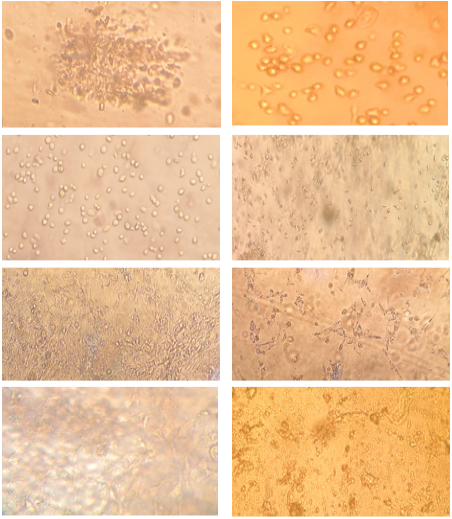
Confluent monolayer CEF cells (fig. 2) were split using 0.25% trypsin, and inoculated with vNDV for adaptation. Three successive passages were conducted to adapt vNDV on CEF that induced full CPE within 24 hr. Then, the adapted vNDV was aliquoted and stored at -80 oC until use.
BM-MSCs proliferation and maturation
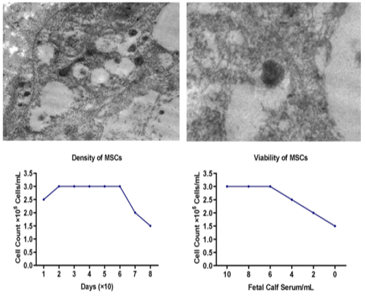
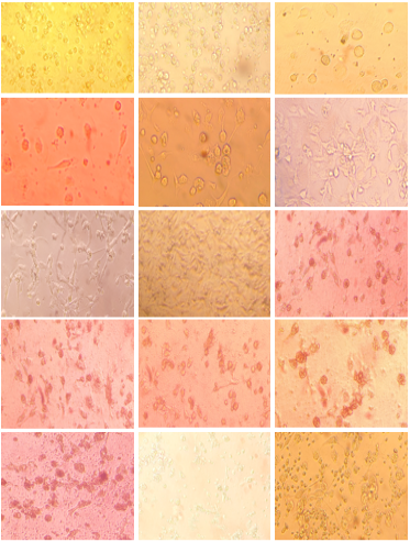
Separated mononuclear cells from bone marrow were cultured in DMEM with 5% FCS (fig. 1). A week later, cells began to proliferate and increase in density. After another week, cells started to elongate (fig. 3C) and form a monolayer by the third week. Cell morphology was spindle or fibroblastoid in shape (fig. 3D). After four weeks, the number of circular non-elongated cells reduced (fig. 3E). By the end of the fifth week, fully elongated mononuclear cells were obtained (fig. 3F), reached a fixed density of 3*105/ ml after 3rd week of incubation and maintained their viability for 70 days before declining to 1.5*105. By 90 days (fig. 1C), cells began to change in shape and enter the stage of apoptosis. Fetal calf serum (FCS) levels lower than 5% failed to maintain maximum cell viability and count for up to 7 successive passages (fig. 1D), while higher FCS levels were better.
CD105+ MSC magnetic separation and vNDV infection
Highly viable and differentiated CD105+ mesenchymal stem cells were obtained after magnetic separation, then were seeded with a density 3*103 cells/ 75 CM2 culture vessel. CEF adapted vNDV was used to infect the CD105+ cells, where five rounds of infection (passages) were applied to induce complete CPE and high cytokine production.
Within the first passage, infected MSCs-CD105 showed loss of elongation and degradation into colonies attached to the surface of tissue culture flask, CPE in infected cells
caused lysis and morphological changes. MSCs - CD105 appeared as rounded cells and fusion of cells led to some cells containing many nuclei (syncytium) scored (1 CPE) that constituted 25% of studied plate area (fig. 3I). This is an immune reaction of cells to the virus to limit its spread from infected cells to protect uninfected cells . In the second passage, MSCs started to die through changes in cell morphology, these changes in the shape of virus-infected cells are considered one of the most important characteristics of virus infection. They are used in laboratory diagnosis of the virus and the CPEs scored 2 that constituted 50% of studied plate area (fig. 3J).
In the third passage, the formation of dendritic-shaped cells was observed in MSCs-CD105 monolayer, this effect was shown under inverted microscopy and the CPEs scoring 3 that constituted 75% of studied plate area (fig. 3K), moreover confirmed by TEM assay. In the fourth passage, a gradual change appeared in the internal parts of the cell, such as the size and shape of the nucleus on the monolayer CPEs scored 3 that constituted 85% of studied plate area (fig. 3L). In the fifth passage, a complete cytopathic effect (CPE) of vNDV on CEF has been shown as plaque formation in MSCs - CD105 monolayer, the CPEs scored 4 that constituted 95% of studied plate area (fig. 3M) (Table 1).
Table 1: CPEs scoring in several passages of infected MSCs-CD105 by vNDVs.
CPE score
Cell death percentage
Figure number
First passage
1
damage 25%
Figure 3I
Second passage
2
damage 50%
Figure 3J
Third passage
3
damage 75%
Figure 3K
Fourth passage
3
damage 85%
Figure 3L
Fifth passage
4
damage 95%
Figure 3M
Stereomicroscope and TEM results
Stereomicroscope analysis enabled visualization of cellular
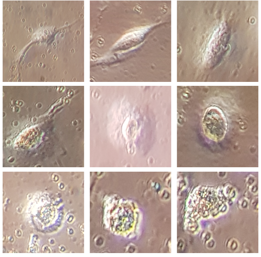
changes during the infection process at the level of cell surface. From the beginning, vNDV infection caused the disappearance of the limb stem cells (Fig. 4). The cell began to increase in size and inflate in the middle area until it became circular in shape, followed by the appearance of small bumps on the outer edges, which led to increase in the cell size and enlargement. The appearance of zigzags on the surface of the cell after shiny surface was attributed to virus replication in the cell, which led to increase in cell size until cell explosion. The virus has been released so it can infect other cells.
Stereomicroscope examination showed that vNDV was able to inhabit MSCs-CD105. Not all cells died; there were some highly resistant MSCs-CD105 able to grow under infected vNDV conditions, which showed elongated and fibroblast shape and grew although vNDV is present (fig. 3N) (fig. 3O).
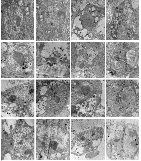
TEM enabled the visualization of internal cellular changes during vNDV infection. In general, there was a disruption in the cellular membranes, and the appearance of vacuoles in most parts of the cell and destruction in general cell organelles. Changes in the shape of the nucleus were also evident; its size was enlarged after infection with the virus by constriction shapes in nucleus and cytoplasm organelles. Moreover, nuclear changes appeared during cell division. This effect is caused by the virus attacking in the cytoplasm or the nucleus as phagocytes. In addition to cell fibrosis by formation of fibers in the string forms in more than one sector. Apoptotic cells containing nuclear fragments were also noticed. The ultra-structural cellular alterations were observed as dilatation of endoplasmic reticulum, damaged mitochondria, dilatation of nuclear envelope, cytoplasmic vacuolization, and nuclear attribution (Fig. 5).
Evaluation of cytokine production
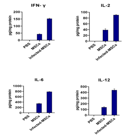
Compared to non-infected cells, and PBS as a blank. Infected MSCs -CD105 cells produced approximately double to triple amount of the cytokines IFN-γ, IL-2, IL-6, and IL-12. The concentrations had increased significantly compared to the normal MSCs-CD105 (Table 2) (Fig. 6).
The difference between negative control (PBS), positive control (MSCs-CD105, uninfected) and treated group was determined by one-way ANOVA at statistic software SPSS. This high percentage observed in infected-MSCs-CD105 compared with uninfected MSCs-CD105 was due to virus infection that stimulated MSCs - CD105 to increase secretion as an immunomodulation effect. These have been observed varyingly in the cytokines secreted from MSCs
Table 2: Cytokine production levels of IFN-γ, IL-2, IL-6 and IL-12 by PBS, bone marrow mesenchymal stem cells (BM-MSCs-CD105) and infected (BM-MSCs-CD105) by vNDVs, measured by ELISA, the data shown are averages ± standard deviation, letters indicate significant differences, one way ANOVA, P<0.05.
IFN- γ
pg/mg protein
IL-2
pg/mg protein
IL-6
pg/mg protein
IL-12
pg/mg protein
PBS
0c
0c
0c
0c
MSCs
42.95 ± 2.55b
38.76 ± 4.29b
344.05 ± 15.71b
138.67 ± 9.72b
Infected-MSCs
152.51 ± 3.29a
90.46 ± 1.66a
786.28 ± 5.95a
439.79 ± 22.47a
as highest value for IL-6 and relatively lower elevation in IL-2 in untreated cells and induced cells. Nevertheless, IL-
12 and IFN-γ were secreted a moderately levels in untreated cells and induced cells.
Cytokine levels increased after virus inoculation on MSCs-CD105. This in-vitro study revealed that induced bone marrow mesenchymal stem cells could produce high levels of interleukin 6 and 12 and a medium amount of interferon-gamma, and a small amount of interleukin 2 .
DISCUSSION
Stem cells are one of the biotechnology tools that can be applied to treat incurable diseases. According to virus effect on stem cells, cellular changes can be diagnosed. Stem cells have an immune effect on the virus, where keeping normal cellular organelles after infection. Different types of primitive cells were extracted from poultry and the embryonic cells were most common for virus propagation such as CEF. Multiple Avian orthoavulavirus-1 genotypes are present and NDV genotype VII is one of the velogenic strains in Egypt . Despite vaccination programs, the virus still causes many obstacles and challenges in the Egyptian poultry industry in general and no doubt that the NDV is one of the most dangerous and destructive viruses. Currently, it’s on the top list of OIE diseases and is considered one of the viruses discovered early in Egypt. The activity and identity of viruses can be measured using HA and HI tests for avian paramyxoviruses (OIE, 2012). The tests showed that the vNDV titer used in the infection had reached 108.2 EID 50/ml. The genotype VII strains are widespread in Egypt in poultry farms, although the vaccination efforts exist in high quality and accuracy . Currently, the use and role of the electron microscope plays a role in detection and classification based on the structure and morphology of viruses for understanding their life cycle and pathogenesis .
Chicken Interferon gamma (chIFNγ) is a natural compound that helps improve the vaccine effectiveness. It plays an additional role with antibody responses to the hepatitis virus and an additional role in antibody and cellular responses to enhanced in the protection of vNDV . ChIFNγ can be used in treatment based on its immune role as another direction of traditional vaccination methods. It was observed that the expression of interferon increased with the increase of the protective role of the virulent NDV in poultry, considering that this cytokine possesses an endogenous antiviral activity .
Transmission electron microscopy (TEM) was an imaging technique that allowed viruses visualization and discovery using high-resolution magnification . It showed that CEFs have immune proteins at the levels in vitro and in vivo by pro-inflammatory cytokines in NDV infected spleen and lung tissues . CEF also showed an immune response to the NDV by secreting high levels of interferon series, which guarantees the protection against the virus by immune modulation in poultry . The CPEs showed shapes in Vero cell line through virus infection as changed in monolayer conformation . Furthermore hematopoietic stem cells separated from the bone marrow to give antimicrobial resistance in gram-negative rods .
The examination showed the cellular changes induced by the virus, whether at the nuclear or cytoplasmic level, by changing the shape, number and shape of the nucleus and the obvious damage to different parts of the cytoplasm such as mitochondria, Golgi bodies, endoplasmic reticulum and plasma membrane, indicate the toxic impact of virus and the immune effect of the cell and its ability to respond to the virus and the ability of cells to return to normal construction by the capabilities of cells on the immune property and resistance to the virus.
The results showed that the serological tests have proved that stem cells have an immunological and therapeutic effect on viral diseases that affect poultry, especially chickens. The study showed an increase in the production of cytokines in infected CD105+ MSC with vNDV, where the proportion of IFN-γ, IL-2, IL-6, and IL-12 increased compared to the uninfected cells, due to a noticeable increase in protecting the infected cells, which showed by CPE distinguished changes in the formation to fibroblast and elongation shape, stopping the virus replications inside the cells by the immunological role of interferon and interleukins, this has been proven by the ability of induced stem cell with antiviral properties . For example, pluripotent cell line had an induced protection against NDV as protective role ;. Human cytomegalovirus exerts its immunity response through stimulation of CD4 T cell in vaccinated transgenic mice , in addition to induced pluripotent stem cells showed more strong resistance to NDV . Moreover, these stem cells had antimicrobial effect through secretion of peptides and antimicrobial proteins . Besides, stem cells enhance the efficiency of the immune system responses to systemic therapy .
Thin tissue section and ultrastructure have been considered the most important techniques for examining virus effect on the host cell. The recommended research has shown that vNDV infection has reduced poultry production in several levels up to death of the whole herd. Moreover, the vNDV biological and immune response represented in cytokines secretion as interleukin 2, interleukin 6 and interleukin 12, so that interferon-gamma stimulated by an immune response to defend and protect cells against virus infection, this is due to the cellular immune effect of cytokines interleukin and interferon which in turn leads to the immune effect of veterinary vaccines, without the need to know the type and composition of the virus and its sequences overcoming the problem of mutations within viruses, as it is possible to potential use compounds from the immune effect of stem cells in against possible infections with any strain of vNDVs.
Vaccines could be also produced through pluripotent cell line to combat the NDV in poultry and stop reliance on embryonated hen’s eggs . Moreover, mice-MSCs enhanced innate immune responses when infected by bacterial for a long duration . It was found that interleukin-2 is a safe substance to improve the poultry immunity with the vaccination against the NDV and immune response by cytokine production as IFN-γ, IL-2 and IL-12 in peripheral blood mononuclear cells by NDV induction . The immune effect of IL-2 has been scientifically proven by reducing the number of NDV by decreasing replication and attenuating pathogenicity of virus total number in in-vivo infection in poultry ; . Finally, according to cytological examination results using inverted microscope, stereo microscope, and transmission electron microscopy, serological tests as enzyme linked immunosorbent assay; had provided an evidence of the cytopathic effect of bone marrow MSCs-CD105 infected with vNDV in poultry and its induction to produce higher cytokines like Il2, Il6, Il12, and IFN-γ.
CONCLUSION
The current results pointed for the first time to stem cells as a thrilling solution against NDV infection. The use of MSCs and or its secretions (secretome) to protect poultry against disastrous commercial effects of virus infection, through several ways by secreting immune modulating chemicals as interferon-gamma and various interleukins, which can be used in the poultry industry after further in vivo and clinical levels confirmation.
ACKNOWLEDGMENTS
The presented article is the PhD thesis work of Tarek A. Wrshana. Authors are thankful to all colleagues in Al-Azhar University Faculty of Agriculture, Cairo University Department of Virology, El-Sadat City Genetic Engineering and Biotechnology Research Institute, and the Veterinary Sera and Vaccines Research Institute Newcastle Disease Department, for their priceless help during this work. Authors are thankful to Dr. Mohammed Rohaim, for critically editing and improving this manuscript.
conflict of interest
Authors declare that they have no competing interests.
novelty statement
This study sheds a beam of light on the possible use of conditioned mesenchymal stem cell secretome in combating virulent newcastle disease virus in poultry, the concept that can be refined and expanded to other viruses.
authors contribution
Bahgat Elfiky conceptualized the study, provided preliminary plan, supervised the work, and revised the initial draft. Yousry Dowidar and Ali El-Rify approved the concept, refined the plan, supervised the work, and revised initial draft. Tarek wrshana executed the experimental work and drafted the mansucript. Walaa El-Sayed and Basem Ahmed, provided some materials, participated in experiment execution and revised initial draft. Basem Ahmed anlayzed the data and prepared submission files. all authors have approved the final manuscript.
Figures
Figure 1. Figure 1.
A & B) NDV by TEM examination by thin and ultrastructure examinations in 15000 nm and 3000nm magnification power, C) the density of stem cells with the progression in time, D) viability of stem cells and the effect of FCS on MSCs-CD105.
Figure 2.
A) First trypsinization, B) second trypsinization, C) third separation shows cell elongation, D) CEFs, E) CEFs maturation, F) CEFs propagation, G) CEFs shows a full cells elongation and forming mono layer, H) CEFs after virus inoculation that from infected CEFs cell, magnification power equal 40x.
Figure 3.
A) Mononuclear cells after separation, B) mononuclear cells after one week shows cell proliferation and cell shape was rounded. C) mononuclear cells after two weeks from separation shows the onset of cell elongation and beginning of the appearance of the bumps entertainment, D) mononuclear cells after three weeks from separation shows a full cells elongation and forming mono layer from MSCs cell which attached on surface and cells was spindle or fibroblastoid, E) mononuclear cells after four weeks from separation shows a full cells elongation and forming monolayer from MSCs cell which attached on surface and cells was spindle or fibroblastoid, F) mononuclear cells after five weeks from separation shows a full cells elongation and forming mono layer from MSCs cell which complete attached and conformation shapes on surface and cells was spindle or fibroblastoid. G) full differentiation to MSCs-CD-105 showed by elongation and connective conjunction between cells like a colonies attached on down surface to adhesive propriety in tissue culture flask, H) Un infected MSCs-CD105 showed by elongation and connective or conjunction between cells like a colonies attached on down surface to adhesive propriety in tissue culture flask with normal growth without any CPEs in MSCs-CD105, I) after inoculation of the virus and attacking cells for beginning of the emergence of the immune and internal response to the virus as first passage infected MSCs-CD105 by NDVs, J) attacking stem cells, and the emergence of giant cells caused by the virus as second passage at infected MSCs-CD105 by NDVs, K) the middle stage of infection with the virus and the spread of infection to most cells as third passages and formation of dendritic shaped cells in MSCs-CD105, L) tourth passage showed complete cythopatic effect (CPE) plaque formation in MSCs-CD105 monolayer, M) the final stage of infection with the virus and the deterioration of the cytological and physiological status of stem cells as fifth passages lysis and degradation, N) restore validity, O) MSCs-CD105 after death by inhibition with NDVs and restore proliferation to generate new resistances cells to NDVs, magnification power equal 40x.
Figure 4.
Micro-photos for vNDV effect on MSCs-CD105 during inhibition process, A) single cell shown, a complete external membrane without any damage without virus treatment, B) the beginning of a decrease in the terminal arms of cells immediately after treatment with the virus, C) the beginning of cells are vesicles, D) nods appearing on the outside of the cells, E) swelling and increase in cell size and giant cell formation, F) increasing in swelling, G) beginning in explosion, H) unstable cell, I) cell lysis and degradation, magnification power equal 100x.
Figure 5.
Micrographs for TEM examination by thin and ultrastructure examinations in infected MSCs-CD105 with vNDVs after 24 hours in 15000 nm and 3000nm magnification power.
Figure 6.
Means of cytokine production levels of IFN-γ, IL-2, IL-6 and IL-12 and evaluated by ELISA in PBS, bone marrow mesenchymal stem cells (BM-MSCs-CD105) and infected BM-MSCs-CD105 by vNDV.
References
Abdel-Latif S. A. A., Atef A., Abdel-Aleem A. M. A., Dahshan A. M., Ali A (2020). Characterization of avian influenza H9N2 and newcastle disease virus isolated from vaccinated chickens in upper egypt. J. Vet. Med. Res., 27(1): 90-108. https://doi.org/10.21608/JVMR.2020.88897[CrossRef]
Agyare C., Boamah V. E., Zumbi C. N., Osei F. B. (2018). Antibiotic use in poultry production and its effects on bacterial resistance. Antimicrobial Resist., A Global Threat. Intech Open. https://doi.org/10.5772/intechopen.79371[CrossRef]
Ahamed T., Hossain K. M., Billah M. M., Islam K. M. D., Ahasan M. M., Islam M. E. (2004). Adaptation of Newcastle disease virus (NDV) on vero cell line. International J. Poult. Sci., 3(3): 153-156. https://doi.org/10.3923/ijps.2004.153.156[CrossRef]
Al-Dhamin Z., Liu L., Li D., Zhang S., Dong S., Nan Y. (2020). Therapeutic Efficiency of Bone Marrow Derived Mesenchymal Stem Cells for Liver Fibrosis: A Systematic Review of in vivo Studies. World J. Gastroenterol., 26(47): 7444-7469. https://doi.org/10.3748/wjg.v26.i47.7444[CrossRef]
Alcayaga-Miranda F., Cuenca J., Khoury M. (2017). Antimicrobial activity of mesenchymal stem cells: Current status and new perspectives of antimicrobial peptide-based therapies. Front. Immunol., 8(1)(Article 339): 1-15. https://doi.org/10.3389/fimmu.2017.00339[CrossRef]
Alexander, D. J. (2017). Newcastle disease, other avian paramyxoviruses. Diseases of Poultry, Thirteenth Edition. doi:https://doi.org/10.1002/9781119421481.ch3[CrossRef]
Amin B. H., Ahmed H. Y., Abd El-Aziz M. M. (2018). In Vitro Anticancer Activity Of Fungal Secondary Metabolites Of Stemphylium Lycopersici. Egyptian J. Microbiol., 50: 80-97.
Amin B. H., Abou‐Dobara M. I., Diab M. A., Gomaa E. A., El‐Mogazy M. A., El‐Sonbati A. Z., El‐Ghareib M. S., Hussien M. A., Salama H. M. (2020). Synthesis, characterization, and biological investigation of new mixed‐ligand complexes. J. Appl. Organometallic Chem., 34 (8). doi:https://doi.org/10.1002/aoc.5689[CrossRef]
Averbuch D., Tridello G., Hoek J., Mikulska M., Akan H., Yanez S. L. (2017). Antimicrobial resistance in gram-negative rods causing bacteremia in hematopoietic stem cell transplant recipients: Intercontinental prospective study of the infectious diseases working party of the european bone marrow transplantation group. Clin. Infect. Dis., 65(11):1819-1828. doi:https://doi.org/10.1093/cid/cix646[CrossRef]
Bouffi C., Bony C., Courties G., Jorgensen C., Noël D. (2010). IL-6-dependent PGE2 secretion by mesenchymal stem cells inhibits local inflammation in experimental arthritis. PLoS One.., 5(12): e14247. doi:https://doi.org/10.1371/journal.pone.0014247[CrossRef]
Chen X., Liu S., Goraya M., Maarouf M., Huang S., Chen J. (2018). Host immune response to influenza a virus infection. Front. Immunol., 9(05 March), 320. https://doi.org/10.3389/fimmu.2018.00320[CrossRef]
Dhupkar P., Gordon N. (2017). Interleukin-2: Old and new approaches to enhance immune-therapeutic efficacy. Adv. Experimen. Med. Biol., 995(2): 33-51. https://doi.org/10.1007/978-3-319-53156-4_2[CrossRef]
FAO. (2013). FAO Statistical Yearbook: World food and agriculture. Food and Agriculture Organization of the United Nations, Rome, 2013. doi:http://www.fao.org/docrep/018/i3107e
Farzaneh M., Attari F., Mozdziak P. E., Khoshnam S. E. (2017a). The evolution of chicken stem cell culture methods. Brit. Poult. Sci., 58(6): 681-686. https://doi.org/10.1080/00071668.2017.1365354[CrossRef]
Farzaneh M., Hassani S. N., Mozdziak P., Baharvand H. (2017b). Avian embryos and related cell lines: A convenient platform for recombinant proteins and vaccine production. Biotechnol. J., 12(5): 1-10. https://doi.org/10.1002/biot.201600598[CrossRef]
Hamza T., Barnett J. B., Li B. (2010). Interleukin 12 a key immunoregulatory cytokine in infection applications. Int. J. Molecul. Sci., 11(1): 789-806. https://doi.org/10.3390/ijms11030789[CrossRef]
Hayretdağ C., Coşkunpinar E. M. (2019). Stem cells and regenerative medicine. Turkish J. Immunol., 7(1): 50−56. https://doi.org/10.25002/tji.2019.856[CrossRef]
Hernandez R., Brown D. (2010). Growth and maintenance of chick embryo fibroblasts (CEF). Curr. Protocols Microbiol. Appendix. 4: 1-41. https://doi.org/10.1002/9780471729259.mca04is17[CrossRef]
Intarapat, S., & Stern, C. (2013). Sexually Dimorphic and Sex-Independent Left-Right Asymmetries in Chicken Embryonic Gonads. PLoS One., 8(7): e69893. doi:https://doi.10.1371/journal.pone.0069893
Johnson V., Webb T., Norman A., Coy J., Kurihara J., Regan D., Dow S. (2017). Activated mesenchymal stem cells interact with antibiotics and host innate immune responses to control chronic bacterial infections. Scient. Rep., 7(1): 1-12. doi:https://doi.org/10.1038/s41598-017-08311-4[CrossRef]
Kagami H. (2016). Perspectives on avian stem cells for poultry breeding. Anim. Sci. J., 87(9): 1065-1075. doi:https://doi.org/10.1111/asj.12620[CrossRef]
Kang Y., Feng M., Zhao X., Dai X., Xiang B., Gao P., Li Yu, Li Ya, Ren T. (2016). Newcastle disease virus infection in chicken embryonic fibroblasts but not duck embryonic fibroblasts is associated with elevated host innate immune response. Virol. J., 13(1): 1-41. doi:https://doi.org/10.1186/s12985-016-0499-1[CrossRef]
Kang Y., Yuan R., Xiang B., Zhao X., Gao P., Dai X., Liao M., Ren T. (2017). Newcastle disease virus-induced autophagy mediates antiapoptotic signaling responses in vitro and in vivo. Oncotarget., 8(43): 73981-73993. https://doi.org/10.18632/oncotarget.18169.[CrossRef]
Khatri M., O’Brien T. D., Sharma J. M. (2009). Isolation and differentiation of chicken mesenchymal stem cells from bone marrow. Stem Cells Develop. 18(10): 1485–1492. doi:https://doi.org/10.1089/SCD.2008.0223[CrossRef]
Lam H. Y., Yusoff K., Yeap S. K., Subramani T., Abd-Aziz S., Omar A. R., Alitheen N. B. (2014). Immunomodulatory effects of Newcastle disease virus AF2240 strain on human peripheral blood mononuclear cells. Int. J. Med. Sci., 11(12): 1240-1247. https://doi.org/10.7150/ijms.8170[CrossRef]
Li T. S., Cheng K., Malliaras K., Smith R. R., Zhang Y., Sun B., Matsushita N., Blusztajn A., Terrovitis J., Kusuoka H., Marban L., Marban E. (2012). Direct comparison of different stem cell types and subpopulations reveals superior paracrine potency and myocardial repair efficacy with cardiosphere-derived cells. J. American College Cardiol., 59(10): 942-953. doi: https://doi.org/10.1016/j.jacc.2011.11.029[CrossRef]
Li X., Wang M., Jing X., Guo W., Hao C., Zhang Y., Gao S., Chen M., Zhang Z., Zhang S., Shen S., Zhang B., Xian S., Wang Z., Wang Y., Sui X., Wang A., Peng J., Lu S., Liu S., Guo Q. (2018). Bone marrow and adipose tissue derived mesenchymal stem cells: characterization, differentiation and applications in cartilage tissue engineering. Crit. Rev. Eukaryotic Gene Expression., 28(4): 285-310. doi:https://doi.org/10.1615/CritRevEukaryotGeneExpr.2018023572[CrossRef]
Mah A. Y., Cooper M. A. (2016). Metabolic regulation of natural killer cell IFN-γ production. Critical reviews in immunology, 36(2), pp. 131-147. doi:https://10.1615/CritRevImmunol.2016017387
Majid N. N., Omar A. R., Mariatulqabtiah A. R. (2020). Negligible effect of chicken cytokine IL-12 integration into recombinant fowlpox viruses expressing avian influenza virus neuraminidase N1 on host cellular immune responses. J. General Virol., 101(7): 772-777. doi:https://doi.org/10.1099/jgv.0.001428[CrossRef]
Megahed M. M., Eid A. A. M., Mohamed W., Hassanin O. (2018). Genetic characterization of egyptian newcastle disease virus strains isolated from flocks vaccinated against newcastle disease virus, 2014-2015. Slovenian Vet. Res., 55(3): 17–29. doi:https://doi.org/10.26873/SVR-623-2018[CrossRef]
Nimal S., McCormick A., Thomas M., Heath A. (2005). An interferon gamma-GP120 fusion delivered as a DNA vaccine induces enhanced priming. Vaccine., 23(30): 3984-3990. doi:https://doi.org/10.1016/j.vaccine.2005.01.160[CrossRef]
OIE. (2012). Newcastle disease. Journal of Manual of Standards for Diagnostic Tests and Vaccines for Terrestrial Animals, pp. 555–573.
OIE. (2021). Newcastle disease infection with newcastle disease virus. Terrestrial manual, In OIE terrestrial manual, 1: 1–22.
Peng J., Jia Z., Yin X., Zhang B., Liu L., Chen M., Mackay A. M., Zhou C. (2008). Comparative analysis of mesenchymal stem cells from bone marrow, cartilage, and adipose tissue. Stem Cells Develop., 17(19): 761–774. doi:https://doi.org/10.1089/scd.2007.0217[CrossRef]
Pösel C., Möller K., Fröhlich W., Schulz I., Boltze J., Wagner C. (2012). Density gradient centrifugation compromises bone marrow mononuclear cell yield. PLoS One., 7(12): e50293. doi: https://doi.org/10.1371/journal.pone.0050293[CrossRef]
Radwan M. M., Darwish S. F., El-Sabagh I. M., El-Sanousi A. A., Shalaby M. A. (2013). Isolation and molecular characterization of newcastle disease virus genotypes II and VIId in Egypt between 2011 and 2012. Virus Genes., 47(56), pp. 311-316. doi:https://doi.org/10.1007/s11262-013-0950-y[CrossRef]
Reed L. J., Muench H. (1938). A simple method of estimating fifty per cent endpoints. American J. Hyg., 27(3): 439-497.
Reiser M., Wieland A., Plachter B., Mertens T., Greiner J., Schirmbeck R. (2011). The immunodominant CD8 T cell response to the human cytomegalovirus tegument phosphoprotein pp65 (495-503) epitope critically depends on CD4 T cell help in vaccinated HLA-A0201 transgenic mice. J. Immunolol., 187(5): 2172-2180. doi:https://doi.org/10.4049/jimmunol.1002512[CrossRef]
Roingeard P., Raynal P. I., Eymieux S., Blanchard E. (2019). Virus detection by transmission electron microscopy: Still useful for diagnosis and a plus for biosafety. Rev. Med. Virol., 29(1): 1-9. doi:https://doi.org/10.1002/rmv.2019[CrossRef]
Sawant P. M., Verma P. C., Subudhi P. K., Chaturvedi U., Singh M., Kumar R., Tiwari A. K. (2011). Immunomodulation of bivalent Newcastle disease DNA vaccine induced immune response by co-delivery of chicken IFN-gamma and IL-4 genes. J. Vet. Immunol. Immunopathol., 144(1-2):36-44. doi:https://doi.org/10.1016/j.vetimm.2011.07.006[CrossRef]
Schramlová J., Arientová S., Hulínská D. (2010). The role of electron microscopy in the rapid diagnosis of viral infections-review. Folia Microbiolog., 55(1): 88-101. doi:https://doi.org/10.1007/s12223-010-0015-8[CrossRef]
Shahsavandi S., Jamshidi-Navroud Z., Firouzi M., Ebrahimi M. M. (2017). Examining responses of chicken embryonic neural stem cell to infectious laryngotracheitis virus infections. Comparat. Clin. Pathol., 26(36):493–498. doi:https://doi.org/10.1007/s00580-017-2405-7[CrossRef]
Shittu I., Zhu Z., Lu Y., Hutcheson J. M., Stice S. L., West F. D., Donadeu M., Dungu B., Fadly A. M., Zavala G., Ferguson-Noel N., Afonso C. L. (2016). Development characterization and optimization of a new suspension chicken induced pluripotent cell line for the production of newcastle disease vaccine. Biologicals., 44(1): 24-32. doi: https://doi.org/10.1016/j.biologicals.2015.09.002[CrossRef]
Susta L., Cornax I., Diel D. G., Garcia S. C., Miller P. J., Liu X., Hu S., Brown C. C., Afonso C. L., (2013). Expression of interferon gamma by a highly virulent strain of Newcastle disease virus decreases its pathogenicity in chickens. J. Microbial. Pathogen., 61-62: 73-83. doi:https://doi.org/10.1016/j.micpath.2013.05.009[CrossRef]
Susta L., Diel D. G., Courtney S., Cardenas-Garcia S., Sundick R. S., Miller P. J., Brown C. C., Afonso C. L. (2015). Expression of chicken interleukin-2 by a highly virulent strain of Newcastle disease virus leads to decreased systemic viral load but does not significantly affect mortality in chickens. Virol. J., 12(122):. 1-17. doi:https://doi.org/10.1186/s12985-015-0353-x[CrossRef]
Susta L., He Ying, Hutcheson J. M., Lu S., West F. D., Stice S. L., Yu P., Abdo Z., Afonso C. L. (2016). Derivation of chicken induced pluripotent stem cells tolerant to newcastle disease virus-induced lysis through multiple rounds of infection. Virol. J., 13(205): 1-15. doi:https://doi.org/10.1186/s12985-016-0659-3[CrossRef]
Talk.ictvonline.org. 2022. Paramyxoviridae - Negative Sense RNA Viruses - Negative Sense RNA Viruses (2011) - ICTV. [online] Available at: [Accessed 17 June 2022].
Tanaka T., Narazaki M., Kishimoto T. (2014). IL-6 in inflammation, immunity, and disease. Cold Spring Harbor Perspect. Biol., 6(10): a016295-a016295. doi:https://doi.org/10.1101/cshperspect.a016295[CrossRef]
Temesgen Z. (2006). Interleukin-2 for the treatment of human immunodeficiency virus infection. Drugs Today (Barc), 42(12): 791-801. doi:https://doi.org/10.1358/dot.2006.42.12.1025703[CrossRef]
Trefil P., Mica´k. A., Mucksova J., Hejnar J., Poplstein M., Bakst M. R., Kalina J., Brillard J. (2006). Restoration of spermatogenesis and male fertility by transplantation of dispersed testicular cells in the chicken. Biol. Reprod., 75: 575–581. doi:https://doi.org/10.1095/biolreprod.105.050278[CrossRef]
Trevisan M., Sinigaglia A., Desole G., Berto A., Pacenti M., Palù G., Barzon, L. (2015). Modeling viral infectious diseases and development of antiviral therapies using human induced pluripotent stem cell-derived systems. Viruses., 7(7): 3835-3856. doi:https://doi.org/10.3390/v7072800[CrossRef]
Umar S., Arif M., Shah M. A., Munir M. T., Yaqoob M., Ahmed S., Khan M. I., Younus M., Shahzad M. (2019). Application of avian cytokines as immuno modulating agents. World’s Poult. Sci. J., 71(4): 643-654. doi: https://doi.org/10.1017/s004393391500238x[CrossRef]
Vaheri A., Ruoslahti E., Sarvas M., Nurminen M. (1973). Mitogenic effect by lipopolysaccharide and pokeweed lectin on density-inhibited chick embryo fibroblasts. J. Experimen. Med., 138(6): 1356–1364. doi:https://doi.org/10.1084/JEM.138.6.1356[CrossRef]
Wambura P., Meers J., Spradbrow P. (2006). Development of a cell culture method for quantal assay of strain I-2 of Newcastle disease virus. Vet. Res. Commun., 30(6): 689–696. doi:https://doi.org/10.1007/S11259-006-3299-Z[CrossRef]
Wu X., Dao L., Huang Y., Billerbeck E., Saha D., Hoffmann H., Wang Y., Silva V., Sarbanes S., Sun T., Andrus L., Yu Y., Quirk C., Li M., MacDonald R., Schneider M., An X., Rosenberg R., Rice M. (2018). Intrinsic immunity shapes viral resistance of stem cells. Cell., 172(3): 423-438.e425. doi:https://doi.org/10.1016/j.cell.2017.11.018[CrossRef]
Yu P., Lu Y., Jordan B. J., Liu Y., Yang J. Y., Hutcheson J. M., Ethridge C. L., Mumaw J. L., Kinder H. A., Beckstead R. B., Stice S. L., West F. D. (2014). Nonviral minicircle generation of induced pluripotent stem cells compatible with production of chimeric chickens. Cellul. Reprogram., 16(5): 366-378. doi:https://doi.org/10.1089/cell.2014.0028[CrossRef]
Yvernogeau L., Robin C. (2017). Restricted intra embryonic origin of bona fide hematopoietic stem cells in the chicken. Development., 177: 2352-2363. doi: https://doi.org/10.1242/dev.151613[CrossRef]
Zakrzewski W., Dobrzyński M., Szymonowicz M., Rybak Z. (2019). Stem cells: past, present, and future. Stem Cell Res. Therap., 10 (68): 1-22. doi:https://doi.org/10.1186/s13287-019-1165-5mn[CrossRef]