
Classical swine fever is a deadly and economically significant pig disease caused by the classical swine fever virus (CSFV). This disease is one of the most contagious viral infections affecting pig herds. Since late 2014 China has had a rising number of C-strain vaccinated swine farms and experienced substantial damages due to outbreaks of the recently discovered subgenotype 2.1d. We aimed to sequence and characterize untranslated regions (5’-and 3’-UTR) of eight new CSFV strains. For this purpose, we did a phylogenetic analysis of new CSFV strains and built neighborhood-based phylogenetic trees. We found that the Heilongjiang isolates HL2018-0494, HL2018-016 HL2016-0205 and Inner Mongolia isolates NM2016-0323 were grouped into subgenotype 2.1d, while the remaining isolates HL2018-0490, HL2018-0462, NM2016-0333 and (Shengdong) SD2018-0461 were grouped into subgenotype 2.1b. We used the support vector machine (artificial intelligence tool) method for more validation and obtained similar results. We demonstrated that monoclonal antibody (mAb) 5B8-2 strongly reacted with all CSFV strains. The new isolates showed molecular alterations in the 5’- and 3’-UTR regions. The findings exposed the genetic diversity and molecular features of CSFV, currently circulating in China, and provided insight into the new pattern of epidemiology. The present study could be useful for building new vaccines and updated CSF diagnostic strategies.
Research Article · Volume 58, Issue 1, 1 January 2026 · Open Access
Identification of New Isolates of Classical Swine Fever Virus Based on Untranslated Regions with Traditional and Machine Learning Classification
· · · · · · · ·
Affiliations & Notes ⌄
- 1Department of Microbiology, Women University Swabi, Kotha Campus, Swabi 23430 2State Key Laboratory of Veterinary Biotechnology, Harbin Veterinary Research Institute, Chinese Academy of Agricultural Sciences, 678 Haping Road, Harbin 150069, China 3Department of Neurology, The Affiliated Yong-chuan Hospital of Chongqing Medical University, Chongqing 402160, China. 4College of Big Data and Internet, Shenzhen Technology University, Shenzhen 518118, China 5CAAS-Michigan State University Joint Labora
Article Info ⌄
- Journal:
- Pakistan Journal of Zoology
- Published:
- 1 January 2026

From the article
- The obtained sequence data sets of UTRs regions were subjected to phylogenetic analysis and investigated systematically to gain the discriminatory ability of the individual genomic regions.
- In total, 25 and 30 genome sequences including the 8 obtained strains together with 21 and 22 reference sequences were used in the construction of the phylogenetic trees based on the 5’- and 3’-UTRs.
- In conclusion, the present findings provide that genetic typing based on targeting UTRs of CSFV may be useful for evaluating methods to detect mutations in the virus population and characterize the strains involved in outbreaks.
- This could help to establish a national program on prevention and control strategies to protect pigs against CSFV infection.
Selected from the article's own text.
Abstract
Introduction
Classical swine fever (CSF) is a communicable infectious disease of pigs, including both domestic and wild types, with a devastating impact on the swine industry, as it causes large economic losses . China provides more than half the world’s pigs, therefore being the largest producer in the world . CSF is caused by the classical swine fever virus (CSFV), which has a single-stranded, positive-sense RNA genome of 12.3 kb. It is a member of the Pestivirus genus and belongs to the Flaviviridae family . On either side of the nucleic acid, there are 5’ and 3’ untranslated regions (UTRs) and a single open reading frame (ORF). It encodes eight nonstructural Npro, p7, NS2, NS3, NS4A, NS4B, NS5A, and NS5B and four structural proteins i-e. C, Erns, E1, and E2 ; . The structural E2 protein aids in the generation of neutralizing antibodies through virus attachment and entrance into the cell, hence protecting pigs from a virulent challenge . The 5’- and 3’-UTRs are important regulatory components of the genome. 5’-UTR is involved in polyprotein translation and replication . It has been shown that the 5’-terminal nucleotides (nt) 1–29 had an inhibitory effect on the translation of CSFV that is facilitated by IRES (the internal ribosome entry site) . The 3’-UTR is involved in negative-strand RNA synthesis by generating many structural elements . Furthermore, the 3’-UTR affects translation by interacting with the 5’-UTR or in certain other ways . The conserved sequences at the UTRs suggest the signals of viral RNA replication ; .
CSFV genotypes 1, 2, and 3 have a total of 11 sub-genotypes i.e., including 1.1, 1.2, 1.3, and 1.4; 2.1, 2.1a, 2.1b, 2.1c, 2.1d, 2.2, 2.3; and 3.1, 3.2, 3.3, and 3.4 ; . Since the 1980s, genotype 2.1 has been circulating, particularly the 2.1b strain was dominant and endemic in most regions of China .
All RNA viruses that include CSFV have higher mutation rates than DNA viruses, the reason behind this is that RNA-dependent RNA polymerase (RdRp) lacks proof-reading ability, resulting in poor fidelity replication ; . Nonetheless, the high mutation frequency might be advantageous for the viruses, allowing them to protect from the host immune responses .
Recently, sequencing based on untranslated and coding regions of CSFV has been a potent method to trace changes in the virus population over time or to characterize strains responsible for outbreaks in combination with epidemiological surveys ; . Moreover, several authors hypothesized that mutations (nucleotide substitutions or insertions/deletions) may accumulate in CSFV strains due to vaccination pressure and thus, in the near future, C-strain-based vaccines may lose the efficacy of protecting pigs against all strains of CSFV ; ; .
Although extensive vaccination strategies have been implemented in China, CSF outbreaks are still common, as demonstrated by recent cases in pig herds vaccinated with C-strain ; . Therefore, the present study aims to identify all CSFV genotypes and sub-genotypes circulating in different regions of China based on analysis of the untranslated regions.
Materials and Methods
CSFV strains
The present study includes eight strains of CSFV Heilongjiang (HL2018-0494, HL2018-016, HL2016-0205, HL2018-0490, HL2018-0462), Inner Mongolia (NM2016-0323, NM2016-0333) and Shengdong (SH2018-0461), which have previously been separated from tissue samples (lymph node, spleen, kidney, and tonsil). These samples were taken from deceased or ill pigs suspected of having CSF at various swine farms in different locations in eastern China. Furthermore, these strains were also identified and characterized earlier by PCR.
RT-PCR, cloning and sequencing
Genomic material (RNA) of CSFV strains extracted from cell cultures and synthesis of cDNA performed and used to amplify the 5’-UTR and 3’-UTR regions by RT-PCR using two pairs of designed primers CSFV-FP-UTR (1-21)-5’-GTATACGAGGTTAGCTCATCC-3’, CSFV-RP-UTR (988-1008)-3’-CTCTACCACAATCGTAGCATC-5’; CSFV-FP-UTR (11054-11075)-5’-CTATGCACATGTCAGAAGTACC-3’, CSFV-RP-UTR (12259-12279)-3’-ACCTTAGTCCAACTATGGACG-5’. The resultant product was stained with gene SafeViewTM (Applied Biological Material, Richmond, BC, Canada) and electrophoresed on 1.5% gel of agarose before being seen using the Gel Doc XR+ system. The amplified product was cloned into the pMD18-T vector before it was incorporated into E. coli (DH5 α) cells (TaKaRa, Dalian, China). The colonies were grown before being delivered to Comate Bioscience in Jiangsu, China, for sequencing.
Phylogenetic analysis and molecular characterization
Multiple sequence alignments of these eight strains of CSFV were generated using the Laser gene (Version 7.1., DNASTAR Inc., Madison, WI, USA) and MEGA6 Software (Center for Evolutionary Functional Genomics, the Biodesign Institute, Tempe, AZ, USA). The neighborhood-joining (NJ) based phylogenetic trees were constructed with MEGA6 .
Immunofluorescence assay
Virus confirmation and titration in the cell line were determined by immunofluorescence assay (IFA) based on the EU Diagnostic Manual (EU Reference Laboratory for CSF (EURL), 2020).
Support vector machines and genetic type prediction
To further validate our results, we employed a Support Vector Machine (SVM) model to perform class predictions on 8 new chains. SVM is a supervised learning algorithm, which realizes classification by finding an optimal hyperplane. SVM is particularly suitable for complex datasets and high-dimensional feature spaces, making it an ideal choice for our research. Its ability to find the best decision boundary ensures accurate classification of the new chains. At first, we used k-mers as features to convert the CSFV gene sequence data into feature vector representations. This method, as described by Rahman et al. (2018), can transform sequence data (such as DNA sequences or protein sequences) into k consecutive character Subsequence, called k-mers. These k-mers can be used as feature representations for sequence data. By calculating the frequency of each k-mers in the sequence, a feature vector can be constructed, where each element represents a different k-mers and represents the number or frequency of occurrences of that k-mers in the sequence. Then, we trained on previous data sets using the SVM model and classified the new 8 CSFV chains. In the process of classifying CSFV, a linear kernel function is used for classification and a one-to-many strategy is used to handle multi-class classification problems. Furthermore, the performance of the model is evaluated through indicators such as Accuracy, Recall, and F1-score. Finally, to gain insights into the classification results, we employed PCA dimensionality reduction to visualize the distribution of the sequences additionally. PCA is a dimensionality reduction technique that allows us to represent the data in a lower-dimensional space while preserving its essential characteristics. In our study, we applied PCA to reduce the dimensionality of the feature vectors extracted from the CSFV gene sequences. By reducing the dimensionality to three components, we could visualize the classification results in a three-dimensional scatter plot.
Results and Discussion
The samples were detected CSFV with IFA and processed next (Fig. 1). All selected monoclonal antibodies were specifically generated against the epitopes of the E2 protein. Epitopes of structural E2 proteins are important not only for the early stage of CSFV infection but also for serological differentiation of CSFV from other ruminant pestiviruses, given strong cross-reactivity with sheep and goat sera in ELISA and neutralization assay . The obtained sequence data sets of UTRs regions were subjected to phylogenetic analysis and investigated systematically to gain the discriminatory ability of the individual genomic regions. In total, 25 and 30 genome sequences including the 8 obtained strains together with 21 and 22 reference sequences were used in the construction of the phylogenetic trees based on the 5’- and 3’-UTRs. The isolates (HL2018-0494, NM2016-0323, HL2018-016 and HL2016-0205) were clustered into sub-genotype 2.1d, while the rest isolates (HL2018-0490, SD2018-0461, HL2018-0462 and NM2016-0333) were clustered into sub-genotype 2.1b (Fig. 2).
Table I. Sample collection and GenBank database information of the UTRs regions.
| No | Isolate name | Sample collection date | GenBank accession no. for 3'UTR | GenBank accession no. for 5'UTR |
| 1 | HL2016-0205 | 2018-6-30 | OQ150757 | OQ150765 |
| 2 | HL2018-0416 | 2018-6-30 | OQ150758 | OQ150766 |
| 3 | HL2018-0462 | 2016-6-16 | OQ150759 | OQ150767 |
| 4 | HL2018-0490 | 2016-3-18 | OQ150760 | OQ150768 |
| 5 | HL2018-0494 | 2018-6-29 | OQ150761 | OQ150769 |
| 6 | NM2016-0323 | 2018-4-29 | OQ150762 | OQ150770 |
| 7 | NM2016-0333 | 2018-6-30 | OQ150763 | OQ150771 |
| 8 | SD2018-0461 | 2018-6-29 | OQ150764 | OQ150772 |
Table II. Results of SVM model predictions. The SVM model successfully predicted the category of each chain, regardless of whether the 5’-UTR or the 3’-UTR data was used for training.
| Gene sequence | 8 Strains ID | Prediction |
| 5'-UTR/3'-UTR | HL2018-0462 | Subgenotype 2.1b |
| HL2018-0490 | ||
| NM2016-0333 | ||
| SD2018-0461 | ||
| NM2016-0323 | Subgenotype 2.1d | |
| HL2018-0494 | ||
| HL2018-0416 | ||
| HL2016-0205 |
The classification results of the SVM model on our dataset achieved perfect Accuracy, Recall, and F1-score, as indicated in Table II. Whether using only the 5’-UTR or the 3’-UTR data for training the SVM model, we were able to successfully predict the categories of the new eight chains, and the prediction results were consistent with our phylogenetic tree analysis. That is, chains HL2018-0462, HL2018-0490, NM2016-0333, and SD2018-0461 were predicted as subgenotype 2.1b, while chains NM2016-0323, HL2018-0494, HL2018-0416, and HL2016-0205 were predicted as subgenotype 2.1d. Figure 3 illustrates the distinct separation of different categories in the PCA-reduced and visually represented classification results, indicating that the SVM model effectively captures
the underlying patterns and discriminative features within the data. The integration of SVM in our study adds a layer of validation and enhances the reliability of our results. The present result agrees with several studies indicating sub-genotype 2.1 strains have been dominant in China since 2000, although other sub-genotypes (1.1, 2.2 and 2.3) were also found in the field ; ; ; . We examined the mutations and deletions on domain Ia, which spans nucleotide (nt), 1 to 29 at the 5’-terminal sequence of the CSFV genome and formed a stem-loop structure. By comparison of other vaccine strains and sub-genotypes (used as reference strains), the 5’-UTRs of the newly isolated strains had a nucleotide T deletion at positions 15 and 21 (T15/T21) and HL18-462, HL18-490, and NM16-333 strains had a nucleotide G insertion at position 18 (Fig. 4A). The mutations in domain la were responsible for high IRES-mediated translation but were less important for CSFV replication . In addition, CSFV isolated strains showed nucleotide C and A deletions at 44 (C44) and 45 (A45) positions, while HL2016-0205, HL2018-0416, HL2018-0494 and NM2016-0323 strains showed mutations at 67(G67), 122(A122), 125(C125), 163(T163), 250(G250) and 293(C293), clearly distinguishing isolated strains as two sub-genotypes (2.1d and 2.1b) (Fig. 4A). Deletions in the 5’-UTR may change structural characteristics and integrity, some studies showed that CSFV virulence varied due to the secondary structure of the 5’-UTR, with differing numbers and shapes of the pseudoknot loop ;. Compared with the new isolates, the 3’-UTR of all vaccine strains have a continuous 12-nucleotide (CTTTTTTCTTTT) insertion at positions 45 to 69. The HL2016-0205, HL2018-0416, HL2018-0494 and NM2016-0323 strains showed mutations at 48(C48), and HL2018-0462, HL2018-0, NM2016-0333 and SD2016-0461 strains showed mutations at positions 99 (T99), 120 (A120) and 124 (A124) (Fig. 4B). The 3’-UTR was the most variable region in the genome and poly (T) deletion was an important virulence factor in that region . Previously, several isolates, including RUCSFPLUM, Brescia, Kolovos, Margarita, Shimen, and Rovac, with different virulence, were found with deletions in the same region . Furthermore, it has been reported that the CSFV Pinar del Rio strain had a unique poly(T) tract in the 3’-UTR, while other CSFV strains had no deletions ; . The reason that unique poly(T) insertion affects virulence is still unknown. Some studies mentioned that 3’-UTR, together with the NS2-3, NS5A, and NS5B genes, can control RNA expression and synthesis ; . The 3’-UTR of the new isolates and sub-genotype 2.1 strains exhibited some discontinuous nucleotide deletions compared with 1.1 isolates. Moreover, the interaction between 3’-UTR nucleotide deletions and other genes needs further investigation.
Conclusion
In conclusion, the present findings provide that genetic typing based on targeting UTRs of CSFV may be useful for evaluating methods to detect mutations in the virus population and characterize the strains involved in outbreaks. This could help to establish a national program on prevention and control strategies to protect pigs against CSFV infection.
Declarations
Acknowledgments
Prof. John Morris (KMITL, Bangkok, Thailand) is thanked for his help in translating the manuscript and the authors extend their appreciation to Dalia Fouad for supporting this research through Researchers Supporting Project number (RSPD2024R965), King Saud University, Riyadh, Saudi Arabia.
Funding
This work was supported by the National Natural Science Foundation of China (Nos. 3157240 and 31402194), Natural Science Foundation of Heilongjiang Province of China (No. ZD201410), and the State Key Laboratory of Veterinary Biotechnology, Harbin Veterinary Research Institute, CAAS, China (no. SKLVBP2015013).
Data availability statement
The genomic sequences of the 5’- and 3’-UTR regions of 8 isolates have been deposited in the NCBI GenBank with accession numbers OQ150757-OQ150772.
Statement of conflicts of interest
The authors have declared no conflict of interest.
Figures
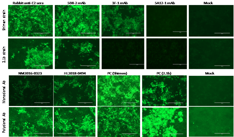
Virus confirmation for reference control strains (Shimen and 2.1b) and representative new strains (NM2016-0323 and HL2018-0494) by indirect fluorescence assay (IFA) in PK-15 cells. The figure showed that mAb (monoclonal antibody) 5B8-2 was effective against both reference control strains. mAb 3F-1 was compatible with the Shimen strain but did not react appropriately with the 2.1b control strain. No fluorescence was observed for the 5A12-1 antibody with all strains. Mock= negative control; PC= positive control. The scale bar is 200 µm.
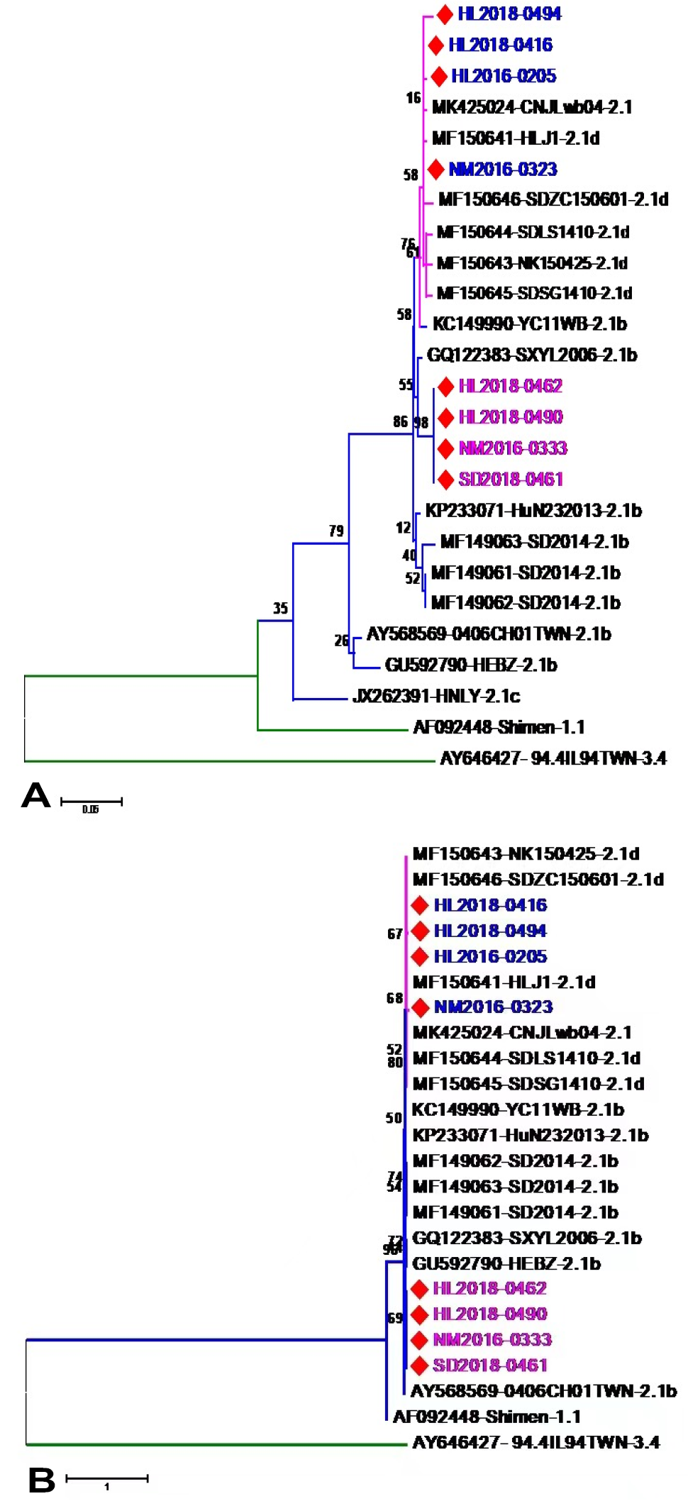
Phylogenetic analysis of the novel CSFV strains based on 5’- 3’-UTRs regions. (A) Phylogenetic analysis of the 8 novel strains and 17 reference strains based on 5’-UTR. All novel CSFV isolates were designated by red diamonds. (B) Phylogenetic analysis of the 8 novel strains and 16 reference strains based on 3’-UTR. All novel CSFV isolates were designated by red diamonds. The phylogenetic tree is built using the neighbor-joining (NJ) method, with 1000 bootstrap replicates.
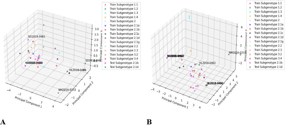
PCA (Principal component analysis) visualization of sequence classification results. A, the SVM model predicts HL2018-0462, HL2018-0490, NM2016-0333, and SD2018-0461 as Subgenotype 2.1b, while NM2016-0323, HL2018-0494, HL2018-0416, and HL2016-0205 are predicted as Subgenotype 2.1d. This pattern is consistent with the predictions in B, where the classification results based on the 3’-UTR region also indicate HL2018-0462, HL2018-0490, NM2016-0333, and SD2018-0461 as Subgenotype 2.1b, and NM2016-0323, HL2018-0494, HL2018-0416, and HL2016-0205 as Subgenotype 2.1d. The overlapping labels or data points are again a result of the high sequence similarity.
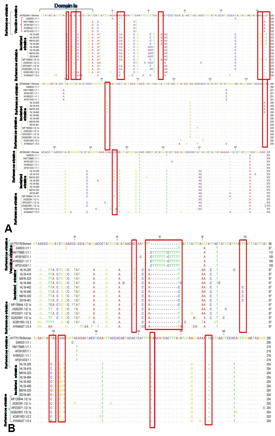
Sequence alignment of isolated, reference and vaccine strains. A, 5’-UTR amino acid sequence alignment of the 8 studied novel CSFV strains and 12 reference isolates downloaded from NCBI (National Center for Biotechnology Information). B, 3’-UTR amino acid sequence alignment of the 8 studied novel CSFV strains and 12 reference isolates downloaded from NCBI. Most of the important mutations are highlighted in red boxes.
References
- Beer, M., Goller, K.V., Staubach, C. and Blome, S., 2015. Genetic variability and distribution of Classical swine fever virus. Anim. Hlth. Res. Rev., 16: 33–39. https://doi.org/10.1017/S1466252315000109[CrossRef]
- Chen, Y., Xiao, J., Xiao, J., Sheng, C., Wang, J., Jia, L., Zhi, Y., Li, G., Chen, J. and Xiao, M., 2012. Classical swine fever virus NS5A regulates viral RNA replication through binding to NS5B and 3’UTR. Virology, 432: 376–388. https://doi.org/10.1016/j.virol.2012.04.014[CrossRef]
- Coronado, L., Liniger, M., Muñoz-González, S., Postel, A., Pérez, L. J., Pérez-Simó, M., Perera, C. L., Frías- Lepoureau, M. T., Rosell, R., Grundhoff, A., Indenbirken, D., Alawi, M., Fischer, N., Becher, P., Ruggli, N. and Ganges, L., 2017. Novel poly-uridine insertion in the 3’UTR and E2 amino acid substitutions in a low virulent classical swine fever virus. Vet. Microbiol., 201: 103–112. https://doi.org/10.1016/j.vetmic.2017.01.013[CrossRef]
- Deng, R. and Brock, K.V., 1993. 5′ and 3′ untranslated regions of pestivirus genome: Primary and secondary structure analyses. Nucl. Acids Res., 21: 1949–1957. https://doi.org/10.1093/nar/21.8.1949[CrossRef]
- Drake, J.W. and Holland, J.J., 1999. Mutation rates among RNA viruses. PNAS, 96: 13910–13913. https://doi.org/10.1073/pnas.96.24.13910[CrossRef]
- EU Reference Laboratory for CSF (EURL). 2020. Eu diagnostic manual for classical swine fever (Csf) diagnosis: Technical part (Fourth Draft March 2020) (Issue March).
- Fletcher, S.P. and Jackson, R.J., 2002. Pestivirus internal ribosome entry site (IRES) structure and function: Elements in the 5′ untranslated region important for IRES function. J. Virol., 76: 5024–5033. https://doi.org/10.1128/JVI.76.10.5024-5033.2002[CrossRef]
- Gong, W., Li, J., Wang, Z., Sun, J., Mi, S., Lu, Z., Cao, J., Dou, Z., Sun, Y., Wang, P., Yuan, K., Zhang, L., Zhou, X., He, S. and Tu, C., 2019. Virulence evaluation of classical swine fever virus subgenotype 2.1 and 2.2 isolates circulating in China. Vet. Microbiol., 232: 114–120. https://doi.org/10.1016/j.vetmic.2019.04.001[CrossRef]
- Greiser-Wilke, I., Zimmermann, B., Fritzemeier, J., Floegel, G. and Moennig, V., 2000. Structure and presentation of a World Wide Web database of CSF virus isolates held at the EU reference laboratory. Vet. Microbiol., 73: 131–136. https://doi.org/10.1016/S0378-1135(00)00140-1[CrossRef]
- Groth, D., Hartmann, S., Klie, S. and Selbig, J., 2013. Principal components analysis. Methods mol. Biol., 930: 527–547. https://doi.org/10.1007/978-1-62703-059-5_22[CrossRef]
- Hao, G., Zhang, H., Chen, H., Qian, P. and Li, X., 2020. Comparison of the pathogenicity of classical Swine fever virus subgenotype 2.1c and 2.1d strains from China. Pathogens, 9: 1–11. https://doi.org/10.3390/pathogens9100821[CrossRef]
- Hsu, W.L., Chen, C.L., Huang, S.W., Wu, C.C., Chen, I.H., Nadar, M., Su, Y.P. and Tsai, C.H., 2014. The untranslated regions of classic Swine fever virus RNA trigger apoptosis. PLoS One, 9. https://doi.org/10.1371/journal.pone.0088863[CrossRef]
- Hu, D., Lv, L., Gu, J., Chen, T., Xiao, Y. and Liu, S., 2016. Genetic diversity and positive selection analysis of classical Swine fever virus envelope protein gene E2 in East China under C-Strain vaccination. Front. Microbiol., 7. https://doi.org/10.3389/fmicb.2016.00085[CrossRef]
- Huang, Y.L., Meyer, D., Postel, A., Tsai, K.J., Liu, H.M., Yang, C.H., Huang, Y.C., Berkley, N., Deng, M.C., Wang, F.I., Becher, P., Crooke, H. and Chang, C.Y., 2021. Identification of a common conformational epitope on the glycoprotein E2 of classical Swine fever virus and border disease virus. Viruses, 13. https://doi.org/10.3390/v13081655[CrossRef]
- Hughes, A.L. and Hughes, M.A.K., 2007. More effective purifying selection on RNA viruses than in DNA viruses. Gene, 404: 117. https://doi.org/10.1016/j.gene.2007.09.013[CrossRef]
- Ito, T., Tahara, S.M. and Lai, M.M.C., 1998. The 3′-untranslated region of hepatitis C virus RNA enhances translation from an internal ribosomal entry site. J. Virol., 72: 8789. https://doi.org/10.1128/JVI.72.11.8789-8796.1998[CrossRef]
- Ji, W., Guo, Z., Ding, N.Z. and He, C.Q., 2015. Studying classical swine fever virus: Making the best of a bad virus. Virus Res., 197: 35–47. https://doi.org/10.1016/j.virusres.2014.12.006[CrossRef]
- Li, X., Xu, Z., He, Y., Yao, Q., Zhang, K., Jin, M., Chen, H. and Qian, P., 2006. Genome comparison of a novel classical swine fever virus isolated in China in 2004 with other CSFV strains. Virus Genes, 33: 133–142. https://doi.org/10.1007/s11262-005-0048-2[CrossRef]
- Luo, Y., Ji, S., Liu, Y., Lei, J.L., Xia, S.L., Wang, Y., Du, M.L., Shao, L., Meng, X.Y., Zhou, M., Sun, Y. and Qiu, H.J., 2017. Isolation and characterization of a moderately virulent classical swine fever virus emerging in China. Transb.. Emerg. Dis., 64: 1848–1857. https://doi.org/10.1111/tbed.12581[CrossRef]
- Luo, Y., Li, S., Sun, Y. and Qiu, H.J., 2014. Classical swine fever in China: A minireview. Vet. Microbiol., 172: 1–6. https://doi.org/10.1016/j.vetmic.2014.04.004[CrossRef]
- Paton, D.J., McGoldrick, A., Greiser-Wilke, I., Parchariyanon, S., Song, J.Y., Liou, P.P., Stadejek, T., Lowings, J.P., Björklund, H. and Belák, S., 2000. Genetic typing of classical swine fever virus. Vet. Microbiol., 73: 137–157. https://doi.org/10.1016/S0378-1135(00)00141-3[CrossRef]
- Pérez, L.J., Díaz de Arce, H., Perera, C.L., Rosell, R., Frías, M.T., Percedo, M.I., Tarradas, J., Dominguez, P., Núñez, J.I. and Ganges, L., 2012. Positive selection pressure on the B/C domains of the E2-gene of classical swine fever virus in endemic areas under C-strain vaccination. Infect. Genet. Evol., 12: 1405–1412. https://doi.org/10.1016/j.meegid.2012.04.030[CrossRef]
- Postel, A., Schmeiser, S., Bernau, J., Meindl-Boehmer, A., Pridotkas, G., Dirbakova, Z., Mojzis, M. and Becher, P., 2012. Improved strategy for phylogenetic analysis of classical swine fever virus based on full-length E2 encoding sequences. Vet. Res., 43: 50. https://doi.org/10.1186/1297-9716-43-50[CrossRef]
- Rahman, A., Hallgrímsdóttir, I., Eisen, M. and Pachter, L., 2018. Association mapping from sequencing reads using k-mers. ELife, 7. https://doi.org/10.7554/eLife.32920[CrossRef]
- Rümenapf, T., Unger, G., Strauss, J.H. and Thiel, H.J., 1993. Processing of the envelope glycoproteins of pestiviruses. J. Virol., 67: 3288–3294. https://doi.org/10.1128/jvi.67.6.3288-3294.1993[CrossRef]
- Sánchez, O., Barrera, M., Rodríguez, M.P., Frías, M.T., Figueroa, N.E., Naranjo, P., Montesino, R., Farnos, O., Castell, S., Venereo, A., Ganges, L., Borroto, C. and Toledo, J.R., 2008. Classical swine fever virus E2 glycoprotein antigen produced in adenovirally transduced PK-15 cells confers complete protection in pigs upon viral challenge. Vaccine, 26: 988–997. https://doi.org/10.1016/j.vaccine.2007.11.014[CrossRef]
- Sanjuán, R., Nebot, M.R., Chirico, N., Mansky, L.M. and Belshaw, R., 2010. Viral mutation rates. J. Virol., 84: 9733–9748. https://doi.org/10.1128/JVI.00694-10[CrossRef]
- Shapshak, P., Somboonwit, C., Sinnott, J.T., Menezes, L.J., Kangueane, P., Balaji, S. and Chiappelli, F., 2019. Global virology III: Virology in the 21st century, pp. 1–680. https://doi.org/10.1007/978-3-030-29022-1[CrossRef]
- Shen, H.Y., Wang, J.Y., Dong, X.Y., Zhao, M.Q., Kang, Y., Li, Y.G., Pei, J.J., Liao, M., Ju, C.M., Yi, L., Hu, Y. and Chen, J.D., 2013. Genome and molecular characterization of a CSFV strain isolated from a CSF outbreak in South China. Intervirology, 56: 122–133. https://doi.org/10.1159/000343917[CrossRef]
- Sheng, C., Wang, J., Xiao, J., Xiao, J., Chen, Y., Jia, L., Zhi, Y., Li, G. and Xiao, M., 2012. Classical swine fever virus NS5B protein suppresses the inhibitory effect of NS5A on viral translation by binding to NS5A. J. Gen. Virol., 93: 939–950. https://doi.org/10.1099/vir.0.039495-0[CrossRef]
- Tamura, K., Peterson, D., Peterson, N., Stecher, G., Nei, M. and Kumar, S., 2011. MEGA5: molecular evolutionary genetics analysis using maximum likelihood, evolutionary distance, and maximum parsimony methods. Mol. Biol. Evol., 28: 2731–2739. https://doi.org/10.1093/molbev/msr121[CrossRef]
- Wang, Y., Wang, Q., Lu, X., Zhang, C., Fan, X., Pan, Z., Xu, L., Wen, G., Ning, Y., Tang, F. and Xia, Y., 2008. 12-nt insertion in 3’ untranslated region leads to attenuation of classic swine fever virus and protects host against lethal challenge. Virology, 374: 390–398. https://doi.org/10.1016/j.virol.2008.01.008[CrossRef]
- Xiao, M., Gao, J., Wang, W., Wang, Y., Chen, J., Chen, J. and Li, B., 2004. Specific interaction between the classical swine fever virus NS5B protein and the viral genome. Europ. J. Biochem., 271: 3888–3896. https://doi.org/10.1111/j.1432-1033.2004.04325.x[CrossRef]
- Xiao, M., Wang, Y., Zhu, Z., Ding, C., Yu, J., Wan, L. and Chen, J., 2011. Influence of the 5’-proximal elements of the 5’-untranslated region of classical swine fever virus on translation and replication. J. Gen. Virol., 92: 1087–1096. https://doi.org/10.1099/vir.0.027870-0[CrossRef]
- Xiao, M., Wang, Y., Zhu, Z., Yu, J., Wan, L., and Chen, J., 2009. Influence of NS5A protein of classical swine fever virus (CSFV) on CSFV internal ribosome entry site-dependent translation. J. Gen. Virol., 90: 2923–2928. https://doi.org/10.1099/vir.0.014472-0[CrossRef]
- Yoo, S.J., Kwon, T., Kang, K., Kim, H., Kang, S.C., Richt, J.A. and Lyoo, Y.S., 2018. Genetic evolution of classical swine fever virus under immune environments conditioned by genotype 1-based modified live virus vaccine. Transbound. Emerg. Dis., 65: 735–745. https://doi.org/10.1111/tbed.12798[CrossRef]
- Zhang, H., Leng, C., Feng, L., Zhai, H., Chen, J., Liu, C., Bai, Y., Ye, C., Peng, J., An, T., Kan, Y., Cai, X., Tian, Z. and Tong, G., 2015. A new subgenotype 2.1d isolates of classical swine fever virus in China, 2014. Infect. Genet. Evol., 34: 94–105. https://doi.org/10.1016/j.meegid.2015.05.031[CrossRef]
- Zhang, H., Leng, C., Tian, Z., Liu, C., Chen, J., Bai, Y., Li, Z., Xiang, L., Zhai, H., Wang, Q., Peng, J., An, T., Kan, Y., Yao, L., Yang, X., Cai, X. and Tong, G., 2018. Complete genomic characteristics and pathogenic analysis of the newly emerged classical swine fever virus in China. BMC Vet. Res., 14. https://doi.org/10.1186/s12917-018-1504-2[CrossRef]
- Zhou, B., 2019. Classical swine fever in China. An update minireview. Front. Vet. Sci., 6: 187. https://doi.org/10.3389/fvets.2019.00187[CrossRef]
Services
Article usage statistics
Loading…