
The aim of this experiment was to study the dynamic changes of PK-15 miRNA in porcine kidney passage cells infected by porcine epidemic diarrhea virus (PEDV). We collected samples from PK-15 cells at different time points after PEDV infection, analysed the changes in the course of PEDV infection, extracted total RNA and constructed libraries, performed high throughput sequencing, established miRNA expression profiles of PK-15 cells in control and infected groups and performed differential analysis, and performed Miranda prediction and GO (Gene Ontology, GO) functional analysis for mRNAs with significant differences. GO functional enrichment analysis was performed for the significantly different mRNAs. The results showed that PEDV reached peak viral load on the second day after infection with PK-15 cells, and 280 differentially expressed mRNAs were found after sequencing, among which 96 mRNAs were up-regulated and 184 mRNAs were down-regulated, among which ssc-miR-32 expression was significantly up-regulated. Prediction of ssc-miR-32 showed that it may regulate leucine zipper protein 1 (LUZP1), and GO analysis showed that LUZP1 mainly affects the formation and development of nerve, epithelium, heart and embryo, suggesting that ssc-miR-32 may mainly affect virus replication by regulating the biological process of cells. This study provides a scientific basis for miRNA regulation of the pathogenic mechanism of PEDV porcine epidemic diarrhoea virus.
Short Communication · Volume 56, 23 March 2025 · Open Access
Identification and Functional Analysis of miRNAs in PEDV-Infected PK-15 Cells
· · · · ·
Affiliations & Notes ⌄
- 1Animal Disease Prevention and Control and Healthy Breeding Engineering Technology Research Centre, Mianyang Normal University, Mianyang 621000, China 2Sichuan Engineering Research Centre for Surveillance and Prevention and Control of Major Pig Epidemics, Mianyang 621000, China 3Key Laboratory of Ecological Safety and Protection of Sichuan Province, Mianyang Normal University, Mianyang 621000, Sichuan, China 4Research Centre of Sichuan County Economic Development, Mianyang 621000, China * Corresp
Article Info ⌄
- Journal:
- Pakistan Journal of Zoology
- Published:
- 23 March 2025

From the article
- This suggests that the growth rate of PK-15 cells infected with PEDV reaches its peak at 48 h, reaching the highest level of infection.
- In conclusion, PEDV reached peak viral load on the second day after infection with PK-15 cells, and sequencing revealed 280 differentially expressed miRNAs, of which 96 miRNA expressions were upregulated and 184 miRNA expressions were downregulated, with significant downregulation of ssc-miR-32 expression.
Selected from the article's own text.
Abstract
Materials and methods
PK-15 cells and PEDV strains were maintained by the Animal Disease Prevention and Control and Healthy Breeding Engineering Technology Research Centre of the Mayan Normal University. When the concentration of PK-15 cells reached 95%, the medium was discarded, washed 3 times with PBS solution and inoculated with PEDV, while the cells without PEDV were used as a control group, placed in 37℃, 5% CO2 cell incubator for 90 min (shaking once every 30 min), discarded the adsorbed solution, washed again 3 times with PBS solution and then added 2 ml of DME to each well. medium in a 37°C, 5% CO2 incubator and incubated further, with three replicates in each group
For total RNA extraction and qRT-PCR PK-15 cells were infected at different time points (0, 24, 48, 72, 96, 120, 144 h), digested with pancreatic enzyme, and frozen at -80℃ for use. The treated sample supernatant of 200μL was absorbed, and the viral RNA was extracted by BIO-RAD nucleic acid extractor according to the instructions of TRNzoI Universal Reagent extraction kit. Real-time fluorescence quantitative PCR was used to determine the reaction system. The reaction procedure was as follows: reverse transcription at 50℃, 5min, pre-denaturation for 3s. 95℃, denaturation 5s, 95℃, annealing and extension 35s, 60℃, data collection, 45 cycles, stored at -20 ℃. Finally, Nano Drop 2000 was used to measure the concentration and purity of RNA.
RNA sample libraries were constructed using the Illumine True Small RNA Kit. SE50 sequencing was performed to obtain initial small RNA data using the Hi-Seq sequencing platform. DNSEP software was used for differential expression analysis of known and unknown mRNAs. Target gene prediction of differential mRNAs was performed using Miranda and enrichment analysis of mRNAs was performed using gene ontology (GO) enrichment analysis to further explore the major biological functions exerted by PEDV virus-specific miRNA and to lay the foundation for further studies in the future.
Results and discussion
Non-infected control cells adhered well to the wall without significant changes (Fig. 1A). After 24 h of PEDV infection, the cells were slightly damaged and began to show CPE (Fig. 1B). After 48 h of inoculation, the cells were obviously injured and numerous cells showed CPE and significant aggregation and fragmentation (Fig. 1C). The myopathic phenomenon was attenuated at 72-144 h after inoculation (Fig. 1D-G). From the analysis of myopathic conditions at different time points, the strain could reach the highest infection level at 48 h of cell infection and continued to adapt to stable proliferation, and the viral infection level decreased at 48 h and was in a stable state.
The qRT-PCR results indicate that the CT value of the 48-h infect in treatment is 27.61, which is the highest peak value as against 28.65, 28.69, 30.76, 28.96 and 32.34 after 24, 72, 96, 120 and 144 h, respectively within 144 h infect. This suggests that the growth rate of PK-15 cells infected with PEDV reaches its peak at 48 h, reaching the highest level of infection. After 48 h, there is a decreasing trend.The total RNA of the samples from the inoculated and control groups was measured by Nano Drop 2000 at OD260nm and OD280nm, and the OD260/280 was 2.08 and OD260/280≥1.8, and the total amount met the requirement for library construction.
After analysis, there were 280 differentially expressed mRNAs in the splice group compared to the control group, of which 96 mRNAs were upregulated and 184 mRNAs were downregulated, among which ssc-miR-32 expression was significantly upregulated, and ssc-miR-32 was selected as the focus of this study.
The target gene prediction of the differential miRNA (ssc-miR-32) was performed by Miranda. Ssc-miR-32 mainly acts by regulating leucine zipper protein 1 (LUZP1); LUZP1 is a centriole and action cytoskeleton localisation protein that regulates both angiogenesis and action filament bundling, while the cytoskeleton and cilia are involved in metastasis and tumour suppression .
Impaired angiogenesis caused by loss of LUZP1 or its interacting protein EPL IN may contribute to the pathological alteration of the associated disease state (Table I).
Table I. miRNA target gene prediction.
| miRNA | Target transcript | Score | Energy | Gene id | Gene name |
| ssc-miR-32 | ENSSSCT00000003924 | 159 | -25.46 | ENSSSCG00000003532 | LUZP1 |
| ssc-miR-32 | ENSSSCT00000061504 | 159 | -25.46 | ENSSSCG00000003532 | LUZP1 |
Figure 2 shows functional pathway enrichment analysis of significantly different miRNA target genes. GO molecular enrichment analysis performed for significantly different mRNAs showed that the unregulated ssc-miR-32 gene is functionally involved in neural furrow bending and formation, epithelial furrow morphogenesis, ventricular septal development, arterial development, primary neural tube formation, cardiac septal development, neural tube formation, ventricular development, embryonic epithelial tube formation, epithelial tube formation, embryonic epithelial morphogenesis, heart chamber development, neural tube development, epithelial tube morphogenesis, epithelial morphogenesis, cardiac development, vascular development, vasculature development, cardiovascular system development, tissue formation, embryonic morphogenesis, and chordate embryonic development, suggesting that ssc-miR-32 may influence viral replication primarily by regulating cellular biological processes.
Porcine epidemic diarrhoea virus (PEDV) is one of the most common pathogens causing diarrhoea in piglets in clinical practice, and since its discovery in 1971, it has continued to affect the entire pig industry, causing severe economic losses in China. As no effective treatment has been found, comprehensive prevention and control can only be achieved through vaccination and biosafety management .
In recent years, research on miRNAs has been intensified, and the study of miRNAs has become an important research direction for the treatment of various epidemics. The effect of miRNA-target gene interactions on viruses has also become a biological factor with high specificity and sensitivity in virus diagnosis and efficacy monitoring . A study by Lang (2015) showed that ssc-let-7f had a significant inhibitory effect on the proliferation of PEDV on PK-15 cells. The analysis of differential miRNA expression in PEDV-infected PK-15 cells by Zhang et al. (2021) revealed many more miRNAs with important functions that need to be annotated and discovered. A study by Zheng (2019) showed that miR-221-5p could activate the NF-κB pathway by targeting NFKBIA and SOCS1, two inhibitors of the NF-κB pathway, and thus inhibit PEDV replication. A study by Wang (2020) showed that differential expression of miR-486-5p, miR-339-5p and miR-1271-5 during PEDV infection significantly inhibited viral replication, while miR-33a promoted viral infection. Zhang et al. (2018) first identified novel-miR-877 and provided preliminary validation of the role of novel-miR-877 and ssc-miR-219a in PEDV replication. Novel-miR-877 and ssc-miR-219a may inhibit PEDV replication by directly targeting the viral genome or by acting on other genes in the cell.
In this experiment, by observing the morphology of PK-15 cells at different periods of PEDV infection and verifying this by real-time fluorescence quantitative PCR, we found that the total viral load of PEDV showed a trend of increasing and then decreasing after infection of PK-15 cells, with the viral load peaking on the second day.
The sequence distribution of clean reads of the four samples was determined after high throughput sequencing of the constructed library; in sample 1-2 (the second day of infection), using a sequence length of 21-22 nt as the shortest sRNA length evaluated, the public sequence analysis of sRNAs between samples showed that there was a significant reduction in the number of small RNAs in sample 1-2 and in unique sRNAs. The analysis of miRNA differential expression between samples showed that there were 280 differentially expressed miRNAs in the inoculated group compared to the control group, of which 96 miRNAs were upregulated and 184 miRNAs were downregulated, among which ssc-miR-32 was significantly downregulated. It was predicted that ssc-miR-32 mainly acted by regulating LUZP1. GO analysis showed that the differentially expressed miRNAs mainly affected nerve, epithelium and heart formation and embryo formation and development. LUZP1 is a centriole and actin cytoskeleton localisation protein that regulates both ciliogenesis and actin filament bundling, and the cytoskeleton and cilia are involved in metastasis and tumour suppression . Impaired ciliogenesis caused by loss of LUZP1 or its interacting protein EPLIN may contribute to the pathological changes in associated disease states (Bozal- Basterra et al., 2021). For example, downregulation of ssc-miR-32 results in a downregulated deletion of LUZP1, which can lead to a decrease in cell number, promote cell migration and invasion, decrease cell viability, increase apoptosis and mitotic cell nucleus size, and alter the actin cytoskeleton, thereby affecting viral proliferation.
Conclusion
In conclusion, PEDV reached peak viral load on the second day after infection with PK-15 cells, and sequencing revealed 280 differentially expressed miRNAs, of which 96 miRNA expressions were upregulated and 184 miRNA expressions were downregulated, with significant downregulation of ssc-miR-32 expression. The prediction of ssc-miR-32 showed that it may regulate leucine zipper protein 1 (LUZP1), and GO analysis showed that LUZP1 mainly affects the formation and development of nerve, epithelium, heart and embryo, suggesting that ssc-miR-32 may mainly affect the replication of virus by regulating the biological process of cells and thus. As more and more miRNAs are found to be related to virus proliferation and growth, the regulation of miRNA specific expression for PEDV prevention and control through biological agents and gene editing technologies will be a hot issue for future research, which needs to be explored urgently.
Funding
We acknowledge the financial support of Sichuan Provincial Department of Agriculture and Rural Affairs Project (SCNYTG20-184), Ecological Safety and Protection Key Laboratory of Sichuan Province Project (ESP1505/ESP1810), Research Centre of Sichuan County Economic Development Project (xy2020003/xy2022043).
Figures
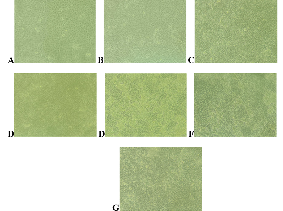
Effect of PEDV infection in PK-15 cells. A, control cells; B, 24 h inoculation; C, 48h after inoculation; D, 72h after inoculation; E, 96h after inoculation; F, 120h after inoculation; G, 144h after inoculation.
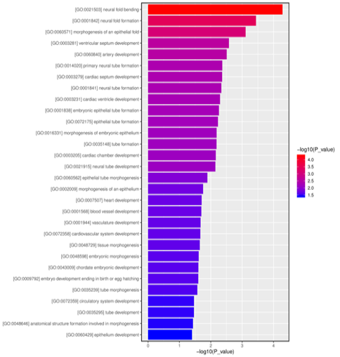
GO functional enrichment histogram of significantly different miRNA target genes.
References
- Bayat H., Pourgholami, M.H., Rahmani, S., Pournajaf, S. and Mowla, S.J., 2023. Mol. Ther. Nucl. Acids, 32: 432-444. https://doi.org/10.1016/j.omtn.2023.04.001[CrossRef]
- Bozal-Basterra, L., Gonzalez-Santamarta, M., Muratore, V., Martín-Martín, N., Ercilla, A., Rodríguez, J.A., Carracedo, A., Sutherland, J.D. and Barrio, R., 2021. Front Cell Dev. Biol., 9: 624089. https://doi.org/10.3389/fcell.2021.624089[CrossRef]
- Gonçalves, J., Sharma, A., Coyaud, É., Laurent, E.M.N., Raught, B. and Pelletier, L., 2020. J. Cell Biol., 219: e201908132. https://doi.org/10.1083/jcb.201908132[CrossRef]
- Gong, S.Y., 2019. Observation on the proliferation pattern of porcine infectious gastroenteritis virus in PK-15 cells domesticated in low serum medium. Sichuan Agricultural University, Sichuan, China.
- Hu, L.J., 2018. Clinical significance of LUZP1 in acute myeloid leukemia. Guangzhou Medical University, Guangzhou, China.
- Hu, Y.Z., Li, Q., Wang, P.F., Li, X.P. and Hu, Z.L., 2023. Front. Oncol., 13: 1140813. https://doi.org/10.3389/fonc.2023.1259236[CrossRef]
- Huang, Z.Y., Sun, J.S. and Huang, J., 2023. Chin. J. Anim. Infect. Dis., 25: 1-12. https://doi.org/10.1186/s40249-023-01051-w[CrossRef]
- Lang, Q.L., 2015. Preliminary study on the changes of miRNA expression profile of PK-15 cells infected with porcine epidemic diarrhea virus and its effect on virus proliferation. Sichuan Agricultural University, Sichuan, China.
- Liu, J., 2020. Molecular epidemiological investigation of PEDV mutant strains and establishment of a dual RT-PCR identification assay for three virulent strains. Shandong Agricultural University, Shandong, China.
- Wang D., Fang, L. and Xiao, S., 2016. Virus Res., 226: 7-13. https://doi.org/10.1016/j.virusres.2016.05.026[CrossRef]
- Wang, X.B., 2020. Analysis of mRNA/miRNA expression profiles of PEDV-infected Vero E6 cells and study on the mechanism of Abl2 affecting virus replication. CAAS, Beijing, China.
- Zhang, C.Y., Chen, J.N. and Wen, J.X., 2018. Chin. J. Prevent. Vet. Med., 40: 1095-1099. (in Chinese).
- Zhang, X., Li, C., Zhang, B., Li, Z., Zeng, W., Luo, R., Cao, J., Cheng, G., Fan, S. and He, Q., 2021. Sci. Rep., 11: 1868. https://doi.org/10.1038/s41598-021-81189-5[CrossRef]
- Zhao, J.Z., Li, H., Liu K., Lin, J.X. and Sun, S.G., 2021. Sci. Bull., 66: 3123-3140. (in Chinese). https://doi.org/10.1360/TB-2020-1581[CrossRef]
- Zheng, H., 2019. MiRNAomic analysis of PEDV-infected porcine small intestinal epithelial cells and the effect of miR-221-5p and miR-615 on viral replication and mechanism. Northwest Agriculture and Forestry University of Science and Technology, XiAn, China.
Services
Article usage statistics
Loading…