
MiRNAs are known as small, non-coding and single stranded RNAs which can regulate cell proliferation, differentiation, apoptosis, and involve in the development of sperm. Meanwhile, DUSP4, PDK4 and FKBP1B were also found to participate in testis development and spermatogenesis. In this study, online software and luciferase reporter gene system were used to predict and verify the target relationship between miR-122 and DUSP4/PDK4, miR-449a and FKBP1B. Thereafter, comparison analysis was done on miR-122, miR-449a, DUSP4, PDK4 and FKBP1B mRNA expression levels in different bull testes tissues (1-day, 12-months-old and 24-months-old). In addition, genes mRNA and protein levels were detected after bull testicular Sertoli cells were transfected with miR-122 and miR-449a. Meanwhile, DUSP4, PDK4 and FKBP1B overexpression experiments were conducted. Eventually, MTT assay was performed to observe the cells proliferation. The results showed that miR-122 and miR-449a were highly-expressed in testis tissues (p<0.01). The expression levels of miR-122 and miR-449a in the 24-month-old testes were higher than those in the neonatal and 12-month-old groups during the testicular maturation process, however, PDK4, DUSP4 and FKBP1B expression levels were decreased (p<0.01). Furthermore, the luciferase in miR-122/449a co-transfected with pmiR-RB-REPORT-DUSP4-WT or pmiR-RB-REPORT-PDK4/FKBP1B-WT group was significantly lower than pmiR-RB-REPORT-DUSP4-mut or PDK4/FKBP1B-mut and negative control groups. Meanwhile, PDK4, DUSP4 and FKBP1B expression levels were down-regulated due to miR-122 and miR-449a overexpression (p<0.01). Moreover, cells proliferation ability was activated after DUSP4, PDK4 and FKBP1B overexpression (p<0.01). Finally, MTT assay results demonstrated that miR-122 or miR-449a could promote bull testicular Sertoli cells proliferation. Collectively, these data suggested that miR-122/PDK4/DUSP4 and miR-449a/FKBP1B pathways could play a vital role in bull testis development.
Research Article · Volume 50, Issue 1, 12 January 2018 · Open Access
The Predicted Target Genes of miR-122/449a Validation and Effects on Sertoli Cells Proliferation
· · · · ·
Affiliations & Notes ⌄
- 1Jilin Academy of Agricultural Sciences, Xixinghua Road 186, Gongzhuling 136100, China 2Agricultural College, Guangdong Ocean University, Zhanjiang 524088, China , * Corresponding authors: a840581260@126.com
- 2yumin_zh@163.com
Article Info ⌄
- Journal:
- Pakistan Journal of Zoology
- Published:
- 12 January 2018

Abstract
Introduction
Sertoli cells (SCs) play a vital role in spermatogenesis by providing support and nutrition for spermatogonia cells. During spermatogenesis, lactate is the preferred substrate of spermatocytes and spermatids. Lactate production from glucose in SCs is an important point for the control of spermatogenesis . SCs also secrete proteins and cytokines, which participate in the control of sperm movement from the testis to the epididymis and in the control of the pH of the seminiferous fluid . Furthermore, adjacent SCs form the blood-testis barrier, which inhibits the entry of molecules exceeding 1000 Da into the seminiferous tubule. This barrier also prevents autoimmunity and maintains a stable microenvironment for germ cell differentiation . The data show that SCs have the support, protection and nutrition effects on the germ cells, which is closely related to spermatogenesis.
MicroRNAs play a regulatory role in a variety of biological processes, such as breast development, milk secretion ; , diabetic nephropathy development , lipid metabolism , testicular development and spermatogenesis . Recently, researchers have found that miRNAs were widely involved in the regulation of male animal reproductive performance. The mitoferrin in the testes of Batcorder dorsalis could be regulated by miR-8-3p . Down-regulation of miR-320a/ 383-sponge-like long non-coding RNA NLC1-C (narcolepsy candidate-region 1 genes) was associated with male infertility and was confirmed to promote testicular embryonic carcinoma cell proliferation . In addition, miR-122 and miR-449a are two important factors in the development of sperm. PTEN, PI3K/AKT and STAT signaling pathways were found to be involved in bull sperm cells apoptosis and this process was affected by miR-122 dysregulation . Meanwhile, miR-449a, miR-34c-5p and miR-122 could be used as biomarkers of germ cell maturation ; , and miR-449a/b/c were essential for normal spermatogenesis and male fertility . Furthermore, the deletion of miR-34bc and miR-449 might led to a mice sterility .
DUSP4 was an early response gene ; ; and its overexpression caused an inhibition of GC cell viability, invasive potential and proliferation ; . Meanwhile, PDK4 was confirmed to participate in lactate production of SCs and this process was associated with spermatogenesis and male fertility . In addition, FKBP1B, a member of the peptidyl-prolyl isomerase family, was widely expressed in many types of cells and FKBP1B function inhibition could cause a Ca2+ dysregulation . However, substantial amounts of Ca2+ were required during testicular development and spermatogenesis. In summary, DUSP4, PDK4 and FKBP1B might participate in bovine testis development and spermatogenesis.
In this study, comparative analysis of miR-122 and miR-449a expression levels in different tissues was conducted. Thereafter, differentially expressed miR-122, miR-449a, DUSP4, PDK4 and FKBP1B were detected in 1-day, 12-months-old and 24-months-old bull testes tissues. Meanwhile, the target relationship were verified and the effects of miR-122 or miR-449a on bull testicular SCs proliferation were examined. It should be helpful for further studies of the role of miR-122, miR-449a and the target genes in the regulation of bull testis development and spermatogenesis.
Materials and methods
Experimental samples and cell line
A total of 9 testicular tissues (3 for 1-day, 3 for 12-months-old and 3 for 24-months-old) were collected from the Agricultural Science Academy of Jilin Province cattle farm. All experimental protocols were approved by Animal Ethics Committee of Agricultural Science Academy of Jilin Province. The bull testis cell line was purchased from the Gaining biology company (Shanghai, China).
RNA extraction and quality analysis
Total RNA was extracted from the collected tissues using trizol reagent (Invitrogen, USA) following the manufacturer’s instructions. The RNA concentration and integrity were detected using the agarose gel electrophoresis and spectrophotometer (Thermo Fisher Scientific, Waltham, America).
QPCR analysis of miRNAs and target genes
Total RNA was extracted from the collected testis tissues using trizol reagent (Invitrogen, USA) following the manufacturer’s instructions. Then the cDNA was synthesized by reverse transcription Kits (Takara, Dalian, China) according to the instruction. Primers of miRNAs and target genes were designed for qPCR according to the genes sequence (Table I). The 20μL qPCR reaction mixture including cDNA (2μL), SYBR Green I (Takara, Dalian, China) (10μL), PCR-F-Primer (0.5μL), PCR-R-Primer (0.5μL) and RNase-free H2O (7µL). The reaction was performed with following procedure: 95°C for 30s; 40 cycles of 95°C for 5s and 60°C for 30s. The expression results of miRNAs and genes were detected by Eppendorf AG-5341 fluorescence quantitative instrument and the data were analyzed using SPSS 22.0 software through 2-ΔΔCT method according to the following formula:
ΔΔCt=
[Ct(positive)-Ct(reference)]-[Ct(control)-Ct(reference)]
Here, 2-ΔΔCt refers to the relative expression ratio.
Table I.- The primer sequence of Real-time PCR.
| Primer name | Primer sequence (5′–3′) |
| miR-122 | RT: GTCGTATCCAGTGCAGGGTCCGA GGTGCACTGGATACGACCAAACAC |
| F: TGCGGTGGAGTGTGACAATGGT | |
| R: CAGTGCAGGGTCCGAGGT | |
| miR-449a | RT: GTCGTATCCAGTGCAGGGTCCGA GGTGCACTGGATACGACACCAGCT |
| F: TGCGGTGGCAGTGTATTGTTAG | |
| R: CAGTGCAGGGTCCGAGGT | |
| U6 | RT: CGCTTCACGAATTTGCGTGTCATD |
| F: GCTTCGGCAGCACATATACTAAAAT | |
| R: CAGTGCAGGGTCCGAGGT | |
| PDK4 | F: TGTTCCATCTCACCTTCACCAT |
| R: ACACCACCTCCTCTGTCTGA | |
| DUSP4 | F: TATTCCGCCGTCATCGTCTA |
| R: AGAACCTCTCATAGCCACCTT | |
| FKBP1B | F: ACAGGAAGTCATCAAGGGTT |
| R: 5GAGCAGCTCCACGTCAAA | |
| GAPDH | F: GTTTGTGATGGGCGTGAAC |
| R: ATGGACCTGGGTCATGAGT |
RT, reverse transcription; F, forward; R, reverse.
Western blot analyzes
Total protein was extracted using RIPA buffer (Boster, Wuhan, China) following the manufacturer’s instructions. Protein concentration was determined using the BCA Protein Assay Kit (Boster, Wuhan, China). Total protein (35 µg per sample) was resolved by SDS-PAGE and transferred onto PVDF membrane (Bio-Red Laboratories Inc., USA). Immunoblotting was conducted using the following primary antibodies with the suggested dilutions from the manufacturer: anti-DUSP4 (Abcam, USA); anti-PDK4 (Abcam, USA); anti-FKBP1B (Abcam, USA); anti-β-actin (Abcam, USA). The antibodies were diluted with 5% BSA (Albumin from bovine serum) and the suggested dilutions were 1:2000. The immunoblots were developed using an ECL Advanced Western Blotting Detection Kit (Invitrogen, USA).
Cell culture and transfection
Bull testicular SCs were purchased from the Gaining biology company. MiR-122 mimics/inhibitor, miR-449a mimics/inhibitor, pmiR-RB-REPORT-DUSP4/PDK4/FKBP1B-WT, pmiR-RB-REPORT-DUSP4/PDK4/FKBP1B-mut and miR-shNC were synthesized by Guangzhou Ribo-Bio company. PBI-CMV3-DUSP4, PBI-CMV3-PDK4, PBI-CMV3-FKBP1B and PBI-CMV3-Si plasmids were synthesized by GENEWIZ in China. 24 h before transfection, the SCs were plated at a concentration of approximately 1×10⁶/well into six-well culture plates with DMEM/F12 (GIBCO, Grand Island, NY, USA) containing 10% fetal bovine serum (FBS; PAA, Pasching, Austria) and 1% Penicillin-Streptomycin. The DMEM/F12 medium was replaced by Opti-MEM serum-free medium (GIBCO, Grand Island, NY, USA) when the cell fusion level reached more than 80%. For luciferase activity detection, 250 µL Opti-MEM serum-free medium (GIBCO, Grand Island, NY, USA) was mixed with 6 µL lipofectamineTM2000 (Invitrogen, USA), 1.8 µg mimics and 1.8µg recombinant plasmid pmiR-RB-REPORT-genes-mut vector or pmiR-RB-REPORT-genes-WT vector, and the transfection mixture was added to each well containing the cells in Opti-MEM serum-free medium after incubation at room temperature for 30 min. The medium was changed to regular cell culture medium after 3-5 h. 24 h after transfection, luciferase activities were detected by multimode reader (PerkinElmer, USA). RNA and protein were extracted. To validate the miR-122 or miR-449a effects on target gene and SCs, 250 µL Opti-MEM serum-free medium was mixed with 6µL lipofectamineTM2000 and 1.8µg mimics, inhibitor or miR-shNC. To investigate the effects of the target genes on SCs, 1.8µg PBI-CMV3-DUSP4, PBI-CMV3-PDK4, PBI-CMV3-FKBP1B and PBI-CMV3-Si plasmids were respectively transfected into cells using LipofectamineTM2000 according to the manufacturer’s instructions.
Bioinformatics analysis and dual luciferase reporter gene system validation
Four bioinformatics software (microRNA.org, miRDB, miRGen, TargetScan) were used to predict the target genes of miR-122 and miR-449a. The luciferase reporter gene system was used to validate the relationship between miR-122, miR-449a and their target genes according to the instructions. The fold change and P-values were calculated and the differential expression was considered true when |log2Ratio| ≥ 1 and P-value ≤ 0.05.
Cell proliferation assay
Bull testicular SCs were respectively transfected with miR-12 mimics/inhibitor, miR-449a mimics/inhibitor and miR-shNC. Then the cells (3×10⁴ cells/well) were cultured in 96-well plates for 72 h. Cell proliferation was examined using an MTT assay according to the manufacturer’s instructions (Sigma, Germany) at 0, 12, 24, 36, 48 and 72 h after transfection. The absorbance was recorded at 450 nm using a microplate spectrophotometer (ACTGene, USA).
Statistical analysis
The data were analyzed using one-way analysis of variance (ANOVA) and independent-samples T test of SPSS 22.0 software. The means and standard deviation were calculated and p<0.05 was considered as a significant difference.
Results
MiR-122 and miR-449a binding sites existed in 3’UTR of target genes
The target genes of the miR-122 and miR-449a were predicted by bioinformatics software (microRNA.org, miRDB, miRGen, TargetScan) based on the matching score of miR-122 and miR-449a seed sequence combined with target sites in genes 3’UTR region. Among all the potential genes, PDK4, DUSP4 and FKBP1B were selected for further verification (Fig. 1A).
Luciferase activities in the group co-transfected with miR-122 and wild-type DUSP4/PDK4 vectors were significantly lower than the group co-transfected with miR-122+mut/Si vector (p<0.01). Meanwhile, the similar expression profile was also observed between miR-449a and FKBP1B (p<0.01) (Fig. 1B). The results above indicated that there was a target relationship between miR-122 with PDK4/DUSP4, and FKBP1B was target gene of miR-449a.
Differential expression of miR-122/449a and target genes in diversities developmental stages of testes tissues
Results showed that miR-122 and miR-449a expression levels were higher in 24-months-old bull testis (p<0.01, Fig. 2A). The mRNA levels result of PDK4, DUSP4 and FKBP1B genes in testicular tissues showed that the expressions of PDK4, DUSP4 and FKBP1B gene in neonatal group were significantly higher than 24-months-old group (p<0.01, Fig. 2B). These results revealed that miR-122 and miR-449a might, respectively, inhibit the mRNA expressions of PDK4/DUSP4 and FKBP1B gene.
MiR-122 and miR-449a efficiently suppressed DUSP4/PDK4 and FKBP1B, respectively
As shown in Figure 3, the following conclusions could be drawn: the expressions of DUSP4/PDK4 and FKBP1B mRNA and protein were inhibited due to a high expression of miR-122/449a (p<0.01), whereas DUSP4/PDK4 and FKBP1B levels were increased when miR-122/449a was suppressed (p<0.01). The results further demonstrated that DUSP4/PDK4 and FKBP1B were the targets of miR-122 and miR-449a.
The SCs proliferation was inhibited by DUSP4, PDK4 or FKBP1B
As shown in Figure 4A and B, DUSP4, PDK4 or FKBP1B has been successfully transfected into SCs. Compared with the control group (PBI-CMV3-Si), DUSP4, PDK4 or FKBP1B was significantly higher in recombinant plasmid transfection group (p<0.01). And higher expression of DUSP4, PDK4 or FKBP1B could attenuate cell proliferation (p<0.01, Fig. 4C).
The SCs proliferation was promoted via miR-122/PDK4/DUSP4 and miR-449a/FKBP1B pathways
Bull testicular SCs were transfected with miR-122 mimics/inhibitor, miR-449a mimics/inhibitor and miR-shNC. MTT assay was performed to observe the cells proliferation. The cell proliferation ability increased in cells that transfected miR-122 mimics/miR-449a mimics. The results demonstrated that miR-122 or miR-449a could promote bull testicular cells proliferation (Fig. 5).
Discussion
MiRNAs have been demonstrated to be involved in reproduction process ; . Previous studies have shown that miR-122 was preferentially expressed in male late-stage germ-cells and miR-449 families were enriched in testis and localized to spermatocytes and spermatids . In this study, miR-122 was found to target PDK4 and DUSP4. The PDK4 protein located in mitochondria and inhibiting the catalyzing function of pyruvate dehydrogenase (PDH) . In testicular tissue, the testis-specific pyruvate dehydrogenase complex, PDH2, has been proved to participate in sperm cells cytopoiesis and PDK4 low activity ensured the energy supplement . Meanwhile, PDK families could disrupt the spindle morphology or promote meiotic maturation . The DUSP4 protein was essential in the process of endoderm generating spermatogonium or oogonium . In sperm cells, DUSP4 protein was mainly found in the tail of spermatozoa . On the other hand, there was a target relationship between miR-449a and FKBP1B. However, the protein function was still unclear. In addition, the immunophilins FKBP family was known to be involved in meiosis, calcium homeostasis, clathrin-coated vesicles, and membrane fusions . Several members of FKBP families such as FKBP23 mRNA was expressed strongly in mouse testis , FKBP38 accumulation phenomenon was detected in Cashmere goat testis and FKBP-like proteins were specific expressed in human testis . These evidences indicated that FKBP family played important roles in testis development. In our results, The SCs was inhibited by DUSP4, PDK4 or FKBP1B overexpression. Indicating that DUSP4, PDK4 or FKBP1B could involve in spermatogenesis.
The comparison analysis was done on miR-122, miR-449a, DUSP4, PDK4 and FKBP1B expression levels in 1-day, 12-months-old and 24-months-old bull testes tissues. The results showed that the expression levels of miR-122 and miR-449a in the 24-month-old testes were higher than those in the neonatal and 12-month-old groups during the testicular maturation process, however, PDK4, DUSP4 and FKBP1B expression levels were decreased (p<0.01). The target relationship was verified in tissues level. These findings suggested that miR-122, miR-449a, PDK4, DUSP4 and FKBP1B might be involved in testicular development and spermatogenesis.
The effects of miR-122 and miR-449a on bull testicular SCs were further detected. The results showed that PDK4/DUSP4 and FKBP1B expression levels were down-regulated respectively by miR-122 and miR-449a in bull testicular SCs and the cell proliferation was promoted by miR-122 and miR-449a. Therefore, we speculate that miR-122/PDK4/DUSP4 and miR-449a/FKBP1B pathways may play a significant role in bull reproductive performance.
Conclusions
In conclusion, meanwhile, PDK4 and DUSP4 were the target genes of miR-122, FKBP1B was the target gene of miR-449a. The bull testicular SCs proliferation could be promoted via miR-122/PDK4/DUSP4 and miR-449a/FKBP1B pathways. Therefore, we consider that miR-122/PDK4/DUSP4 and miR-449a/FKBP1B pathways may play a vital role in bull reproductive performance.
Acknowledgments
This study was supported by a grant from the National High-Tech Research and Development Program of China (863 Program, 2013AA102505-2) and the Natural Science Foundation of Jilin Province (20160204006N Y).
Statement of conflict of interest
The authors declare that there is no conflict of interest existed.
Figures
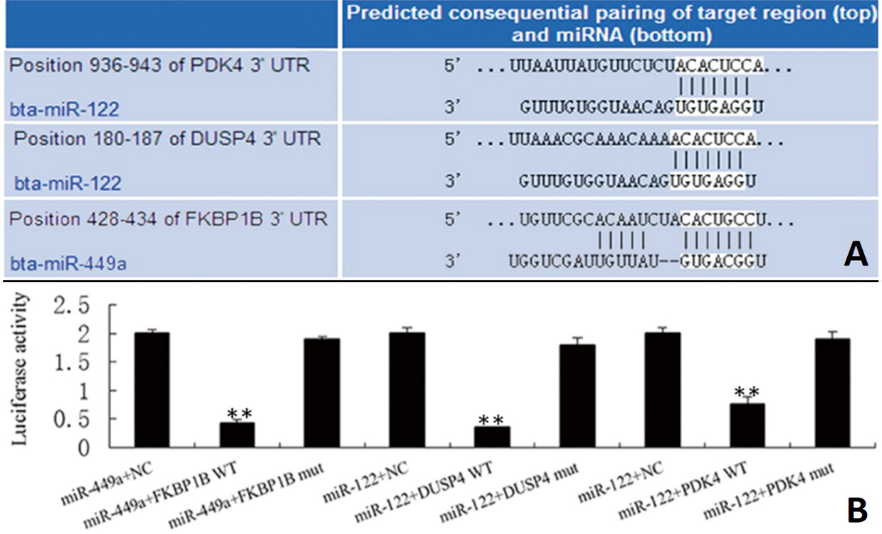
Target sites of miR-122/499a prediction and luciferase assay. A, miR-122 and miR-449a target genes prediction results; B, the target relationship between miR-122 and DUSP4/PDK4, miR-449a and FKBP1B detected by dual-luciferase reporter system. NC, blank vector; WT, wild-type vector; mut, mutant vector (**p<0.01).
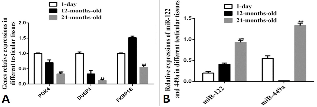
The genes expression levels in different months of testicular tissues. A, relative expressions of miR-122 and miR-449a in 1-day, 12-months-old and 24-months-old bull testes tissues; B, the mRNAs expressions of PDK4, DUSP4 and FKBP1B in 1-day, 12-months-old and 24-months-old bull testes tissues (**p<0.01).
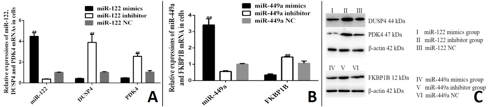
miR-122/449a and their target genes expression levels detection in bull testicular Sertoli cells. A, miR-122 and DUSP4, PDK4 expression levels were detected in Sertoli cells that transfected with miR-122 mimics, miR-122 inhibitor and miR-shNC (**p<0.01); B, miR-449a and target gene FKBP1B expression levels were detected in cells that transfected miR-449a mimics, miR-449a inhibitor and miR-shNC (**p<0.01); C, Western blot analysis for DUSP4, PDK4 and FKBP1B after miR-122 and miR-449a transfection.
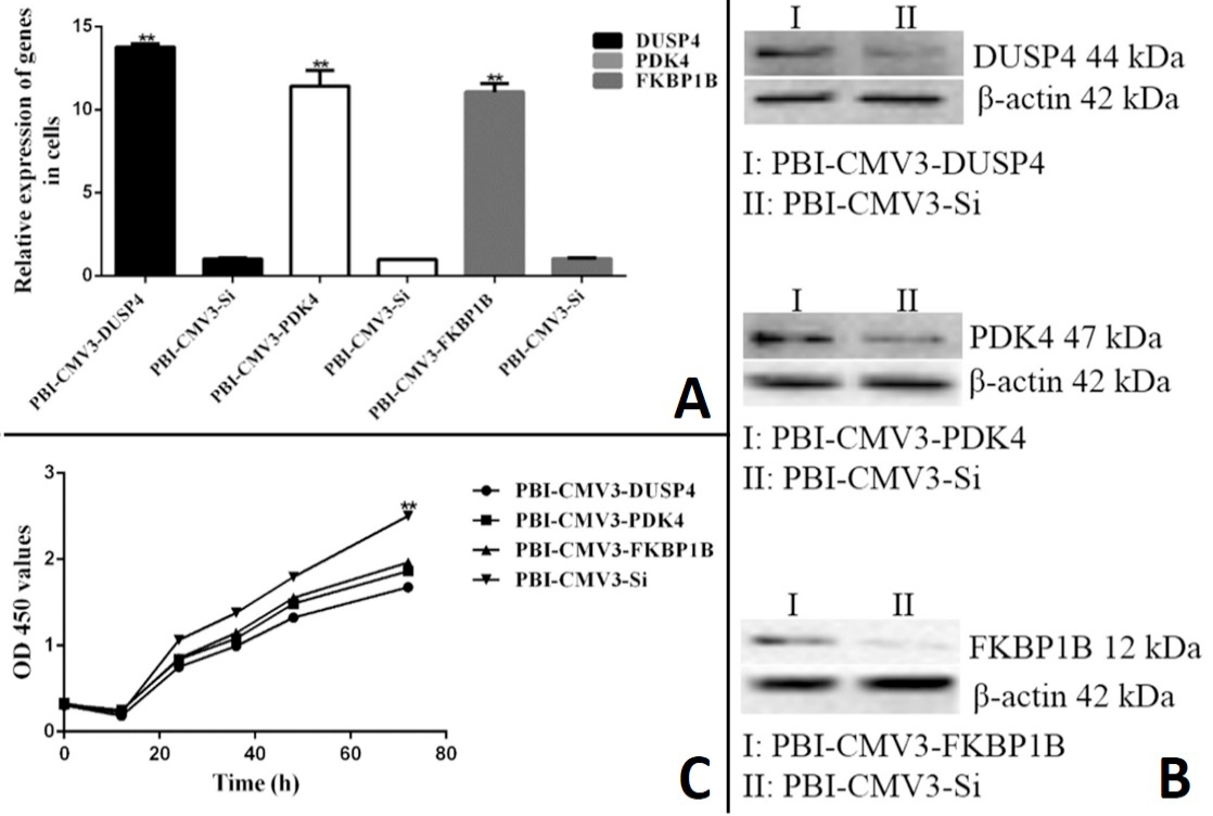
The Sertoli cells proliferation was inhibited by DUSP4, PDK4 or FKBP1B. The DUSP4, PDK4 or FKBP1B recombinant plasmid was transfected into Sertoli cells. A, the mRNA expression levels of DUSP4, PDK4 and FKBP1B; B, Western blot detection results for DUSP4, PDK4 and FKBP1B; C, MTT assay results after recombinant plasmid overexpression (**p<0.01).
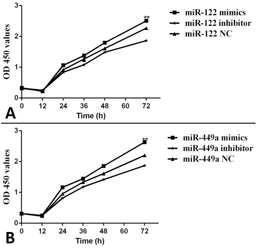
The Sertoli cells proliferation was promoted via miR-122/PDK4/DUSP4 and miR-449a/FKBP1B pathways. A, Sertoli cells proliferation was analyzed by MTT assay after the cells were transfected with miR-122 mimics, miR-122 inhibitor and miR-shNC; B, Sertoli cells proliferation was analyzed by MTT assay after the cells were transfected with miR-449a mimics, miR-449a inhibitor and miR-shNC (**p<0.05, **p<0.01).
References
- Abu-Halima, M., Hammadeh, M., Backes, C., Fischer, U., Leidinger, P., Lubbad, A.M., Keller, A. and Meese, E., 2014. Panel of five micrornas as potential biomarkers for the diagnosis and assessment of male infertility. Fertil. Steril., 102: 989-997. https://doi.org/10.1016/j.fertnstert.2014.07.001[CrossRef]
- Amit, I., Citri, A., Shay, T., Lu, Y., Katz, M., Zhang, F., Tarcic, G., Siwak, D., Lahad, J., Jacob-Hirsch, J., Amariglio, N., Vaisman, N., Segal, E., Rechavi, G., Alon, U., Mills, G.B., Domany, E. and Yarden, Y., 2007. A module of negative feedback regulators defines growth factor signaling. Nat. Genet., 39: 503-512. https://doi.org/10.1038/ng1987[CrossRef]
- Bao, J., Li, D., Wang, L., Wu, J., Hu, Y., Wang, Z., Chen, Y., Cao, X., Jiang, C., Yan, W. and Xu, C., 2012. Microrna-449 and microrna-34b/c function redundantly in murine testes by targeting e2f transcription factor-retinoblastoma protein (e2f-prb) pathway. J. biol. Chem., 287: 21686-21698. https://doi.org/10.1074/jbc.M111.328054[CrossRef]
- Brown, J.L., Snir, M., Noushmehr, H., Kirby, M., Hong, S.K., Elkahloun, A.G. and Feldman, B., 2008. Transcriptional profiling of endogenous germ layer precursor cells identifies dusp4 as an essential gene in zebrafish endoderm specification. Proc. natl. Acad. Sci. U.S.A., 105: 12337-12342. https://doi.org/10.1073/pnas.0805589105[CrossRef]
- Capra, E., Turri, F., Lazzari, B., Cremonesi, P., Gliozzi, T.M., Fojadelli, I., Stella, A. and Pizzi, F., 2017. Small RNA sequencing of cryopreserved semen from single bull revealed altered mirnas and pirnas expression between high- and low-motile sperm populations. BMC Genom., 18: 14. https://doi.org/10.1186/s12864-016-3394-7[CrossRef]
- Chen, H.Y., Zhong, X., Huang, X.R., Meng, X.M., You, Y., Chung, A.C. and Lan, H.Y., 2014. Microrna-29b inhibits diabetic nephropathy in db/db mice. Mol. Ther., 22: 842-853. https://doi.org/10.1038/mt.2013.235[CrossRef]
- Comazzetto, S., di Giacomo, M., Rasmussen, K.D., Much, C., Azzi, C., Perlas, E., Morgan, M. and O’Carroll, D., 2014. Oligoasthenoteratozoospermia and infertility in mice deficient for mir-34b/c and mir-449 loci. PLoS Genet., 10: e1004597. https://doi.org/10.1371/journal.pgen.1004597[CrossRef]
- Eisenberg, I., Kotaja, N., Goldman-Wohl, D. and Imbar, T., 2015. MicroRNA in human reproduction. Adv. Exp. Med. Biol., 888: 353-387. https://doi.org/10.1007/978-3-319-22671-2_18[CrossRef]
- Gant, J.C., Blalock, E.M., Chen, K.C., Kadish, I., Porter, N.M., Norris, C.M., Thibault, O. and Landfield, P.W., 2014. Fk506-binding protein 1b/12.6: A key to aging-related hippocampal ca2+ dysregulation? Europ. J. Pharmacol., 739: 74-82. https://doi.org/10.1016/j.ejphar.2013.10.070[CrossRef]
- Gonzalez-Fernandez, L., Ortega-Ferrusola, C., Macias-Garcia, B., Salido, G.M., Pena, F.J. and Tapia, J.A., 2009. Identification of protein tyrosine phosphatases and dual-specificity phosphatases in mammalian spermatozoa and their role in sperm motility and protein tyrosine phosphorylation. Biol. Reprod., 80: 1239-1252. https://doi.org/10.1095/biolreprod.108.073486[CrossRef]
- Grassian, A.R., Metallo, C.M., Coloff, J.L., Stephanopoulos, G. and Brugge, J.S., 2011. Erk regulation of pyruvate dehydrogenase flux through pdk4 modulates cell proliferation. Genes Dev., 25: 1716-1733. https://doi.org/10.1101/gad.16771811[CrossRef]
- Hirsch, D.D. and Stork, P.J., 1997. Mitogen-activated protein kinase phosphatases inactivate stress-activated protein kinase pathways in vivo. J. biol. Chem., 272: 4568-4575. https://doi.org/10.1074/jbc.272.7.4568[CrossRef]
- Hou, X., Zhang, L., Han, L., Ge, J., Ma, R., Zhang, X., Moley, K., Sched, T. and Wang, Q., 2015. Differing roles of pyruvate dehydrogenase kinases during mouse oocyte maturation. J. Cell Sci., 128: 2319-2329. https://doi.org/10.1242/jcs.167049[CrossRef]
- Korotchkina, L.G., Sidhu, S. and Patel, M.S., 2006. Characterization of testis-specific isoenzyme of human pyruvate dehydrogenase. J. biol. Chem., 281: 9688-9696. https://doi.org/10.1074/jbc.M511481200[CrossRef]
- Liao, Y., Du, X. and Lonnerdal, B., 2010. Mir-214 regulates lactoferrin expression and pro-apoptotic function in mammary epithelial cells. J. Nutr., 140: 1552-1556. https://doi.org/10.3945/jn.110.124289[CrossRef]
- Lu, M., Tian, H., Cao, Y.X., He, X., Chen, L., Song, X., Ping, P., Huang, H. and Sun, F., 2015. Downregulation of mir-320a/383-sponge-like long non-coding RNA nlc1-c (narcolepsy candidate-region 1 genes) is associated with male infertility and promotes testicular embryonal carcinoma cell proliferation. Cell Death Dis., 6: e1960. https://doi.org/10.1038/cddis.2015.267[CrossRef]
- Mazumdar, A., Poage, G.M., Shepherd, J., Tsimelzon, A., Hartman, Z.C., Den Hollander, P., Hill, J., Zhang, Y., Chang, J., Hilsenbeck, S.G., Fuqua, S., Kent Osborne, C., Mills, G.B. and Brown, P.H., 2016. Analysis of phosphatases in er-negative breast cancers identifies dusp4 as a critical regulator of growth and invasion. Breast Cancer Res. Treat., 158: 441-454. https://doi.org/10.1007/s10549-016-3892-y[CrossRef]
- Munoz, X., Mata, A., Bassas, L. and Larriba, S., 2015. Altered mirna signature of developing germ-cells in infertile patients relates to the severity of spermatogenic failure and persists in spermatozoa. Scient. Rep., 5: 17991. https://doi.org/10.1038/srep17991[CrossRef]
- Na, R.S., Sun, W., Sun, X.W., Qiu, X.Y., Chen, L.P. and Huang, Y.F., 2015. Expressional analysis of immune-related mirnas in breast milk. Genet. Mol. Res., 14: 11371-11376. https://doi.org/10.4238/2015.September.25.4[CrossRef]
- Nakamura, T., Yabe, D., Kanazawa, N., Tashiro, K., Sasayama, S. and Honjo, T., 1998. Molecular cloning, characterization, and chromosomal localization of fkbp23, a novel fk506-binding protein with ca2+-binding ability. Genomics, 54: 89-98. https://doi.org/10.1006/geno.1998.5571[CrossRef]
- Oliveira, P.F., Sousa, M., Barros, A., Moura, T. and Rebelo da Costa, A., 2009. Intracellular ph regulation in human sertoli cells: Role of membrane transporters. Reproduction, 137: 353-359. https://doi.org/10.1530/REP-08-0363[CrossRef]
- Peng, X.P., Huang, L. and Liu, Z.H., 2016. Mirna-133a attenuates lipid accumulation via tr4-cd36 pathway in macrophages. Biochimie, 127: 79-85. https://doi.org/10.1016/j.biochi.2016.04.012[CrossRef]
- Rato, L., Alves, M.G., Socorro, S., Duarte, A.I., Cavaco, J.E. and Oliveira, P.F., 2012. Metabolic regulation is important for spermatogenesis. Nat. Rev. Urol., 9: 330-338. https://doi.org/10.1038/nrurol.2012.77[CrossRef]
- Raudsepp, T., McCue, M.E., Das, P.J., Dobson, L., Vishnoi, M., Fritz, K.L., Schaefer, R., Rendahl, A.K., Derr, J.N., Love, C.C., Varner, D.D. and Chowdhary, B.P., 2012. Genome-wide association study implicates testis-sperm specific fkbp6 as a susceptibility locus for impaired acrosome reaction in stallions. PLoS Genet., 8: e1003139. https://doi.org/10.1371/journal.pgen.1003139[CrossRef]
- Regueira, M., Artagaveytia, S.L., Galardo, M.N., Pellizzari, E.H., Cigorraga, S.B., Meroni, S.B. and Riera, M.F., 2015. Novel molecular mechanisms involved in hormonal regulation of lactate production in sertoli cells. Reproduction, 150: 311-321. https://doi.org/10.1530/REP-15-0093[CrossRef]
- Robinson, C.J., Sloss, C.M. and Plevin, R., 2001. Inactivation of jnk activity by mitogen-activated protein kinase phosphatase-2 in eahy926 endothelial cells is dependent upon agonist-specific jnk translocation to the nucleus. Cell Signal, 13: 29-41. https://doi.org/10.1016/S0898-6568(00)00121-2[CrossRef]
- Smith, B.E. and Braun, R.E., 2012. Germ cell migration across sertoli cell tight junctions. Science, 338: 798-802. https://doi.org/10.1126/science.1219969[CrossRef]
- Sunnotel, O., Hiripi, L., Lagan, K., McDaid, J.R., de Leon, J.M., Miyagawa, Y., Crowe, H., Kaluskar, S., Ward, M., Scullion, C., Campbell, A., Downes, C.S., Hirst, D., Barton, D., Mocanu, E., Tsujimura, A., Cox, M.B., Robson, T. and Walsh, C.P., 2010. Alterations in the steroid hormone receptor co-chaperone fkbpl are associated with male infertility: A case-control study. Reprod. Biol. Endocrinol., 8: 22. https://doi.org/10.1186/1477-7827-8-22[CrossRef]
- Tariq, K., Metzendorf, C., Peng, W., Sohail, S. and Zhang, H., 2016. Mir-8-3p regulates mitoferrin in the testes of bactrocera dorsalis to ensure normal spermatogenesis. Scient. Rep., 6: 22565. https://doi.org/10.1038/srep22565[CrossRef]
- Xie, R., Lin, X., Du, T., Xu, K., Shen, H., Wei, F., Hao, W., Lin, T., Lin, X., Qin, Y., Wang, H., Chen, L., Yang, S., Yang, J., Rong, X., Yao, K., Xiao, D., Jia, J. and Sun, Y., 2016. Targeted disruption of mir-17-92 impairs mouse spermatogenesis by activating mtor signaling pathway. Medicine (Baltimore), 95: e2713. https://doi.org/10.1097/MD.0000000000002713[CrossRef]
- Yu, Z., Raabe, T. and Hecht, N.B., 2005. MicroRNA mirn122a reduces expression of the posttranscriptionally regulated germ cell transition protein 2 (tnp2) messenger RNA (mRNA) by mRNA cleavage. Biol. Reprod., 73: 427-433. https://doi.org/10.1095/biolreprod.105.040998[CrossRef]
- Yuan, S., Tang, C., Zhang, Y., Wu, J., Bao, J., Zheng, H., Xu, C. and Yan, W., 2015. Mir-34b/c and mir-449a/b/c are required for spermatogenesis, but not for the first cleavage division in mice. Biol. Open, 4: 212-223. https://doi.org/10.1242/bio.201410959[CrossRef]
- Zhang, R., Wang, G., Zhang, P.F., Zhang, J., Huang, Y.X., Lu, Y.M., Da, W., Sun, Q. and Zhu, J.S., 2016. Sanguinarine inhibits growth and invasion of gastric cancer cells via regulation of the dusp4/erk pathway. J. cell. mol. Med., 21: 1117-1127. https://doi.org/10.1111/jcmm.13043[CrossRef]
- Zhang, X., Li, C., Liu, X., Lu, C., Bai, C., Zhao, Z. and Sun, B., 2015. Differential expression of mir-499 and validation of predicted target genes in the testicular tissue of swine at different developmental stages. DNA Cell Biol., 34: 464-469. https://doi.org/10.1089/dna.2014.2728[CrossRef]
- Zheng, X., Hao, X.Y., Chen, Y.H., Zhang, X., Yang, J.F., Wang, Z.G. and Liu, D.J., 2012. Molecular characterization and tissue-specific expression of a novel fkbp38 gene in the cashmere goat (Capra hircus). Asian-Australas. J. Anim. Sci., 25: 758-763. https://doi.org/10.5713/ajas.2011.11398[CrossRef]
Services
Article usage statistics
Loading…