
The developmental efficiency of cloned embryos from somatic cells remains low, and many factors contribute to overall development of the embryo, including gene expression by the donor cells. The human hematopoietic stem/progenitor cell 117 (HSPC117) protein has been identified as being involved in placental formation, and can be modified by epigenetics. However, whether HSPC117 affects development of cloned embryos mRNA expression is unknown. To investigate the influences of HSPC117 on embryonic development, we generated transgenic porcine embryos by handmade cloning. We then assessed the embryonic developmental rate at cleavage and blastocyst stages. Our results showed that the HSPC117 transgenic embryos had markedly higher cleavage and blastocyst rates when compared to the embryos with pcDNA 3.1 vector, (69.7 ± 3.5% vs. 64.6 ± 1.8%, and 24.8 ± 2.2% vs. 15.9 ± 4.3%, respectively). However, blastocyst cell number was not different between groups. Furthermore, proto-oncogene was reported to play roles in embryo development, thus we assessed c-Fos, c-Jun, Raf-1, and c-Myc mRNA expression in HSPC117 transgenic and pcDNA 3.1 blastocysts. It was revealed that over-expression of HSPC117 mRNA in blastocysts up-regulated c-Fos, c-Jun, Raf-1, and c-Myc mRNA expression. We suggest that over-expressed HSPC117 is an important contributing factor to the development of HMC embryos in vitro viathe regulation of mRNA expression of several proto-oncogenes.
Research Article · Volume 51, Issue 5, 21 June 2019 · Open Access
HSPC117 Improves the Rate of Embryonic Development by Up-Regulating the Transcription of Proto-Oncogenes
· · · ·
Affiliations & Notes ⌄
- 1Heilongjiang Academy of Agriculture Science, Harbin 150086, P.R. China , * Corresponding author: mahong_ok@163.com
- 2liudi1963@163.com
Article Info ⌄
- Journal:
- Pakistan Journal of Zoology
- Published:
- 21 June 2019

From the article
- In conclusion, the experimental results demonstrated that, we generated several HSPC117-transfected porcine fetal fibroblast cells lines and identified HSPC117 mRNA relative expression level in 5 cell lines.
- Then, the cells line with the highest HSPC117 mRNA expression level was used as donor cells to establish transgenic embryos.
- These HSPC117-transgenic embryos showed higher cleavage and blastocysts development rates and proto-oncogenes (c-Fos, c-Jun, Raf-1, and c-Myc) mRNA expression levels than those pcDNA 3.1 embryos.
- We suggest that HSPC117 gene can exert some influence on proto-oncogene gene expression and contribute to blastocysts development further.
Selected from the article's own text.
Abstract
Introduction
Somatic cell nuclear transfer (SCNT) has successfully produced cloned or transgenic animals in many species . However, SCNT has some technical shortcomings due to its relatively low efficiency . Handmade cloning (HMC) has recently been devised, which is a simplified and micromanipulator-free version of SCNT . Compared to those embryos derived from fertilization, development of most cloned embryos is blocked at an early stage in vitro, and as a result, the implantation and birth rates for cloned animals are very low; e.g., the birth rates for the cloned pig usually approximate 1-5% . The human hematopoietic stem/progenitor cell 117 (HSPC117) protein (with an analogous protein in bacteria and archaea, called RtcB) , is an essential component protein complexe as evidenced by its frequent presence in the osmotic response element binding protein KIAA0827, TNF-α mRNA 3’ AU-rich element binding complexes (Ramana and Gupta et al., 2010), and the tRNA ligase complex . Moreover, studies have shown that murine focal adhesion associated protein (FAAP, a homologous protein of HSPC117) induced the expression levels of extracellular signaling related kinase (ERK) dephosphorylation and/or reduced phosphorylation in mice . Recent studies have shown that HSPC117 was influential in mouse pre- and post-implantation embryonic development , and our previous work demonstrates that HSPC117 expression was regulated by epigenetic modification . Although HSPC117 was proven to be useful in mouse in-vivo produced (IVP) and somatic cell nuclear transfer (SCNT) blastocysts, the mechanism underlying its contribution to embryonic pre-implantation development remains poorly understood.
Proto-oncogenes are often involved in different functions in the cell; for example, growth, division, and apoptosis. c-Fos, c-Jun, Raf-1, and c-Myc are members of this group, and many studies have shown that these genes exert important influences in embryonic development ; ; ; . However, there is a paucity of data regarding HSPC117 regulation in the expression of proto-oncogenes during in-vitro embryo development, and it was therefore important for us to understand the function of the HSPC117 gene in early embryonic development. The scope of this study was to evaluate the influence of HSPC117 expression on porcine embryonic development. Additionally, the expression of c-Fos, c-Jun, Raf-1, and c-Myc, when HSPC117 was over-expressed, was assessed in order to examine the association between over-expressed HSPC117 mRNA and embryonic proto-oncogene expression.
Materials and methods
Chemicals
All chemicals were purchased from Sigma-Aldrich Corp. unless otherwise indicated. The pcDNA 3.1 vector was courtesy of Northeast Agricultural University.
Construction of the HSPC117 expression vector
Total RNA from a porcine fibroblast cell line was extracted using the Trizol Reagent (Invitrogen, Grand Island, USA) according to the manufacturer’s instructions. The absorbance at 260 and 280 nm was assessed to evaluate the purity of extracted RNA, and 1 ug of total RNA was used to reverse transcribe to cDNA. According to the porcine HSPC117 cDNA sequences (GenBank: DQ508263.1), we synthesized the HSPC117 specific primers (sense, 5’-AAGCTTATGAGTCGCAGCTATAATGATGAG-3’; antisense, 5’-GGATCCCTAT CCTTTGATCACAGCAA-TTGGTC-3’), and HindIII and BamHI restriction sites were added to the 5’ end of the forward and reverse primers. The PCR products were tested and separated by 2% agarose gel electrophoresis. To generate the pcDNA3.1-HSPC117 plasmid, the PCR products were digested with restriction enzymes HindIII and BamHI, allowing cloning into the empty vector pcDNA3.1, which was digested using the same enzymes. Subsequently, the pcDNA3.1-HSPC117 plasmid was sequenced to verify the sequence of the inserted fragment.
Establishment of a transgenic PFF cell line
We obtained a porcine fetal fibroblast (PFF) cell line from a 40-day-old fetus, and cells were grown in high glucose Dulbecco’s modified Eagle’s medium (DMEM; GIBCO Inc.), supplemented with 15% fetal calf serum, 1% glutamine, 1% NEAA and 2 ng/ml bFGF; and were incubated at 37°C with 5% (v/v) CO2 and 95% humidity. When the cells had reached approximately 80-90% confluency, cells were passaged. Cells at passages 2-5 were frozen in DMEM containing 20% fetal bovine serum (FBS) and 10% dimethylsulfoxide, and stored in liquid nitrogen. The day before transfection, PFF cells were seeded onto 6-well plates. When the cells had reached approximately 70-80% confluency, we performed a plasmid transfection of pcDNA3.1-HSPC117 or pcDNA3.1 using 4 μg of plasmid DNA and 10 μL Lipofectamine 2000 reagent (Invitrogen, Carlsbad, CA, USA), respectively. After 6 h, the transfection medium was replaced with normal growth medium.
For generating stable transfected cell lines, 0.8 mg/ml of G418 was added to the growth medium the next day. Well-isolated single clumps of transfected cells were transferred into a well of a 24-well plate, and we continued culturing the cells in medium with 0.5 mg/ml of G418 for 1-2 additional passages. Cells stably transfected with HSPC117 were thereby contained in each well. Cells stably transfected with HSPC117 or pcDNA3.1 were stored in liquid nitrogen in growth medium with 20% FBS and 10% DMSO and used to generate blastocysts or extract total RNA for future use.
Analysis of HSPC117 expression in fibroblast cells
Twelve cells stably transfected with HSPC117 were analyzed by PCR. Genomic DNA of cells transfected with HSPC117 was amplified using three differently sized specific primer pairs: p1308-Iden1: F, GCTACAGCTCCGATTCAA; R, CTGGGTGTCCAC-AATCAA; p1308-Iden2: F, TGTGGACTGGTCGCTAA; R, GGACTCAGGTGCCTCTT; CMV-F: F, TCCCATA-GTAACGCCAATA, R, CTGGGTGTCCACAATCAA.
The PCR procedure was performed under the following PCR conditions: denaturation at 95°C for 5 min followed by 35 cycles of 95°C for 30 sec, 52°C for 30 sec, and 72°C for 50 sec. To detect HSPC117 transcripts in candidate clones, total RNA from each clone was extracted using TRIzol reagent (Invitrogen), and the cDNA was synthesized using the RevertAid First Strand cDNA Synthesis Kit (MBI Fermentas, Ontario, Canada) according to the manufacturer’s protocol. The positive clones were confirmed using quantitative real-time PCR (qPCR) by determination of HSPC117 mRNA expression, and sequence-specific primers of HSPC117 and GAPDH were then designed. Each qPCR reaction was performed in a 20-μL volume and 10 μL of Power SYBR Green Master Mix was used for all of the reactions: 0.8 μL of primers, 2 μL (50 ng/μL) of template cDNA, 0.4 μL Rox Dye, and 6.8 μL of dd H2O. The qPCR cycling conditions were as follows: a single cycle of 5 min at 95°C followed by 40 cycles of 30 s at 95°C, 1 min at 60°C, and 30 s at 55°C. Each qPCR experiment was replicated at least three times. The cell line stably transfected with HSPC117 with the highest mRNA expression level was selected as the donor cell line, and was stored in liquid nitrogen.
Establishment of reconstructed embryos
HSPC117 and pcDNA 3.1 transgenic porcine blastocysts were created by HMC. Briefly, cumulus-oocyte complexes (COCs) were retrieved from slaughterhouse-derived porcine ovaries and matured in TCM-199 medium supplemented with 10% porcine follicular fluid, 10% (FBS, 10 IU/ml pregnant mare serum gonadotropin (PMSG), 5 IU/ml human chorionic gonadotropin (hCG), and continuous cultured at 38.5°C, in 5% CO2 in humidified air for 41-44 h. The cumulus cells were removed with 1 mg/ml hyaluronidase, and 3.3 mg/ml pronase was used to digest zonae pellucidae. Zona-free oocytes were split, and the polar body was removed manually from the remaining putative cytoplast. Subsequently, the donor cells were trypsinized for use. For fusion, available cytoplasts were transferred to medium supplemented with 1 mg/ml of phytohemagglutinin, and then a single donor fibroblast was quickly attached. After attachment, cytoplast-fibroblast cell pairs were fused in mannitol using a direct current of 2.0 kV/cm for 9 s, and incubated to observe whether fusion had occurred. One h after the first fusion, cytoplast-fibroblast cell pairs were fused with another cytoplast and activated in activation medium (0.14 mM MgSO4, 0.01% polyvinyl alcohol, 0.3 M mannitol and 0.1 mM CaCl2) by a single direct current pulse of 0.85 kV/cm, 80 μs. When fusion was observed, reconstructed embryos were cultured in porcine zygote medium 3(PZM 3) supplemented with 5 mg/mL cytochalasin B and 10 ug/ml cycloheximide at 38.5°C in 5% CO2, 5% O2 and 90% N2 and maximal humidity. After 4 h of incubation, embryos were washed and cultured in PZM3 at 38.5°C in 5% CO2, 5% O2 and 90% N2 and maximal humidity until blastocysts were observed.
Reverse transcription and quantitative real-time PCR
Total RNA isolated from blastocysts was extracted with TRIzol (Invitrogen) reagent, according to the manufacturer. Twenty blastocyts per replicate per treatment were used for RNA extraction. First-strand cDNA was synthesized using a M-MLV First Strand Kit (Invitrogen) reagent according to the manufacturer’s protocol followed by PCR amplification. The specific primer sequences were as follows:
HSPC117: F, 5′CCAAGTAGCCACAGATGC 3′;
R, 5′CATTCCCTTCAGGTAGTCC 3′
c-Jun: F, 5’ AAAGATGGAAACGACCTTC 3’;
R, 5’ GGTTACTGTAGCCGTAGG 3’
c-Fos: F, 5’ CTGCTGAAGGAGAAGGAA 3’;
R, 5’ CAGGTCATCAGGGATCTT 3’
Raf-1: F, 5’ AATAGAAGCCAGTGAAGTGAT 3’;
R, 5’ CAACATCTCCGTGCCATT 3’
c-Myc: F, 5’ CGGAACTCTTGTGCGTAAGG3’;
R, 5’ TCATAGGTGATTGCTCAGGACA 3’
GAPDH: F, 5′CAGTCAAGGCGGAGAACG 3′;
R, 5′ATTTGATGTTGGCGGGAT 3′.
GAPDH mRNA was amplified as an internal control for normalization of each sample. Total gene transcript levels were quantified with real-time RT-PCR in a Stratagene Mx3000P. The relative expression of HSPC117, c-Fos, c-Jun, Raf-1, and c-Myc mRNA was calculated using the 2-ΔΔCt method, and GAPDH was used as the reference gene.
Statistical Analysis
T-test was performed to compare cleavages rates, blastocyst formation rates and cell number between groups using SPSS 15.0 software (SPSS Inc., USA). The data are expressed as the mean ± standard deviation.
Results and discussion
Identification and analysis of transgenic donor cell clones
To determine the integration status of the HSPC117 gene into transgenic cell clones, 12 clonal cell lines were randomly selected; and detection was via PCR with three different pairs of primers. As shown in Figure 1, three differently sized PCR products were detected at the same time in seven clones (1, 2, 3, 6, 9, 11, 12), which shows that these seven cell clones were all positive for the HSPC117 gene. The relative expression of HSPC117 mRNA in five clones was then assessed, as shown in Figure 2. The expression of HSPC117 was also evaluated in a pcDNA3.1-HSPC117 plasmid, which was used as a positive control. As shown in Figure 2, qPCR analysis showed that the HSPC117 mRNA expression levels of 5 candidate donor cell lines were raised to varying degrees, and GAPDH was used as a control. Expression was slightly higher in the first cell line (p > 0.05) compared to the control group; however, the ratios of HSPC117 mRNA expression in cell lines 2, 3, 4, and 5 were significantly higher, and the fourth clone was four times higher (p < 0.05).
The effect of HSPC117 on development of reconstructed embryos
In our study, HSPC117 and pcDNA-3.1 transfected porcine fetal fibroblast cells were used as donor cells to establish transgenic embryos. HSPC117 mRNA expression levels were detected; and, in addition to, cleavage rate, blastocyst formation rate, and blastocyst cell number were counted. The results showed that HSPC117 mRNA expression was up-regulated approximately 2.3-fold in the embryos with HSPC117-transfected donor cells relative to the embryos with pcDNA3.1 transfected donor cell (Fig. 3) (p < 0.05); Moreover, by calculating the cleavage and blastocyst formation rates among these three groups, we assessed the ability of HSPC117 to support embryonic development. As shown in Table I, the over-expression of HSPC117 had a significant impact on embryonic development at the blastocyst stage by increasing the cleavage and blastocyst formation rates compared to the pcDNA-3.1 control group (69.7% vs. 64.6% for cleavage rate; and 24.8% vs. 15.9% for blastocyst formation rate) (p < 0.05), but it did not affect blastocyst cell number (p > 0.05).
Table I.- The effect of HSPC 117 on early embryonic development.
| RE | Cleavage embryos | Blastocysts | Blastocysts cells No. | |
| HSPC117 | 129 | 90 (69.7±3.5a) | 32 (24.8±2.2a) | 39±3.3 |
| pcDNA 3.1 | 133 | 73 (64.6±1.8b) | 18 (15.9±4.3b) | 37±2.7 |
a,bNote: Different letters in the same columns denote significant difference between the treatments (P<0.05); the same letter in the same columns denotes no significant difference between treatments (P>0.05). Values are mean +/- SD. RE, Reconstructed embryos.
Compared with in vitro fertilized embryos, the production of SCNT or HMC embryos is insufficient due to the low cloning efficiency, fetal abnormalities, and placental insufficiency. There are many reasons for these results, including the capabilities of the nuclear donor cell for reprogramming. The cells originating from fibroblasts are often used as donor cells because they are easier to reprogram than cells of epithelial origin with respect to interspecies SCNT, cloning efficiency, epigenetic status, and gene expression patterns .
In our experiment, we chose those fibroblast cells transfected with pcDNA3.1 or HSPC117 as donor cells. Our results showed that the embryos from HSPC117-transfected donor cells had the highest cleavage and blastocyst formation rates compared with those embryos from pcDNA3.1 donor cells. To create the pcDNA3.1 or HSPC117-transfected donor cell lines, fetal fibroblast cells endured longer cultures, an increased number of passages, and augmented gene transfection and drug screening. Numerous studies have shown that long-term in-vitro culture and different treatments changes epigenetic stability of donor cells, and may decrease the success rate of subsequent HMC or SCNT ; ; and compared to the pcDNA3.1 embryos, the HSPC117-transgenic embryos manifested higher in-vitro developmental capability. Thus, we concluded that HSPC117 exerts some positive influence on embryos derived from HMC.
Effect of HSPC117 on the expression of proto-oncogenes in blastocysts
To determine whether higher HSPC117 mRNA expression affected the expression of proto-oncogenes, we analyzed expression levels for c-Fos, c-Jun, Raf-1, and c-Myc mRNA in HSPC117-transgenic blastocysts, and the pcDNA 3.1 blastocysts were used as controls. The mRNA expression of the proto-oncogenes in HSPC117-transgenic blastocysts was significantly increased in comparison to pcDNA 3.1 blastocysts (Fig. 4). Results showed that c-Fos, c-Jun, Raf-1, and c-Myc mRNA levels in HSPC117-transgenic blastocysts were about 15 times (p = 0.01), 6 times (p = 0.0375), 9 times (p = 0.0226), and 17 times (p = 0.0156) higher than those of the pcDNA 3.1-transgenic blastocysts, respectively. These results suggest that HSPC117 gene over-expression causes up-regulation of c-Fos, c-Jun, Raf-1, and c-Myc mRNA levels in HMC blastocysts.
The HSPC117 protein has been identified as being involved in many important functions, including tRNA splicing and other RNA repair reactions . Compared with in-vivo produced (IVP) embryos, HMC embryos run a greater risk of aberrant epigenetic reprogramming. HSPC117 has been suggested to be regulated by epigenetic modification, and possesses important roles in embryonic development. A previous report implied that a large number of HSPC117 RNAi knock-down embryos might be affected during pregnancy when they are transferred into pseudo-pregnant females . To determine whether HSPC117 was associated with embryonic developmental capability, we analyzed the relationship between some proto-oncogenes and HSPC117 mRNA expression levels in HMC embryos using qPCR assays. Our research demonstrated that over-expressed HSPC117 mRNA significantly up-regulated the expression of c-Fos, c-Jun, Raf-1, and c-Myc mRNAs. Previous studies showed that several oncogenes, such as c-Fos, c-Jun, Raf-1, and c-Myc mRNA are expressed by trophoblasts, resulting in regulation of a large number of down-stream target genes involved in cellular proliferation and differentiation ; ; ; . Previous studies have shown that several proto-oncogenes play important roles in embryonic development. However, there is no evidence to suggest that HSPC117 is involved in proto-oncogenenic gene expression. Our results suggest that HSPC117 exerts an influence on proto-oncogene gene expression, either through a direct or indirect pathway.
Conclusions
In conclusion, the experimental results demonstrated that, we generated several HSPC117-transfected porcine fetal fibroblast cells lines and identified HSPC117 mRNA relative expression level in 5 cell lines. Then, the cells line with the highest HSPC117 mRNA expression level was used as donor cells to establish transgenic embryos. These HSPC117-transgenic embryos showed higher cleavage and blastocysts development rates and proto-oncogenes (c-Fos, c-Jun, Raf-1, and c-Myc) mRNA expression levels than those pcDNA 3.1 embryos. We suggest that HSPC117 gene can exert some influence on proto-oncogene gene expression and contribute to blastocysts development further.
Acknowledgements
This work was supported by Heilongjiang Natural Science Foundation (JJ2018ZZ0082), National Natural Science Foundation of China (31872980 and 31101700), and the Heilongjiang Postdoctoral Scientific Research Developmental 239 Fund (LBH-Q15131).
Statement of conflict of interest
All authors declare that there is no conflict of interests regarding the publication of this article.
Figures
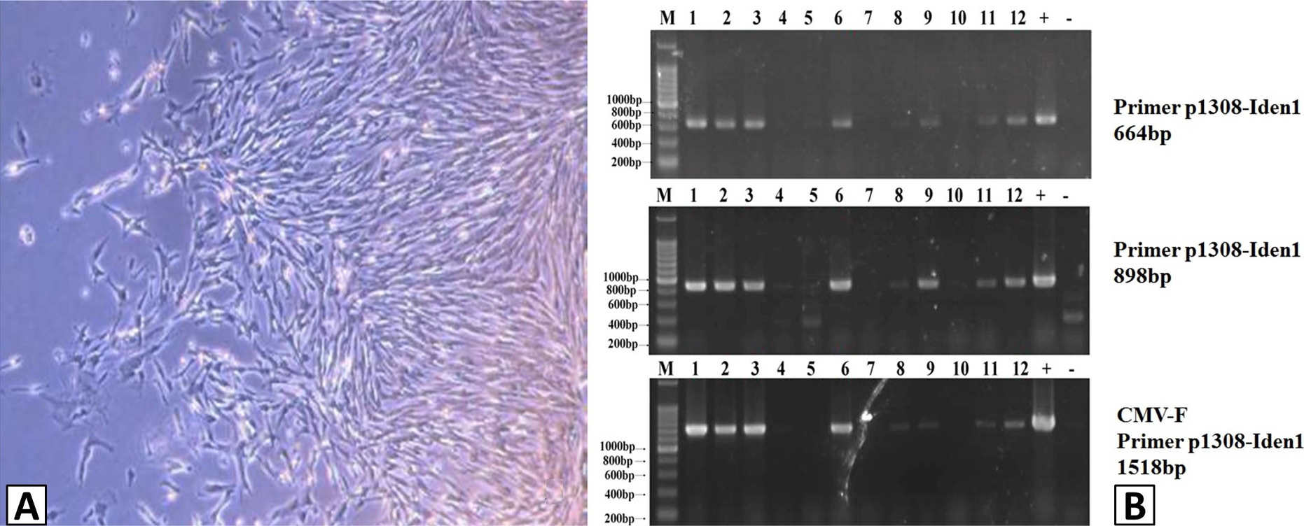
Identification of positive cell lines. A, HSPC117-transfected porcine fetal fibroblast cells line; B, HSPC117-transfected cell line confirmed using three differential size specific primer pairs.
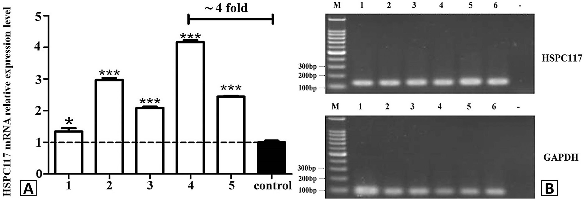
Relative HSPC117 mRNA levels of cell clones. A, detection of HSPC117 mRNA expression in positive cell clones; B, electrophoresis of HSPC117 PCR product of cells.
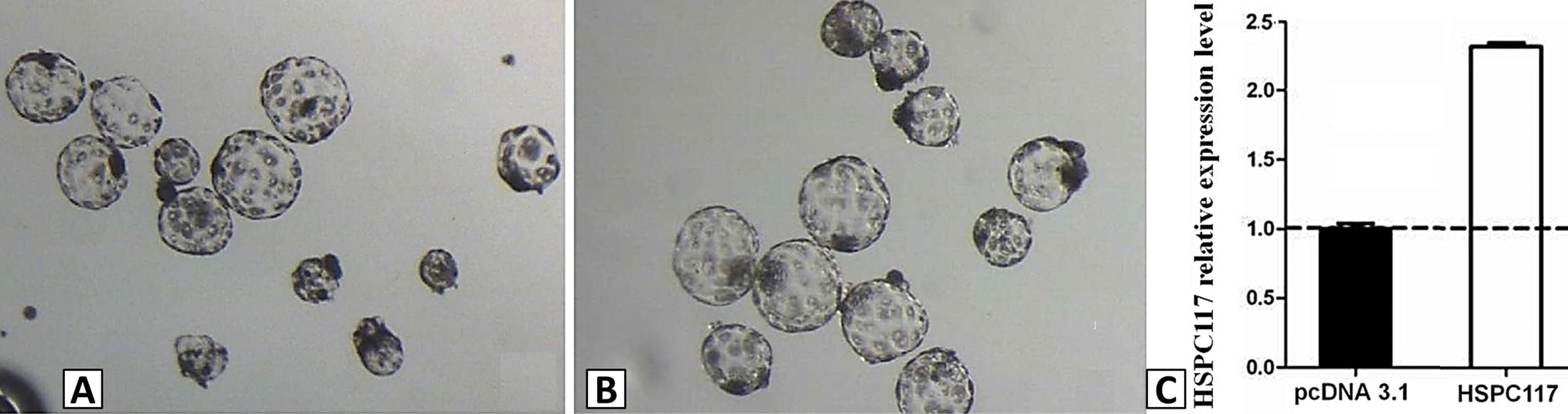
Early embryonic development. A, HSPC117 transgenic embryo; B, pcDNA3.1 embryo (control); C, HSPC117 mRNA relative expression level.
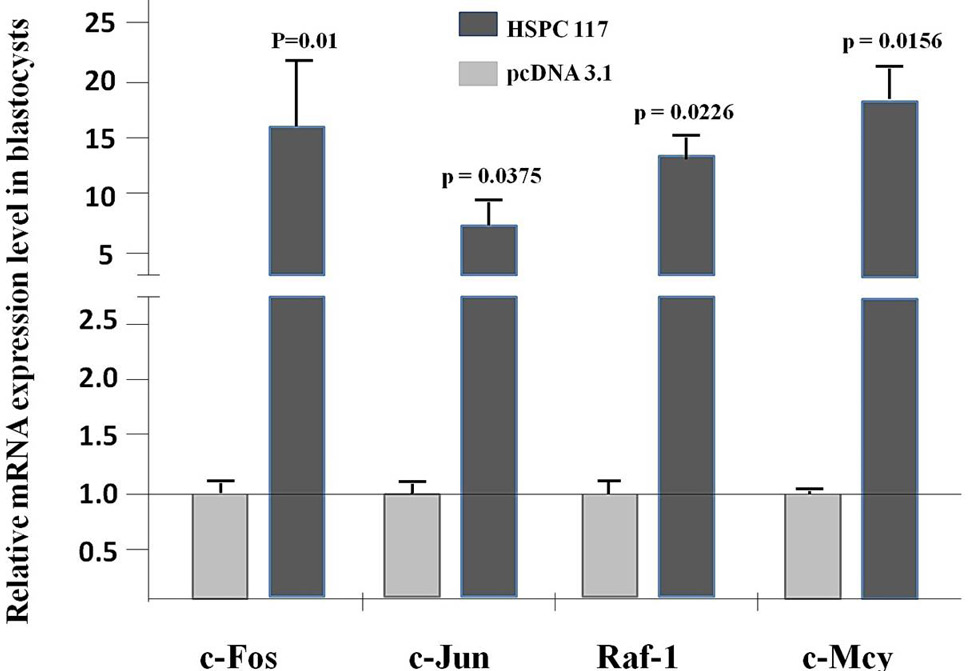
Relative expression levels of c-Fos, c-Jun, Raf-1, c-Myc and control mRNA in HSPC117-transfected reconstructed blastocysts.
References
- Du, Y., Kragh, P.M., Zhang, Y., Li, J., Schmidt, M., Bøgh, I.B., Zhang, X., Purup, S., Jørgensen, A.L., Pedersen, A.M., Villemoes, K., Yang, H., Bolund, L. and Vajta, G., 2007. Piglets born from handmade cloning, an innovative cloning method without micromanipulation. Theriogenology, 68: 1104-1110. https://doi.org/10.1016/j.theriogenology.2007.07.021[CrossRef]
- Edmunds, L.R., Sharma, L., Wang, H., Kang, A., d’Souza, S., Lu, J., McLaughlin, M., Dolezal, J.M., Gao, X., Weintraub, S.T., Ding, Y., Zeng, X., Yates, N., Prochownik, E.V., 2015. c-Myc and AMPK control cellular energy levels by cooperatively regulating mitochondrial structure and function. PLoS One, 10: e0134049. https://doi.org/10.1371/journal.pone.0134049[CrossRef]
- Fakruzzaman, M., Ghanem, N., Bang, J.I., Ha, A.N., Lee, K.L., Sohn, S.H., Wang, Z., Lee, D.S. and Kong, I.K., 2015. Effect of peroxiredoxin II on the quality and mitochondrial activity of pre-implantation bovine embryos. Anim. Reprod. Sci., 159: 172-183. https://doi.org/10.1016/j.anireprosci.2015.06.015[CrossRef]
- Genschik, P., 1998. Characterization of the Escherichia coli RNA 3’-terminal phosphate cyclase and its sigma 54-regulated operon. J. biol. Chem., 273: 25516-25526. https://doi.org/10.1074/jbc.273.39.25516[CrossRef]
- Hu, J., Teng, J., Ding, N., He, M., Sun, Y., Yu, A.C. and Chen, J., 2008. FAAP, a novel murine protein, is involved in cell adhesion through regulating vinculin-paxillinassociation. Front. Biosci., 13: 7123-7131. https://doi.org/10.2741/3215[CrossRef]
- Huan, Y.J., Wu, Z.F., Zhang, J.G., Zhu, J., Xie, B.T., Wang, J.Y., Li, J.Y., Xue, B.H., Kong, Q.R. and Liu, Z.H., 2016. Alteration of the DNA methylation status of donor cells impairs the developmental competence of porcine cloned embryos. J. Reprod. Dev., 62: 71-77. https://doi.org/10.1262/jrd.2015-048[CrossRef]
- Hwang, I.S., Kwon, D.J., Oh, K.B., Ock, S.A., Chung, H.J., Cho, I.C., Lee, J.W., Im, G.S. and Hwang, S., 2015. Production of cloned Korean native pig by somatic cell nuclear transfer. Dev. Reprod., 19: 79-84. https://doi.org/10.12717/DR.2015.19.2.079[CrossRef]
- Li, W., Sun, S., Chen, Y., Yu, H., Chen, Z.Y. and Li, H., 2013. Disrupting the interaction between retinoblastoma protein and Raf-1 leads to defects in progenitor cell proliferation and survival during early inner ear development. PLoS One, 8: e83726. https://doi.org/10.1371/journal.pone.0083726[CrossRef]
- Liu, H., Li, Y., Wei, Q., Liu, C., Bolund, L., Vajta, G., Dou, H., Yang, W., Xu, Y., Luan, J., Wang, J., Yang, H., Staunstrup, N.H. and Du, Y., 2013. Development of transgenic minipigs with expression of antimorphic human cryptochrome. PLoS One, 8: e76098. https://doi.org/10.1371/journal.pone.0076098[CrossRef]
- Ma, H., Qi, M.Y., Zhang, X., Zhang, Y.L., Wang, L., Li, Z.Q., Fu, B., Wang, W.T. and Liu, D., 2014. HSPC117 is regulated by epigenetic modification and is involved in the migration of JEG-3 cells. Int. J. mol. Sci., 15: 10936-10949. https://doi.org/10.3390/ijms150610936[CrossRef]
- Macnicol, A.M., Muslin, A.J., Howard, E.L., Kikuchi, A., Macnicol, M.C. and Lt, W., 1995. Regulation of Raf-1-dependent signaling during early Xenopus development. Mol. Cell Biol., 12: 6686-6693. https://doi.org/10.1128/MCB.15.12.6686[CrossRef]
- Marzioni, D., Todros, T., Cardaropoli, S., Rolfo, A., Lorenzi, T., Ciarmela, P., Romagnoli, R., Paulesu, L., Castellucci, M., 2010. Activating protein-1 family of transcription factors in the human placenta complicated by preeclampsia with and without fetal growth restriction. Placenta, 31: 919-927. https://doi.org/10.1016/j.placenta.2010.08.001[CrossRef]
- Matoba, S., Liu, Y., Lu, F., Iwabuchi, K.A., Shen, L., Inoue, A. and Zhang, Y., 2014. Embryonic development following somatic cell nuclear transfer impeded by persisting histone methylation. Cell, 159: 884-895. https://doi.org/10.1016/j.cell.2014.09.055[CrossRef]
- Miyoshi, K., Kawaguchi, H., Maeda, K., Sato, M., Akioka, K., Noguchi, M., Horiuchi, M. and Tanimoto, A., 2016. Birth of cloned microminipigs derived from somatic cell nuclear transfer embryos that have been transiently treated with valproic acid. Cell Reprogr., 18: 390-400. https://doi.org/10.1089/cell.2016.0025[CrossRef]
- Popow, J., Englert, M., Weitzer, S., Schleiffer, A., Mierzwa, B., Mechtler, K., Trowitzsch, S., Will, C.L., Lührmann, R., Söll, D. and Martinez, J., 2011. HSPC117 is the essential subunit of a human tRNA splicing ligase complex. Science, 331: 760-764. https://doi.org/10.1126/science.1197847[CrossRef]
- Rahat, B., Hamid, A., Ahmad-Najar, R., Bagga, R. and Kaur, J., 2014. Epigenetic mechanisms regulate placental c-Myc and hTERT in normal and pathological pregnancies; c-Myc as a novel fetal DNA epigenetic marker for pre-eclampsia. Mol. Hum. Reprod., 20: 1026-1040. https://doi.org/10.1093/molehr/gau053[CrossRef]
- Ramana, J. and Gupta, D., 2010. FaaPred: A SVM-based prediction method for fungal adhesins and adhesin-like proteins. PLoS One, 5: e9695. https://doi.org/10.1371/journal.pone.0009695[CrossRef]
- Wang, Y., Hai, T., Liu, Z., Zhou, S., Lv, Z., Ding, C., Liu, L., Niu, Y., Zhao, X., Tong, M., Wang, L., Jouneau, A., Zhang, X., Ji, W. and Zhou, Q., 2010. HSPC117 deficiency in cloned embryos causes placental abnormality and fetal death. Biochem. biophys. Res. Commun., 397: 407-412. https://doi.org/10.1016/j.bbrc.2010.05.105[CrossRef]
- Yu, D., Wang, J., Zou, H., Feng, T., Chen, L., Li, J., Qi, X., Li, Z., Duan, X., Xu, C., Zhang, L., Long, X., Lan, J., Chen, C., Wang, C., Xu, X., Ren, J., Zhao, Y., Hu, X., Lian, Z., Men, H., Pan, D., Li, N., Capecchi, M.R., Du, X., Zhao, Y. and Wu, S., 2018. Silencing of retrotransposon-derived imprinted gene RTL1 is the main cause for postimplantational failures in mammalian cloning. Proc. natl. Acad. Sci. U.S.A., 115: 11071-11080. https://doi.org/10.1073/pnas.1814514115[CrossRef]
Services
Article usage statistics
Loading…