The sweat glands in camels are of the simple tubular exocrine variety. They are large glands which always secrete into an adjacent primary hair follicle; epitrichial sweat glands (Figure 1).
The glands are composed of two portions; namely, the secretory portion and the excretory duct.
The secretory portion (adenomeres) of the gland, being of the coiled, tubular type with a widely dilated lumen, is located in the dermis at a level one fold deeper than that of the hair papillae (Figure 1). The secretory portion is lined by high cuboidal or columnar cells. The apical portion of some cells bears a budding appearance (cytoplasmic blebbing). The cytoplasm presents numerous basophilic granules at the apical portion of the cells (Figure 2). The nucleus is rounded or oval and centrally located. The glands have a discontinuous layer of myoepithelial cells between the secretory cells and the basement membrane (Figure 2).
The secretory end-piece leads suddenly to a distinctively narrow excretory duct reaching 1:4 the diameter of that of the secretory portion (Figure 1 and 3A- B). The ducts, pursuing a slightly wavy pattern run parallel to the neighbouring hair follicle in their way to open into the bottom of a peculiar destination device. The latter device is found at the outer most portion of the accompanying primary hair follicle in the vicinity of the epidermis. The ducts may open onto the surface of the skin in an interfollicular location (atrichial sweat gland) by a similar pattern. The ex-cretory duct being lined by two layers of cuboidal epithelium, receives a conical jacket of stratified epithelium towards its way to open into the neighbouring primary hair follicle (Figure 3C-D). The terminal portion of the duct, reaching the apex of the destination device, runs further inwards forming nozzle-shaped extension which projects into the bottom of the destination device (Figure 4, 5 and 6A- B). The tubular terminal portion of the duct is composed of 3-6 flattened or cuboidal cells-long pipe (Figure 6A- B).
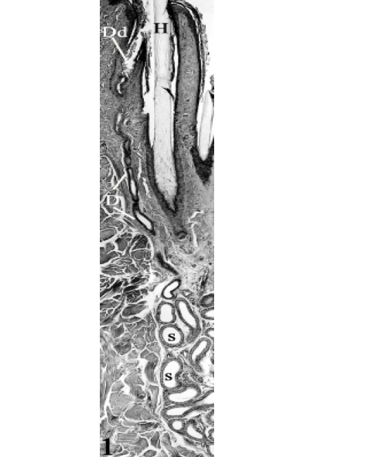
Figure 1: An epitrichial sweat gland at the thigh region of seven years-old camel , consisting of two portions: the secretory portion (S) and the excretory duct (D) opening into the primary hair follicle in the nearby of the epidermis. Dilated destination device (Dd), primary hair (H). H&E, X100.
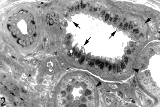
Figure 2: The secretory end-pieces of the epitrichial sweat glands at the thigh region of 6 years-old camel, showing numerous basophilic granules at the apical portion of the cells. Some cells bear a budding appearance (Cytoplasmic blebbing, arrows) apically. Myoepithelial cells (Arrowheads). Toluidine blue, X400.
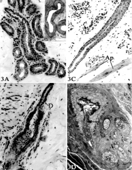
Figure 3(A-D): 3A, The secretory portion of the epitrichial sweat gland at the belly region of 6 months-old camel, showing the commencement of the excretory duct (D), secretory portion (S). Toluidine blue, X160. Inset: The excretory duct at the thigh region of 6 years-old camel, showing a wavy pattern in their way to open intoprimary hair follicle. Toluidine blue, X250. 3B, The transition between the secretory portion (S) and the excretory duct (D) at the neck region, of 3 years-old camel. Toluidine blue X160. 3C,
The excretory duct of the epitrichial sweat gland at the shoulder region, towards its opening into the primary hair follicle (upwards), Ap= Arrector pili muscle. Toluidine blue, X160. 3D, The excretory duct (arrow), receiving a conical jacket of stratified epithelium, in its way to open into the apex of the destination device, sebaceous gland (Sg). Toluidine blue, X 250.
The destination device is represented by a dilatation of various shapes. It may be oval-, flask-, triangular- or rounded-shaped (Figure 6A- B). It joins the hair follicle by its base “mouth” at a level higher than that of the sebaceous gland. It receives the terminal portion of the excretory duct at its apex. The destination device is lined by cornified stratified epithelium, consisting of 5-7 layers in thickness (Figure 4 and 6A- B). The basal layer is composed of one row of cuboidal cells showing numerous melanin pigments within their cytoplasm. The middle layer is composed of about 5 rows of large polyhedral cells which increase in size towards the surface where the largest cells are demonstrated. The cytoplasm is peculiarly pale harbouring large, rounded, central, vesicular nucleus. The cells of the middle layer present fewer amount of intracytoplasmic melanin.
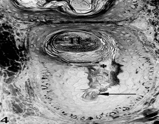
Figure 4: Cross section of the hair follicle at the level of the dilated destination device, at the thigh region, of six years-old camel, showing the nozzle-shaped terminal portion of the excretory duct (arrow) and the opening of this device into a neighbouring hair follicle (*). Notice the histomorphological differences between the epithelial lining of the hair follicle and that of the dilated device. Primary hair (H), Toluidine blue, X250.
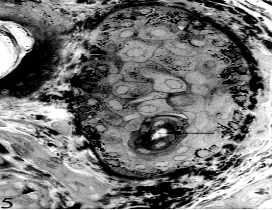
Figure 5: Tangential section of the destination dilated device, showing the peculiar cellular elements of the lining epithelium, at the thigh region of six years-old camel. Excretory duct (arrow), Toluidine blue, X250.
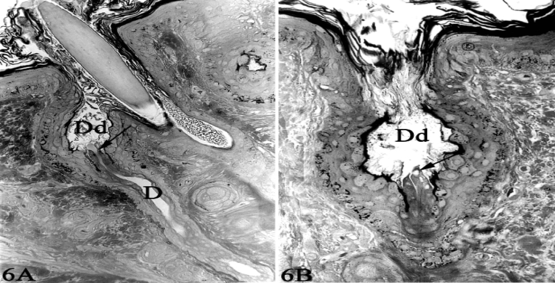
Figure 6 (A & B): The terminal portion of the excretory duct (D) opening into an oval or rounded-shaped dilated destination device (Dd) at the outer most portion of a primary hair follicle (epitrichial sweat gland “A”) or emptying on to the skin surface in an interfollicular location (atrichial sweat gland “B”). Notice the terminal portion of excretory duct (arrow) projecting into the lumen of the dilated destination device. Toluidine blue, X250.
The superficial layer is formed of several layers of keratinized material which fills the lumen and condenses at the aperture of the device into the primary hair follicle and or the surface of the skin. The flask-shaped dilatations are surrounded with a highly vascular connective tissue.