Examining normal skin sections of the Camel under DICM revealed a clear epidermis and dermis layers with all their structures and its cells (Figure 2). Under DICM, the epidermis appeared to comprises of keratinized stratified squamous epithelium involves of several sublayers (from most superficial layer to the deepest), these are stratum corneum, stratum lucidum , stratum granulosum, stratum spinosum and stratum basale (germinativum) (Figure 3). The Stratum corneum (horny layer) appeared as the outermost layer and primarily consisted of dying and dead skin cell filled with mature keratin. This layer appeared to occupy ½ -3/4 of the total epidermal thickness and showed a fully keratinized cells pushed up from basal layers. The Stratum lucidum appeared as a dense eosinophilic layer beneath and very close to the stratum corneum. Stratum granulosum (granular layer) appeared as a single layer of cells in some areas and discontinuous in others. Its nuclei was pycnotic, almost of the cytoplasm has been replaced with keratin; Stratum spinosum (prickle layer) was reduced in thickness but revealed daughter cells of the basal layer and was 1-3 cells thick. These cells appeared as a viable and nucleated and actively synthesize keratin. Stratum germinatium (stratum basale) appeared as the deepest layer of cuboidal or columnar cells, most of which was keratinocytes with a few melanocytes.
The dermis appeared as thick inner compartment of the skin. Variations were recognized in the thickness between different parts of the body skin and revealed a superficial layer composed of loose connective tissue interdigitating with undulations in the epidermis and deep dermis, which was composed of connective tissue rich in elastic and collagen fibers in its subepithelial layer, while reticular fiber predominate deep in the compartment. Moreover, an area
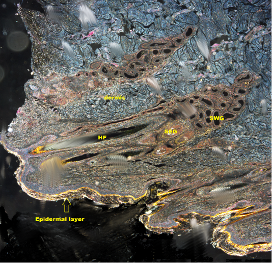
Figure 2: Shows DICM image of the skin of the camel revealing all layers of the skin (X10). HF: Hair follicle; SWG:Sweat glands; SEG: Sebaceous gland
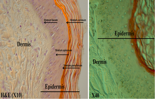
Figure 3: Shows the epidermis layers under DICM
with thick fibers (stratum reticular) was seen. The dermis contains hair follicles, blood and lymph vessels, nerves, and sebaceous and sweat glands. The mid-dermis is characterized by a proliferation of blood vessels, in contrast to that in other domestic animals (Figure 4). Vessel walls are hyalinized. The dermal fibres, its fibroblasts and the dermal ground substance were very clear in all examined sections (Figure 5). Myoepithelial cells surrounding the glands are also seen in the dermis.Hair follicles were observed to be in various stages (anagen, catagen, telogen). The majority of hair shafts showed a small discontinuous medulla. Most primary hairs, especially the giant guard hairs, contain a prominent eosinophilic medulla, while the secondary hairs have variable modulation (Figure 6). The sweat glands were found in all examined sections from the body areas. The glands were always associated with the large cover hair, clustered around the hair bulb in the reticular layer of the dermis with variation in the secretory tubules . The secretory tubules are lined with tall columnar, cuboidal or flat
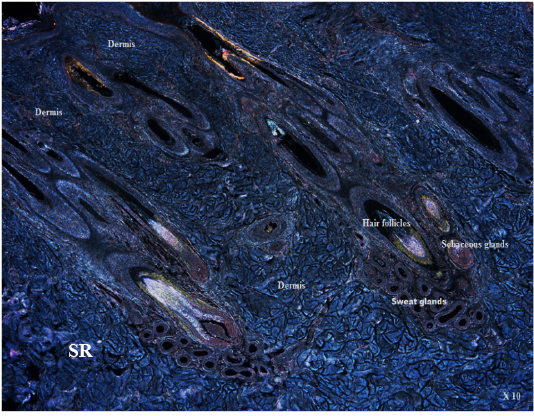
Figure 4: Shows DICM image of the dermis layer , SR: Stratum reticular (X10)
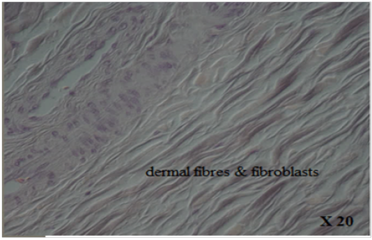
Figure 5: Shows DICM image of the dermal fibres & fibroblasts
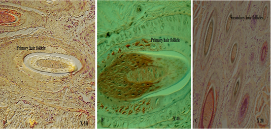
Figure 6: Shows DICM image of primary hair follicle (A. X10; B. X 40; C. X20)
epithelium that differs greatly in size (Figure 7). The sebaceous glands appeared as simple and branched alveolar and associated with hair follicles. The glands consist of cuboidal cells form sebum and their ducts lined by stratified squamous epithelium.
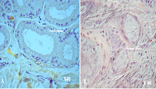
Figure 7: Shows DICM image of A. sweat glands ( X 40); B. Sebaceous glands (X 40)