
A successful pregnancy is a biological paradox as the maternal immune system accepts and tolerates the semi allogeneic fetus. Continued tolerance of the fetus relies on an optimal immune privileged environment that allows the developing fetus to avoid recognition and rejection. Any dysregulation in the crosstalk between the host’s immune system and the fetal allograft would result in rejection of the fetus, subsequently leading to miscarriages. The functioning of human leukocyte antigen (HLA) system at the maternal fetus interface plays a pivotal role in acceptance of the fetus. Alterations in the modulated activity of HLA system influence the fetal tolerance. Therefore, this review will provide an insight on how the working of HLA at the maternal fetal interface contributes towards recurrent pregnancy losses (RPL). Antigen hiding is a phenomenon that contributes towards immune evasion by the fetal cells as the placental barrier lacks HLA expression. However, the HLA molecules that are expressed by the extravillous trophoblast modulate maternal immune activity towards a protective function, leading to acceptance of the fetus. Further studies encompassing the functioning of HLA system at maternal fetal interface would prove to be beneficial for the affected couples.
Review · Volume 54, Issue 4, 24 April 2022 · Open Access
HLA System and its Participation in Recurrent Pregnancy Loss
· · · · · · ·
Affiliations & Notes ⌄
- 1Department of Biochemistry, Kinnaird College for Women, 93 Jail Road, GOR 1, Lahore, Pakistan 2Department of Chemistry and Biochemistry, University of Oklahoma, Norman, Oklahoma, United States 3Internal Medicine, Marshfield Clinic Health System, Marshfield, Wisconsin, United States 4School of Biological Sciences, University of Punjab, Quaid-I-Azam Campus, Lahore, 54590, Pakistan 5Department of Anatomy, Central Park Medical College, Lahore, Pakistan * Corresponding author: raazia.tasadduq@kinnair
Article Info ⌄
- Journal:
- Pakistan Journal of Zoology
- Published:
- 24 April 2022

From the article
- A regulated functioning of the immune system at the maternal fetal interface is a prerequisite for a healthy pregnancy.
- Certain immunomodulatory compounds establish fetal tolerance, in which HLA system plays a pivotal role.
- HLA antigens interact with maternal immune system and modulate its activity to hide and accept the developing fetus.
- Hence, any dysfunction in HLA family leads to fetal rejection and may cause RPL.
Selected from the article's own text.
Abstract
INTRODUCTION
A successful pregnancy requires the mother and fetus, two genetically different entities, to coexist . The mother’s immune system, that is in proximity with the fetal antigens, tolerates the semi allogenic fetus and does not reject it . The maternal immune system is not silent but recognizes the fetal antigens and actively diverges towards a protective response ; ; . Originally, recurrent pregnancy loss (RPL), affecting 1-2% women, had been described as three or more repetitive fetal losses before the completion of twentieth week of gestation . Recent classification defines RPL as loss of two or more sequential pregnancies, thus increasing the percentage of the affected couples to 5% . Despite the various etiological factors for RPL, in many couples the causative factor remains unknown ; . Unexplained recurrent pregnancy losses are believed to occur due to immune responses that induces maternal allogenic immune reaction against the fetus . Dysregulation in the maternal fetal immune crosstalk is associated with gestational complications and if the protective and tolerant immune response fails, pathological pregnancy state such as miscarriage might occur .
Perturbations in the activity of the human leukocyte antigen (HLA) system come under the umbrella term of immune system etiology of RPL since the expression of HLA genes and their interlinked regulatory factors play a vital role at feto maternal interface and may lead to fetal loss . The villous (syncytiotrophoblast) trophoblasts cells at the placenta protect the fetus from possible maternal rejection by acting as an immunological barrier, as these lack the expression of HLA antigens ; . However, alleles of HLA-C, HLA-E and HLA-G manifest on the extravillous trophoblast cells . In the light of the existing evidence, this review explores the functioning of the HLA system at the placental trophoblast that may contribute towards rejection of the fetus.
ROLE OF HLA SYSTEM IN RPL
HLA-C, and its duality at the maternal fetal interface
HLA-C is the only classical HLA class I antigen expressed on trophoblast cells and is believed to support placentation . HLA-C not only interacts with natural killer (NK) cells but also accounts for the autologous identification of the fetal cells. Expressed on the extravillous trophoblasts (EVT), HLA-C binds to the killer immunoglobulin like receptors (KIRs) on NK cells and promotes trophoblast invasion . KIRs regulate the activity of NK cells by conferring either an inhibitory or activating signal. The KIR genes are categorized into two haplotypes, A and B. The most frequently occurring KIR A haplotype consists of inhibitory genes whereas KIR B haplotype, that is more genetically variable, is the activating KIR. Two allotype groups recognized for HLA-C are: HLA-C1 and HLA-C2. HLA-C1 is involved in the activation of KIR2DS2 and inhibition of KIR2DL2/3 receptors whereas HLA-C2 activates KIR2DS1 and inhibits KIR2DL1 receptors by acting as ligands .
The KIR B haplotype is also believed to confer protection to the mother from gestational dysfunctions and hence its absence can increase the risk of complications . This haplotype consists of KIR2DS1 receptor which upon binding to its ligand (HLA-C2) stimulates NK cells to secrete chemokines such as GM CSF that support trophoblast invasion (Fig. 1). In contrast, females with homozygous KIR A haplotype and heterozygous HLA-C1/HLA-C2 with fetal cells having homozygous C2 are at an increased risk of complicated pregnancy . This is because KIR AA genotype encodes two copies of the KIR2DL1, the inhibitory KIR for HLA-C2 that interacts with the fetal HLA-C2 allotype, inhibiting the NK cells and results in defective placentation , 2014). Furthermore, decreased occurrence of KIR2DS1 has been observed in women undergoing repetitive miscarriages . It has been noted that when both the partners in RPL patients have the HLA-C2, KIR2DS1 expression is lowered . These findings implicate the role of KIR2DS1 in activation of NK cells to produce trophoblast supporting cytokines. Furthermore, women with history of preeclampsia and RPL have greater chance of carrying the KIR (AA) genotype with concurrent fetal HLA-C2 expression. Hiby et al. (2010) reported occurrence of RPL due to variations in paternally inherited HLA-C2.
Among the HLAs that are expressed on the trophoblast, HLA-C is considered to have the greatest number of polymorphisms . Some of these polymorphisms have been reported to have deleterious effects on the trophoblast proliferation by inhibiting the NK cells, consequently restricting growth, and leading to RPL ; ; ; .
HLA-C also establishes maternal tolerance by modulating the activity of CD8+T cells and regulatory T cells (Treg cells). The decidual CD8+T cells express significant levels of the co-inhibitory molecules; Cytotoxic T lymphocyte associated protein 4, the programmed cell death 1 and lymphocyte activation gene 3 (van der Zwan et al., 2018). As the maternal uterine CD+8 T cells interact with the paternal HLA-C molecules, these co-inhibitory cellular molecules also interact with their respective co-receptors on fetal cells and downregulate the cytotoxic activity of the CD8+T cells (van der Zwan et al., 2018). HLA-C also induce maternal Treg cells to release anti-inflammatory mediators and thus prevent inflammatory reactions in response to paternal antigens . Decreased number and activity of Treg cells has been found in women suffering from RPL and preeclampsia, both in the decidua and peripheral blood . Moreover, it has been speculated that the Treg differentiation occurs after the maternal T cell recognition of the fetal HLA-C but the mechanisms pertaining to this phenomenon remain unexplored.
HLA-E modulator of NK response
HLA-E is involved in maternal immunosuppression at the fetal-maternal interface and hence might also contribute to RPL . HLA-E is recognized by the CD94/NKG2A inhibitory receptors and the CD94/NKG2C activating receptors. Since all the extravillous trophoblast cells have HLA-E expression and the decidual NK cells express the CD94/NKG2, the interactions between these aids the trophoblast cells in evading the NK cell mediated killing and hence provide protection from maternal immune response . Hence, it is the degree of the functioning of the activating and inhibitory NK receptors that dictate the NK cell response towards fetus ; .
The surface expression of HLA-E is dependent on a conserved nonamer that is derived from the signal sequence of class I HLA . The specificity of this nonamer is important as a highly restricted repertoire can be loaded on HLA-E . HLA-E presents signal peptides of HLA-A, B, C and G to inhibitory receptor NKG2 on NK cells . In other words, HLA-E specifically interacts with NKG2A to inhibit the NK cells . This result in subsequent target cell protection by the interactions of HLA-E/peptide and CD94/NKG2 receptor on NK cells .
It has been elucidated that the NK cells with the CD94/NKG2A have a greater affinity for the HLA-E/peptide than the CD94/NKG2C ; Vales-Gomez et al., 1999). In addition, this affinity is highly dependent on the nonamer peptide complexed with HLA-E . Interactions have postulated that it is the specific CD94/NKG2 mediated recognition of the HLA-E that plays a role in the cellular communications at maternal-fetal interface . In the placenta, the leader peptides for HLA-E are restricted to HLA-G and HLA-C due to its hydrophobic characteristics (O’Challaghan et al., 1998). Moreover, it has been established that the HLA-E preferentially presents the HLA-G leader peptides . Bioimaging of trophoblast cell lines have shown increased co-localization of HLA-G and HLA-E reflecting upon their codependent surface expression . The HLA-E/HLA-G complex inhibits the cytotoxic activity of the NK cells by interacting with CD94/NKG2A and may also activate the NK cells to produce supporting chemokines by interacting with CD94/NKG2C (Fig. 2).
HLA-E is recognized by the CD94/NKG2A and modulates the cytotoxic activity of NK cell. Decreased expression of NKG2A on uNK cells has been associated with recurrent miscarriages . Interaction between HLA-E and NKG2A inhibits the cytolytic pathway of NK cells and instead promote endometrial invasion. On the other hand, lack of this interaction due to loss of NKG2A would result in activation of the cytolytic pathway and lysis of the extravillous trophoblast ; (Fig. 2). Women with repeated pregnancies have increased population of memory uNK cells that express higher levels of NKG2C. Therefore, interaction between NKG2C and HLA-E result in increased secretion of INF-γ and VEGFα that promote vascularization and placentation .
HLA-E is a non-classical HLA class I gene with two alleles of practical importance, E*0101 and E*0103, that are distinguished on the basis of non-synonymous substitution from arginine to glycine at codon 107 ; . Compromised activity of NK cells to protect against maternal immune system has been associated with decreased expression and stability of HLA-E allele 0101 . In Egyptian RPL population, the frequency of allele 0101 had been found to be greater and similar results have been reported from African, Indian, Caucasian and Hispanic populations ; ; . This association is found to be stronger in homozygous RPL women . The increased frequency of the 0101 allele in RPL women can be connected to the varying levels of the protein as the surface levels of HLA-E are lower when cells have the 0101 allele than when they have the 0103 allele . In conjunction is the dependence of HLA-E with the HLA-G nonamer. The E*0103 has a greater affinity for the HLA-G derived nonamer than the E*0101, the complex also having greater thermal stability supporting the decreased expression of HLA-E when the allele E*0101 is present . This links the allelic distribution with the etiology of RPL. However, the greater frequency of 0103 has been documented in Chinese and Japanese populations ; . Hence, the association between HLA-E and fetal loss remains inconclusive due to conflicting results from different populations . Therefore, further investigations are needed to explore the linkage between the different allelic frequencies of HLA-E and recurrent miscarriages (Dhal and Hviid, 2012).
HLA-G, a non-classical mediator of immune tolerance at maternal-fetal interface
HLA-G, another non-classical HLA molecule, is expressed on the fetal placental cells and contributes towards implantation and maternal acceptance of the fetus. HLA-G mRNA levels are associated with increased embryo cleavage rate along with successful implantation of the embryo . A maternal immune response is not generated against the placenta at the feto-maternal interface, even though the cytotrophoblast does not express the class I molecules, HLA-A and HLA-B. Normally the absence of class I HLA molecules is recognized by NK cells which attack the cells lacking the class I molecules (Munz et al., 1997). The HLA-G interacts with the decidual immune cell population and supports fetal tolerance by inhibiting the cytotoxic activity of NK cells and CD8+ T cells; suppress the proliferation of CD4+ T cells; reduces the B cell activity and stimulate the proliferation and activity of T regulatory cells (Fig. 3). HLA-G is able to execute these functions by communicating with cells harboring the receptors LILRB1 (inhibitory receptors on APC), CD160 (receptor on T lymphocytes, NK and endothelial cells) and KIR2DL4 (receptor on NK cells) ; .
The uNK cells of women with repeated pregnancies are characterized by increased expression of immunoglobin like transcript 2 (ILT-2), which is alternatively known as LILRB1 . In such women increased secretion of INF-γ and VEGFα has been observed upon interaction of ILT-2, an inhibitory receptor on NK cells, with HLA-G and NKG2C with HLA-E . Increased production of INF-γ and VEGFα by uNK cells in multigravid women support vascularization and maintenance of the decidua . Hence it has been demonstrated that the ILT-2 acts as an ‘activating’ receptor in the decidua and its interaction with HLA-G supports pregnancy due to increased expression of INF-γ and VEGFα .
Another fascinating way in which HLA-G induce tolerance is via its interaction with inhibitory receptors, immunoglobin like transcript 2 (ILT-2) and 4 (ILT-4). These receptors are present on dendritic cells, monocytes, and macrophages . HLA-G binds preferentially with these inhibitory receptors and thus results in prevention of cytotoxic activity of CD8+ T cells, as CD8 T cells and ILT-2/4 engage in competition for binding with HLA-G . Moreover, both membrane bound and soluble HLA-G inhibit the alloproliferation of T cells through interactions with ILT-2 and ILT-4 .
Soluble HLA-G is produced by fetal trophoblast cells which is taken up by the NK cells and transported to the endosome containing KIR2DL4. Binding of soluble HLA-G and KIR2DL4 in endosome leads to cellular senescence of NK cells and concurrent release of cytokines that promote vascular remodeling . The significance of the KIR2DL4 can be deduced by the findings that the uNK cells of fertile women have a higher protein level of this receptor than the women experiencing RPL . Interaction of HLA-G and KIR2DL4 does not stimulate the cytotoxic activity of NK cells but activates the production and release plethora of chemokine and cytokine such as IL-6, IL-8, IL-1β, INF-γ and TNF-α that support vascularization ; ; .
Decreased levels of HLA-G expression and polymorphisms associated with HLA-G loci have been implicated with pregnancy complications such as recurrent miscarriages and preeclampsia. Mothers with successful pregnancies have been found to have a greater amount of soluble HLA-G in the serum in comparison to women with preeclampsia and those experiencing unexplained pregnancy losses having downregulated HLA-G and decreased population of HLA-G+ cells ; . It has been reported that women with decreased concentrations of soluble HLA-G are at a greater risk of developing placental abruption during pregnancy .
As HLA-G gene expression is implicated in successful implantation, various polymorphisms of the gene that result in decreased production of HLA-G have been studied as a possible cause of RPL ; ; . A homozygous 14-bp insert in exon 8 in HLA-G gene results in unstable mRNA leading to lower production of soluble HLA-G ; . This polymorphism has been associated with repetitive fetal loss and unsuccessful IVF cycles . Moreover, decreased expression of HLA-G is reported to suppress the decidual NK cells and could lead to RPL . Greater fetal loss rates have been associated with polymorphisms 725C/G, 1573T/C and 1746C/A in the promoter region of the HLA-G ; . Moreover, polymorphisms of HLA-G with concurrence of HY antibodies in male offspring can lead to not only reduced birth weight but RPL as well . Considering this, observing HLA-G protein levels of can be used as a diagnostic tool for pathogenesis of gestational complications.
HLA-D
Class II HLA constitute HLA-DR, HLA-DQ and HLA-DP and are involved in allograft rejection . Class II antigens present peptides to CD4+ T cells and are predominantly expressed by macrophages, dendritic cells, and B cells . As the HLA class II antigens are absent at the feto-maternal interface, this allows continued survival of the fetus as the mother’s immune system is unable to recognize it as a foreign entity.
In normal pregnancies, the trophoblast cells not only lack HLA-DR4 antigens but also the HLA-DR4 enhancing interferons . In contrast, trophoblast cells have been reported to express the HLA-DR4 in complicated pregnancies such as preeclampsia . Enhanced production of cytokines such as IL-4, INFγ and TNF have also been observed due to the dysfunctional endothelial state which augment the class II HLA expression .
Association between class II HLA and retarded intrauterine growth has been established with occurrence of HLADR4 and HLAD1w2 haplotypes . In women with RPL, increased occurrence of HLA-DRBI allele, HLA-DP and HLA-DQB1 has been observed indicating possible association between pregnancy loss and HLA-D ; ; . HLA-DR has also been observed in the syncytiotrophoblast cells in placentae of preeclampsia women . HLA-DQB1 allele 03:03:02 has been implicated with a greater risk of miscarriages in South Indian women . Moreover, increased expression of HLA-DQ2/DQ8 has been noted in RPL women. The HLA-DQ2/DQ8 haplotype code proteins that are involved with peptide presentation to T-lymphocytes .
An intriguing finding has been reported by the studies that linkage disequilibrium exists between HLA-G*010102 and HLA-DR3. An increased risk for RPL had been established with HLA-DR3/G*010102 and HLA-DR1/G*010102 haplotypes than with the independent occurrence of HLA-DR3, HLA-DR1 and HLA-G*010102 (14-bp insertion in the 3’UTR) alleles . Moreover, family studies indicate the occurrence of linkage disequilibrium between the genes of HLA-DR1 and HLA-DR3 or genes in linkage disequilibrium to these effect the risk of RPL . This can be explained by the occurrence of increased secretion of cytokines such as TNF-α (harmful for normal pregnancy), predisposed by the HLA class II alleles due to linkage disequilibrium with TNF-α alleles ; .
Patients with family history of pregnancy losses are at a higher risk of suffering from RPL . Moreover, siblings of patients suffering from idiopathic recurrent miscarriages share genetic factors such as HLA class II alleles and HLA-G 14-bp insert, that increase their risk of miscarriage . Female relatives of patients having the alleles HLA-DRB1*01 or HLA-DRB1*03 are considered at a risk for pregnancy losses especially because HLA-DRB1*03 has been associated with RPL, with the disposition worsened in women with secondary RPL or higher pregnancy losses ; . Therefore, a significant association of certain HLA-D alleles has been linked with gestational complications.
Role of HLA-antibodies
The extravillous trophoblast, that is in direct contact with the maternal cells expresses the non-classical HLA-E and HLA-G along with classical HLA-C. The chimeric cells in the peripheral circulation direct the production of HLA antibodies as these express all class I and class II HLA antigens. About 30% of healthy women form HLA antibodies during gestation and the titer of antibodies increase till 28 weeks of pregnancy. In addition, the antibodies may persist till the conception of the next pregnancy ; . The presence of these antibodies has been reported to be higher during the first trimester among multiparous women compared to nulliparous women .
The occurrence of HLA antibodies in early pregnancy is related to decreased probability of live birth in women with RPL . Antibody dependent rejection of the fetus might occur because of antibody mediated complement fixation against the paternal HLA antigens. In women with RPL, a greater deposition of C4d, a marker of the antibody dependent complement activation, has been reported at the maternal fetal interface . Circulating maternal anti-HLA class I antibodies have been associated with C4d appearing in fetal umbilical cord endothelium in women with premature births . Moreover, the frequency of complement fixing antibodies is greater in patients with a history of complicated birth . Therefore, it can be inferred that the concurrent increase in complement fixation mediated by HLA-antibodies at the trophoblast may lead to the repetitive pregnancy losses .
It has been reported that the probability of secondary recurrent miscarriage (RM) is higher for pregnancies preceded by the delivery of a boy. Other studies have reported a higher amount of HLA antibodies in women who had delivered a boy prior to the miscarriage , 2010). Mounting of an abnormal immune reaction against male specific minor histocompatibility antigens (H-Y antigens) has been described as one of the contributing factors towards fetal loss .
Patients suffering from secondary RM have increased allogeneic or immunologically incompatible fetal cells appearing in the circulation of the mother. The higher frequency of the antibodies in individuals having a firstborn son and/or history of miscarriages can be linked to the increased fetal cell microchimerism . The fetal cells persist in mother’s circulation for years after delivery and their number has been found to be greater in secondary RM patients with a prior boy .
The occurrence of HLA antibodies is not implicated with any adverse effects in normal fertile population . However, the prevalence of class I HLA antibodies has been found to be higher in patients who suffer from placental abruption and have been associated with increased activity of monocytes in the fetal circulation. This leads to the heightened immune response against the developing fetus .
The detrimental effects of HLA antibodies in RPL patients have been attributed to the dysfunctions of different components of the immune system. The placental mesenchyme can be stimulated to express the class II HLA molecules in response to INFγ. As a result, maternal HLA antibodies can target the HLA-C or the HLA-DR/HLA-DQ . Inflammatory response due to repetitive pregnancy loss at the maternal-fetal interface damages the trophoblast barrier, allowing the anti-HLA antibodies to target the fetal tissue . It is probable that women with RPL might have different subtypes of anti-HLA antibodies in contrast to normal obstetric women . The maternal immune response diverges from fetal tolerance to fetal rejection as more amount of pro-inflammatory mediators were reported to be present in RPL patients when fetal antigens were presented to maternal APCs . Increased microchimerism can also cause engraftment of cells leading to increase in mother’s anti-HLA cell activity which supports the cytotoxic T cells ; . The association of HLA-antibodies with RPL considering specific immunogenic dysfunctions needs to be further investigated to establish a concrete relation.
HLA SHARING: A WAY OF PREVENTING HOMOZYGOSITY
For successful implantation, the maternal immune system needs to be adapted for pregnancy and this is aided by HLA sharing ; . Studies have backed the notion that HLA compatibility among couples lead to greater RPL rates as inbred populations have been found to be more prone to RPL ; . HLA alleles have been shown to possess positive linkage disequilibrium. Increased incidence of identical HLA-A and HLA-B alleles were found in families with increased occurrence of RPL .
During pregnancy, the mother’s immune system initiates an alloimmune response to maintain pregnancy the activation of which is mediated by HLA molecules. In the case when both the partners share the same HLA antigens possible tissue rejection sequence against the fetus is activated and hence it is believed that repeated miscarriages occur in these situations .
Maternal hypo responsiveness is invoked against paternal antigens in pregnancy, a consequence of excessive HLA antigenic sharing among couples and resulting in pregnancy failure . The lack of fetal tolerant inducing response has been linked to failure in maternal recognition of the fetal tissues, a consequence of the compatibility of the HLA system among couples ; . Hence, HLA incompatibility and not histocompatibility as presumed in earlier studies, confers a certain reproductive advantage ; .
An excessive HLA sharing has been observed in primary and secondary RM patients with the primary RM patients sharing the HLA-A and HLA-DQ antigens and both the primary and secondary RM patients sharing three or more HLA-A, HLA-B, HLA-DR and HLA-DQ antigens . It has been observed that partners who share HLA-DQA1 alleles have a significant chance of miscarriage early in pregnancy . The HLA-DQ might interfere with implantation and hence results in loss of fertility. This is due to the persistent exposure of the fetal cells to the maternal uNK cells that have enough cytotoxic activity to be detrimental to the trophoblast (van der Zwan et al., 2018).
CONCLUSION
A regulated functioning of the immune system at the maternal fetal interface is a prerequisite for a healthy pregnancy. Certain immunomodulatory compounds establish fetal tolerance, in which HLA system plays a pivotal role. HLA antigens interact with maternal immune system and modulate its activity to hide and accept the developing fetus. Hence, any dysfunction in HLA family leads to fetal rejection and may cause RPL. For couples with undetermined etiology of RPL, HLA typing and testing at cellular and molecular levels will be beneficial and provide better prognosis. Future laboratory and clinical investigations must consider HLA driven mechanisms and variables that promote fetal tolerance and could be manipulated to affect the pregnancy outcome.
ACKNOWLEDGEMENT
We would like to acknowledge BioRender.com that aided in the creation of the illustrations presented in this review.
Statement of conflicts of interest
The authors have declared no conflict of interest.
Figures
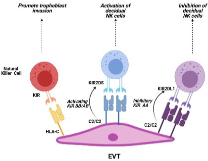
Modulation of maternal immune response by HLA-C is influenced by its interaction with the activating or inhibitory KIR.
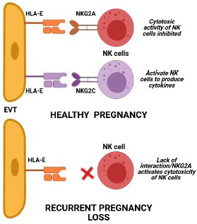
Effect of interactions between HLA-E and NKG2 in normal pregnancy and pregnancy loss.
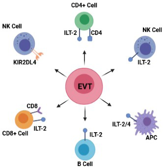
HLA-G, a mediator of cross-talk between EVT and maternal immune cells.
References
- Aghaeepour, N., Ganio, E.A., Mcilwain, D., Tsai, A.S., Tingle, M., and Van Gassen, S., 2017. An immune clock of human pregnancy. Sci. Immunol., 2: eaan2946. https://doi.org/10.1126/sciimmunol.aan2946[CrossRef]
- Aruna, M., Nagaraja, T., Andal, B.S., Tarakeswari, S., Reddy, A., and Thangaraj, K., 2011. Novel alleles of HLA-DQ and -DR loci show association with recurrent miscarriages among South Indian women. Hum. Reprod., 26: 765-774. https://doi.org/10.1093/humrep/der024[CrossRef]
- Athanassakis, I., Paflis, M., Ranella, A., and Vassiliadis, S., 1999. Detection of soluble HLA-G levels in maternal serum can be predictive for a successful pregnancy. Transpl. Proc., 31: 1834-1837. https://doi.org/10.1016/S0041-1345(99)00181-5[CrossRef]
- Baek, K.H., Lee, E.J., and Kim, Y.S., 2007. Recurrent pregnancy loss: the key potential mechanisms. Trends mol. Med., 13: 310-317. https://doi.org/10.1016/j.molmed.2007.05.005[CrossRef]
- Bari, R., Bell, T., Leung, W.H., Vong, Q.P., Chan, W.K., and Das-Gupta, N., 2009. Significant functional heterogeneity among KIR2DL1 alleles and a pivotal role of arginine 245. Blood, 114: 5182–5190. https://doi.org/10.1182/blood-2009-07-231977[CrossRef]
- Beer, A.E., Quebbeman, J.F., Ayers, J.W., and Haines, R.F., 1981. Major histocompatibility complex antigens, maternal and paternal immune responses, and chronic habitual abortions in humans. Am. J. Obstet. Gynecol., 141: 987–999. https://doi.org/10.1016/S0002-9378(16)32690-4[CrossRef]
- Benson, D.M., and Caligiuri, M.A., 2018. Natural killer cell immunity. In: Hematology (eds. R. Hoffman, E.J. Benz, L.E. Silberstein, H.E. Heslop, J.I. Weitz, J. Anastasi, M.E. Salama and S.A. Abutalib). Elsevier. pp. 240-246.e3. https://doi.org/10.1016/B978-0-323-35762-3.00022-6[CrossRef]
- Beytamouni, T.S., and Ghanem, E., 2016. Properties of uterine natural killer cells in human pregnancy major receptors involved, and routes of trophoblast invasion. Single Cell Biol., 5: 133. https://doi.org/10.4172/2168-9431.1000133[CrossRef]
- Branch, D.W., Gibson, M., and Silver, R.M., 2010. Clinical practice. Recurrent miscarriage. N. Engl. J. Med., 363: 1740-1747. https://doi.org/10.1056/NEJMcp1005330[CrossRef]
- Braud, V.M., Allan, D.S., O’callaghan, C.A., Söderström, K., D’andrea, A., and Ogg, G.S., 1998. HLA-E binds to natural killer cell receptors CD94/NKG2A, B and C. Nature, 391: 795–799. https://doi.org/10.1038/35869[CrossRef]
- Carretero, M., Palmieri, G., Llano, M., Tullio, V., Santoni, A., and Geraghty, D.E., 1998. Specific engagement of the CD94/NKG2-A killer inhibitory receptor by the HLA-E class Ib molecule induces SHP-1 phosphatase recruitment to tyrosine-phosphorylated NKG2-A: Evidence for receptor function in heterologous transfectants. Eur. J. Immunol., 28: 1280–1291. https://doi.org/10.1002/(SICI)1521-4141(199804)28:04<1280::AID-IMMU1280>3.0.CO;2-O[CrossRef]
- Cecati, M., Giannubilo, S.R., Emanuelli, M., Tranquilli, A.L., and Saccucci, F., 2011. Hla-G and pregnancy adverse outcomes. Med. Hypoth., 76: 782-784. https://doi.org/10.1016/j.mehy.2011.02.017[CrossRef]
- Christiansen, O.B., 2013. Reproductive immunology. Mol. Immunol., 55: 8-15. https://doi.org/10.1016/j.molimm.2012.08.025[CrossRef]
- Christiansen, O.B., Andersen, H.H., Højbjerre, M., Kruse, T.A., Lauritzen, S.L., and Grunnet, N., 1995. Maternal HLA class II allogeno types are markers for the predisposition to fetal losses in families of women with unexplained recurrent fetal loss. Eur. J. Immunogenet., 22: 323–334. https://doi.org/10.1111/j.1744-313X.1995.tb00248.x[CrossRef]
- Christiansen, O.B., Nielsen, H.S., and Kolte, A.M., 2006. Inflammation and miscarriage. Semin. Fetal. Neonatal Med., 11: 302–308. https://doi.org/10.1016/j.siny.2006.03.001[CrossRef]
- Christiansen, O.B., Riisom, K., Lauritsen, J.G., Grunnet, N., and Jersild, C., 1989. Association of maternal HLA haplotypes with recurrent spontaneous abortions. Tissue Antigens, 34: 190–199. https://doi.org/10.1111/j.1399-0039.1989.tb01736.x[CrossRef]
- Christiansen, O.B., Ring, M., Rosgaard, A., Grunnet, N., and Gluud, C., 1999. Association between HLA-DR1 and -DR3 antigens and unexplained repeated miscarriage. Hum. Reprod. Update, 5: 249-255. https://doi.org/10.1093/humupd/5.3.249[CrossRef]
- Colbern, G.T., Chiang, M.H., and Main, E.K., 1994. Expression of the nonclassic histocompatibility antigen HLA-G by preeclamptic placenta. Am. J. Obstet. Gynecol., 170: 1244-1250. https://doi.org/10.1016/S0002-9378(13)90442-7[CrossRef]
- Colonna, M., Samaridis, J., Cella, M., Angman, L., Allen, R.L., and O’callaghan, C.A., 1998. Cutting edge: Human myelomonocytic cells express an inhibitory receptor for classical and nonclassical MHC class I-molecules. J. Immunol., 160: 3096-3100.
- Colucci, F., 2019. The immunological code of pregnancy. Science, 365: 862-863. https://doi.org/10.1126/science.aaw1300[CrossRef]
- Dahl, M., and Hviid, T.V., 2012. Human leucocyte antigen class Ib molecules in pregnancy success and early pregnancy loss. Hum. Reprod. Update, 18: 92–109. https://doi.org/10.1093/humupd/dmr043[CrossRef]
- De Luca Brunori, I., Battini, L., Simonelli, M., Brunori, E., Valentino, V., and Curcio, M., 2003. HLA-DR in couples associated with preeclampsia: Background and updating by DNA sequencing. J. Reprod. Immunol., 59: 235-243. https://doi.org/10.1016/S0165-0378(03)00050-0[CrossRef]
- D’ippolito, S., Gasbarrini, A., Castellani, R., Rocchetti, S., Sisti, L., and Scambia, G., 2016. Human leukocyte antigen (HLA) DQ2/DQ8 prevalence in recurrent pregnancy loss women. Autoimmun. Rev., 15: 638-643. https://doi.org/10.1016/j.autrev.2016.02.009[CrossRef]
- Durmanova, V., Homolova, M., Drobny, J., Shawkatova, I., and Buc, M., 2013. Role of HLA-G and other immune mechanisms in pregnancy. Open Life Sci., 8: 226-239. https://doi.org/10.2478/s11535-013-0130-4[CrossRef]
- Ford, H.B., and Schust D.J., 2009. Recurrent pregnancy loss: Etiology, diagnosis, and therapy. Rev. Obstet. Gynecol., 2: 76-83.
- Fritz, M.A., and Speroff, L., 2010. Clinical gynecologic endocrinology and infertility. 4th ed. Lippincott Williams and Wilkins, Philadelphia.
- Gamliel, M., Goldman-Wohl, D., Isaacson, B., Gur, C., Stein, N., and Yamin, R., 2018. Trained memory of human uterine NK cells enhances their function in subsequent pregnancies. Immunity, 48: 951–962.e5. https://doi.org/10.1016/j.immuni.2018.03.030[CrossRef]
- Gharesi-Fard, B., Askarinejad-Behbahani, R., and Behdin, S., 2014. The effect of HLA-DRB1 sharing between the couples with recurrent pregnancy loss on the pregnancy outcome after leukocyte therapy. Iran. J. Immunol., 11: 13–20.
- Grimsley, C., and Ober, C., 1997. Population genetic studies of HLA-E: Evidence for selection. Hum. Immunol., 52: 33–40. https://doi.org/10.1016/S0198-8859(96)00241-8[CrossRef]
- Guo, W., Fang, L., Li, B., Xiao, X., Chen, S., and Wang, J., 2017. Decreased human leukocyte antigen-G expression by miR-133a contributes to impairment of proinvasion and proangiogenesis functions of decidual NK cells. Front. Immunol., 8: 741. https://doi.org/10.3389/fimmu.2017.00741[CrossRef]
- Hauck, W.W., and Ober, C., 1991. Statistical analysis of outcomes from repeated pregnancies: Effects of HLA sharing on fetal loss rates. Genet. Epidemiol., 8: 187–197. https://doi.org/10.1002/gepi.1370080305[CrossRef]
- Hiby, S.E., Apps, R., Chazara, O., Farrell, L.E., Magnus, P., and Trogstad, L., 2014. Maternal KIR in combination with paternal HLA-C2 regulate human birth weight. J. Immunol., 192: 5069-5073. https://doi.org/10.4049/jimmunol.1400577[CrossRef]
- Hiby, S.E., Apps, R., Sharkey, A.M., Farrell, L.E., Gardner, L., and Mulder, A., 2010. Maternal activating KIRs protect against human reproductive failure mediated by fetal HLA-C2. J. clin. Invest., 120: 4102-4110. https://doi.org/10.1172/JCI43998[CrossRef]
- Hiby, S.E., Walker, J.J., O’shaughn-Essy, K.M., Redman, C.W., Carrington, M., and Trowsdale, J., 2004. Combinations of maternal KIR and fetal HLA-C genes influence the risk of preeclampsia and reproductive success. J. exp. Med., 200: 957–965. https://doi.org/10.1084/jem.20041214[CrossRef]
- Ho, H.N., Gill, T.J., Nsieh, R.P., Hsieh, H.J., and Lee, T.Y., 1990. Sharing of human leukocyte antigens in primary and secondary recurrent spontaneous abortions. Am. J. Obstet. Gynecol., 163: 178–188. https://doi.org/10.1016/S0002-9378(11)90696-6[CrossRef]
- Hunt, J., and Orr, H., 1993. HLA and maternal-fetal recognition. Fed. Am. Soc. exp. Biol. J., 6: 2344-2348. https://doi.org/10.1096/fasebj.6.6.1544544[CrossRef]
- Hviid, T. and Christiansen, O., 2005. Linkage disequilibrium between human leukocyte antigen (HLA) class II and HLA-G possible implications for human reproduction and autoimmune disease. Hum. Immunol., 66: 688-699. https://doi.org/10.1016/j.humimm.2005.03.003[CrossRef]
- Hviid, T., Hylenius, S., Lindhard, A., and Christiansen, O., 2004. Association between human leukocyte antigen-G genotype and success of in vitro fertilization and pregnancy outcome. Tissue Antigens, 64: 66-69. https://doi.org/10.1111/j.1399-0039.2004.00239.x[CrossRef]
- Ishitani, A., Sageshima, N., and Hatake, K., 2006. The involvement of HLA-E and -F in pregnancy. J. Reprod. Immunol., 69: 101-113. https://doi.org/10.1016/j.jri.2005.10.004[CrossRef]
- Jabeen, A., Miranda-Sayago, J.M., Obara, B., Spencer, P.S., Dealtry, G.B., and Hayrabedyan, S., 2013. Quantified colocalization reveals heterotypic histocompatibility class I antigen associations on trophoblast cell membranes: Relevance for human pregnancy. Biol. Reprod., 89: 94. https://doi.org/10.1095/biolreprod.113.111963[CrossRef]
- Jin, L.P., Chen, Q.Y., Zhang, T., Guo, P.F., and Li, D.J., 2009. The CD4+CD25 bright regulatory T cells and CTLA-4 expression in peripheral and decidual lymphocytes are down-regulated in human miscarriage. Clin. Immunol., 133: 402-410. https://doi.org/10.1016/j.clim.2009.08.009[CrossRef]
- Karami, N., Boroujerdnia, M.G., Nikbakht, R., and Khodadadi, A., 2012. Enhancement of peripheral blood CD56(dim) cell and NK cell cytotoxicity in women with recurrent spontaneous abortion or in vitro fertilization failure. J. Reprod. Immunol., 95: 87-92. https://doi.org/10.1016/j.jri.2012.06.005[CrossRef]
- Kimkong, I., Mutirangura, A., and Pimtanothai, N., 2003. Distribution of human leukocyte antigens-E alleles in Thailand. J. med. Assoc. Thail., 86(Suppl. 2): S230–S236.
- Kolte, A.M., Nielsen, H.S., Moltke, I., Degn, B., Pedersen, B., and Sunde, L., 2011. A genome-wide scan in affected sibling pairs with idiopathic recurrent miscarriage suggests genetic linkage. Mol. Hum. Reprod., 17: 379–385. https://doi.org/10.1093/molehr/gar003[CrossRef]
- Krieg, S., and Westphal, L., 2015. Immune function and recurrent pregnancy loss. Semin. Reprod. Med., 33: 305–312. https://doi.org/10.1055/s-0035-1554917[CrossRef]
- Kruse, C., Steffensen, R., Varming, K., and Christiansen, O.B., 2004. A study of HLA-DR and -DQ alleles in 588 patients and 562 controls confirms that HLA-DRB1*03 is associated with recurrent miscarriage. Hum. Reprod., 19: 1215-1221. https://doi.org/10.1093/humrep/deh200[CrossRef]
- Labarrere, C.A., and Faulk, W.P., 1995. Intercellular adhesion molecule-1 (ICAM-1) and HLA-DR antigens are expressed on endovascular cytotrophoblasts in abnormal pregnancies. Am. J. Reprod. Immunol., 33: 47–53. https://doi.org/10.1111/j.1600-0897.1995.tb01137.x[CrossRef]
- Le Gars, M., Seiler, C., Kay, A., Bayless, N., Starosvetsky, E., and Moore, L., 2019. Pregnancy induced alterations in NK cell phenotype and function. Front. Immunol., 10: 2469. https://doi.org/10.3389/fimmu.2019.02469[CrossRef]
- Leber, A., Teles, A., and Zenclussen, A., 2021. Regulatory T cells and their role in pregnancy. Am. J. Reprod. Immunol., 63: 445-459. https://doi.org/10.1111/j.1600-0897.2010.00821.x[CrossRef]
- Lee, J., Romero, R., Xu, Y., Kim, J.S., Topping, V., and Yoo, W., 2011. A signature of maternal anti-fetal rejection in spontaneous preterm birth: Chronic chorioamnionitis, anti-human leukocyte antigen antibodies, and C4d. PLoS One, 6: e16806. https://doi.org/10.1371/journal.pone.0016806[CrossRef]
- Lee, N., Goodlett, D.R., Ishitani, A., Marquardt, H., and Geraghty, D.E., 1998. HLA-E surface expression depends on binding of TAP-dependent peptides derived from certain HLA class I signal sequences. J. Immunol., 160: 4951-4960.
- Li, C., Houser, B., Nicotra, M., and Strominger, J., 2009. HLA-G homodimer-induced cytokine secretion through HLA-G receptors on human decidual macrophages and natural killer cells. Proc. natl. Acad. Sci., 106: 5767-5772. https://doi.org/10.1073/pnas.0901173106[CrossRef]
- Llano, M., Lee, N., Navarro, F., García, P., Albar, J.P., and Geraghty, D.E., 1998. HLA-E-bound peptides influence recognition by inhibitory and triggering CD94/NKG2 receptors: preferential response to an HLA-G-derived nonamer. Eur. J. Immunol., 28: 2854–2863. https://doi.org/10.1002/(SICI)1521-4141(199809)28:09<2854::AID-IMMU2854>3.0.CO;2-W[CrossRef]
- Meuleman, T., Cohen, D., Swings, G.M., Veraar, K., Claas, F.H., and Bloemenkamp, K.W., 2016. Increased complement C4d deposition at the maternal-fetal interface in unexplained recurrent miscarriage. J. Reprod. Immunol., 113: 54–60. https://doi.org/10.1016/j.jri.2015.12.001[CrossRef]
- Meuleman, T., Van Beelen, E., Kaaja, R., Van Lith, J., Claas, F., and Bloemenkamp, K., 2016. HLA-C antibodies in women with recurrent miscarriage suggests that antibody mediated rejection is one of the mechanisms leading to recurrent miscarriage. J. Reprod. Immunol., 116: 28-34. https://doi.org/10.1016/j.jri.2016.03.003[CrossRef]
- Moffett, A., Hiby, S.E., and Sharkey, A.M., 2015. The role of the maternal immune system in the regulation of human birthweight. Philos. Trans. R. Soc. Lond. B Biol. Sci., 370: 20140071. https://doi.org/10.1098/rstb.2014.0071[CrossRef]
- Moghraby, J., Tamim, H., Anacan, V., Al Khalaf, H., and Moghraby, S., 2010. HLA sharing among couples appears unrelated to idiopathic recurrent fetal loss in Saudi Arabia. Hum. Reprod., 25: 1900-1905. https://doi.org/10.1093/humrep/deq154[CrossRef]
- Mosaad, Y., Abdel-Dayem, Y., El-Deek, B., and El-Sherbini, S., 2011. Association Between HLA-E *0101 Homozygosity and Recurrent Miscarriage in Egyptian Women. Scand. J. Immunol., 74: 205-209. https://doi.org/10.1111/j.1365-3083.2011.02559.x[CrossRef]
- Münz, C., Holmes, N., King, A., Loke, Y.W., Colonna, M., and Schild, H., 1997. Human histocompatibility leukocyte antigen (HLA)-G molecules inhibit NKAT3 expressing natural killer cells. J. Exp. Med., 185: 385–391. https://doi.org/10.1084/jem.185.3.385[CrossRef]
- Naji, A., Durrbach, A., Carosella, E.D., and Rouas-Freiss, N., 2007. Soluble HLA-G and HLA-G1 expressing antigen-presenting cells inhibit T-cell alloproliferation through ILT-2/ILT-4/FasL-mediated pathways. Hum. Immunol., 68: 233–239. https://doi.org/10.1016/j.humimm.2006.10.017[CrossRef]
- Nielsen, H.S., Andersen, A.M., Kolte, A.M., and Christiansen, O.B., 2008. A firstborn boy is suggestive of a strong prognostic factor in secondary recurrent miscarriage: A confirmatory study. Fertil. Steril., 89: 907–911. https://doi.org/10.1016/j.fertnstert.2007.04.029[CrossRef]
- Nielsen, H.S., Steffensen, R., Varming, K., Van Halteren, A.G., Spierings, E., and Ryder, L.P., 2009. Association of HY-restricting HLA class II alleles with pregnancy outcome in patients with recurrent miscarriage subsequent to a firstborn boy. Hum. Mol. Genet., 18: 1684–1691. https://doi.org/10.1093/hmg/ddp077[CrossRef]
- Nielsen, H.S., Witvliet, M.D., Steffensen, R., Haasnoot, G.W., Goulmy, E., and Christiansen, O.B., 2010. The presence of HLA-antibodies in recurrent miscarriage patients is associated with a reduced chance of a live birth. J. Reprod. Immunol., 87: 67-73. https://doi.org/10.1016/j.jri.2010.05.006[CrossRef]
- Obeagu, E.I., 2015. Hemolytic disease of the newborn: A review. Int. J. Pharmacother., 5: XX-XX.
- Ober, C., 1998. C. HLA and pregnancy: The paradox of the fetal allograft. Am. J. Hum. Genet., 62: 1–5. https://doi.org/10.1086/301692[CrossRef]
- Ober, C., Aldrich, C.L., Chervoneva, I., Billstrand, C., Rahimov, F., and Gray, H.L., 2003. Variation in the HLA-G promoter region influences miscarriage rates. Am. J. Hum. Genet., 72: 1425–1435. https://doi.org/10.1086/375501[CrossRef]
- Ober, C., Steck, T., Van Der Ven, K., Billstrand, C., Messer, L., and Kwak, J., 1993. MHC class II compatibility in aborted fetuses and term infants of couples with recurrent spontaneous abortion. J. Reprod. Immunol., 25: 195–207. https://doi.org/10.1016/0165-0378(93)90063-N[CrossRef]
- O’callaghan, C.A., Tormo, J., Willcox, B.E., Braud, V.M., Jakobsen, B.K., and Stuart, D.I., 1998. Structural features impose tight peptide binding specificity in the nonclassical MHC molecule HLA-E. Mol. Cell., 1: 531–541. https://doi.org/10.1016/S1097-2765(00)80053-2[CrossRef]
- Omu, A., Al-Qattan, F., and Bukhadour, N., 1998. Human leucocyte antigens in pregnant women with pre-eclampsia associated with intrauterine growth retardation and in normal controls. Arch. Gynecol. Obstet., 261: 129–137. https://doi.org/10.1007/s004040050212[CrossRef]
- Omu, A.E., Makhseed, M., and Al-Qattan, F., 1995. The comparative value of interleukin-4 in sera of women with preeclampsia and cord sera. Nutrition, (Suppl. 5): 688–691.
- Owen, J., Punt, J., Stranford, S., Jones, P., and Kuby, J., 2013. The major histocompatbibilty complex and antigen presentation. Kuby Immunol., 261-296.
- Persson, G., Jørgensen, N., Nilsson, L.L., Andersen, L., and Hviid, T., 2020. A role for both HLA-F and HLA-G in reproduction and during pregnancy? Hum. Immunol., 81: 127–133. https://doi.org/10.1016/j.humimm.2019.09.006[CrossRef]
- Pociot, F., Briant, L., Jongeneel, C.V., Mölvig, J., Worsaae, H., and Abbal, M., 1993. Association of tumor necrosis factor (TNF) and class II major histocompatibility complex alleles with the secretion of TNF-alpha and TNF-beta by human mononuclear cells: A possible link to insulin-dependent diabetes mellitus. E J. Immunol., 23: 224–231. https://doi.org/10.1002/eji.1830230135[CrossRef]
- Rajagopalan, S. and Long, E.O., 2012. KIR2DL4 (CD158d): An activation receptor for HLA-G. Front. Immunol., 3: 258. https://doi.org/10.3389/fimmu.2012.00258[CrossRef]
- Rajagopalan, S., 2014. HLA-G-mediated NK cell senescence promotes vascular remodeling: Implications for reproduction. Cell Mol. Immunol., 11: 460–466. https://doi.org/10.1038/cmi.2014.53[CrossRef]
- Regan, L., Braude, P.R., and Hill, D.P., 1991. A prospective study of the incidence, time of appearance and significance of anti-paternal lymphocytotoxic antibodies in human pregnancy. Hum. Reprod., 6: 294–298. https://doi.org/10.1093/oxfordjournals.humrep.a137325[CrossRef]
- Rocklin, R.E., Kitzmiller, J.L., and Garvoy, M.R., 1982. Maternal-fetal relation. II. Further characterization of an immunologic blocking factor that develops during pregnancy. Clin. Immunol. Immunopathol., 22: 305-315. https://doi.org/10.1016/0090-1229(82)90047-2[CrossRef]
- Shiroishi, M., Tsumoto, K., Amano, K., Shirakihara, Y., Colonna, M., and Braud, V.M., 2003. Human inhibitory receptors Ig-like transcript 2 (ILT2) and ILT4 compete with CD8 for MHC class I binding and bind preferentially to HLA-G. Proc. natl. Acad. Sci. U. S. A., 100: 8856–8861. https://doi.org/10.1073/pnas.1431057100[CrossRef]
- Sibai, M.B., 1991. Immunologic aspects of preeclampsia. Clin. Obstet. Gynecol., 34: 27-34. https://doi.org/10.1097/00003081-199103000-00007[CrossRef]
- Singh, M., Rajak, J., Kadam, S., and Rajadhyaksha, S.B., 2019. Alloimmunization and role of HLA in pregnancy: Complications of pregnancy. IntechOpen, https://doi.org/10.5772/intechopen.84211[CrossRef]
- Steinborn, A., Seidl, C., Sayehli, C., Sohn, C., Seifried, E., and Kaufmann, M., 2004. Anti-fetal immune response mechanisms may be involved in the pathogenesis of placental abruption. Clin. Immunol., 110: 45–54. https://doi.org/10.1016/j.clim.2003.09.004[CrossRef]
- Steinman, R.M., Hawiger, D., and Nussenzweig, M.C., 2003. Tolerogenic dendritic cells. Annu. Rev. Immunol., 21: 685–711. https://doi.org/10.1146/annurev.immunol.21.120601.141040[CrossRef]
- Strauss-Albee, D., Horowitz, A., Parham, P., and Blish, C., 2014. Coordinated regulation of NK receptor expression in the maturing human immune system. J. Immunol., 193: 4871-4879. https://doi.org/10.4049/jimmunol.1401821[CrossRef]
- Strong, R., Holmes, M., Li, P., Braun, L., Lee, N., and Geraghty, D., 2003. HLA-E allelic variants. J. Biol. Chem., 278: 5082-5090. https://doi.org/10.1074/jbc.M208268200[CrossRef]
- Takakuwa, K., Hataya, I., Arakawa, M., Kikuchi, A., Higashino, M., and Yasuda, M., 1999. Possible susceptibility of the HLA-DPB1*0402 and HLA-DPB1*04 alleles to unexplained recurrent abortion: Analysis by means of polymerase chain reaction-restricted fragment length polymorphism method. Am. J. Reprod. Immunol., 42: 233–239. https://doi.org/10.1111/j.1600-0897.1999.tb00096.x[CrossRef]
- Tasadduq, R., Ajmal, L., Batool, F., Zafar, T., Babar, A., and Riasat, A., 2021. Interplay of immune components and their association with recurrent pregnancy loss. Hum. Immunol., 82: 162-169. https://doi.org/10.1016/j.humimm.2021.01.013[CrossRef]
- Tersigni, C., Redman, C.W., Dragovic, R., Tannetta, D., Scambia, G., and Di Simone, N., 2018. HLA-DR is aberrantly expressed at feto-maternal interface in pre-eclampsia. J. Reprod. Immunol., 129: 48–52. https://doi.org/10.1016/j.jri.2018.06.024[CrossRef]
- Tilburgs, T., Roelen, D.J., Van Der Mast, B.J., Van Schip, J.J., Kleijburg, C., and De Groot-Swings, G.M., 2006. Differential distribution of CD4(+) CD25 (bright) and CD8(+) CD28(-) T-cells in decidua and maternal blood during human pregnancy. Placenta, 27 (Suppl): S47-S53. https://doi.org/10.1016/j.placenta.2005.11.008[CrossRef]
- Tripathi, P., Naik, S., and Agrawal, S., 2006. HLA-E and immunobiology of pregnancy. Tissue Antigens, 67: 207-213. https://doi.org/10.1111/j.1399-0039.2005.00550.x[CrossRef]
- Trowsdale, J. and Moffett, A., 2008. NK receptor interactions with MHC class I molecules in pregnancy. Semin. Immunol., 20: 317-320. https://doi.org/10.1016/j.smim.2008.06.002[CrossRef]
- Valés-Gómez, M., Reyburn, H.T., Erskine, R.A., López-Botet, M., and Strominger, J.L., 1999. Kinetics and peptide dependency of the binding of the inhibitory NK receptor CD94/NKG2-A and the activating receptor CD94/NKG2-C to HLA-E. Eur. Mol. Biol. Org. J., 18: 4250–4260. https://doi.org/10.1093/emboj/18.15.4250[CrossRef]
- Van Der Zwan, A., Bi, K., Norwitz, E.R., Crespo, Â.C., Claas, F.H.J., and Strominger, J.L., 2018. Mixed signature of activation and dysfunction allows human decidual CD8+ T cells to provide both tolerance and immunity. Proc. natl. Acad. Sci. U. S. A., 115: 385-390. https://doi.org/10.1073/pnas.1713957115[CrossRef]
- Van Kampen, C., Versteeg-Van Der Voort Maarschalk, M., Langerak-Langerak, J., Van Beelen, E., Roelen, D., and Claas, F., 2001. Pregnancy can induce long-persisting primed CTLs specific for inherited paternal HLA antigens. Hum. Immunol., 62: 201-207. https://doi.org/10.1016/S0198-8859(01)00209-9[CrossRef]
- Vianna, P., Mondadori, A.G., Bauer, M.E., Dornfeld, D., and Chies, J.A., 2016. HLA-G and CD8+ regulatory T cells in the inflammatory environment of pre-eclampsia. Reproduction, 152: 741–751. https://doi.org/10.1530/REP-15-0608[CrossRef]
- Wang, S., Zhao, Y.R., Jiao, Y.L., Wang, L.C., Li, F.J., and Cui, B., 2007. Increased activating killer immunoglobulin-like receptor genes and decreased specific HLA-C alleles in couples with recurrent spontaneous abortion. Biochem. Biophys. Res. Commun., 360: 696-701. https://doi.org/10.1016/j.bbrc.2007.06.125[CrossRef]
- Yan, W.H., Lin, A., Chen, B.G., Zhou, M.Y., Dai, M.Z., and Chen, X.J., 2007. Possible roles of KIR2DL4 expression on uNK cells in human pregnancy. Am. J. Reprod. Immunol., 57: 233–242. https://doi.org/10.1111/j.1600-0897.2007.00469.x[CrossRef]
- Yazdani, N., Shekari Khaniani, M., Bastami, M., Ghasemnejad, T., Afkhami, F., and Mansoori, D.S., 2018. HLA-G regulatory variants and haplotypes with susceptibility to recurrent pregnancy loss. Int. J. Immunogenet., 45: 181–189. https://doi.org/10.1111/iji.12364[CrossRef]
- Zhang, B.Y., Wei, Y.S., Niu, J.M., Li, Y., Miao, Z.L., and Wang, Z.N., 2010. Risk factors for unexplained recurrent spontaneous abortion in a population from southern China. Int. J. Gynaecol. Obstet., 108: 135–138. https://doi.org/10.1016/j.ijgo.2009.09.019[CrossRef]
Services
Article usage statistics
Loading…