Research Article · Volume 13, Issue 6, 22 May 2025 · Open Access
Histopathological Effects of Streptomycin Treatment on Macrophages in Lymph Nodes, Spleen, Liver and Kidneys of Rats
· · · · ·
Affiliations & Notes ⌄
1Department of Pathology and Poultry Diseases, College of Veterinary Medicine, University of Basrah, Basrah, Iraq
2Department of Surgery and Obstetrics, College of Veterinary Medicine, University of Basrah, Basrah, Iraq. *Correspondence | Mohammed R. Abduljaleel, Department of Surgery and Obstetrics, College of Veterinary Medicine, University of Basrah, Basrah, Iraq
This study investigates the histopathological effects of streptomycin treatment on macrophages in various organs (lymph nodes, spleen, liver, and kidneys) of mature rats. Fifteen rats were divided into three groups, with two groups receiving different doses of streptomycin (100 mg and 150 mg) and one control group. After one month, necropsy was performed, and histopathological examinations were conducted. The results showed vacuolation of macrophages in the lymph nodes, splenomegaly, liver enlargement, and kidney damage. The study concludes that streptomycin, while effective against serious infections, can cause significant histopathological changes in organs, particularly in the liver and kidneys, specify streptomycin-induced oxidative stress.
Streptomycin is the primary antibiotic aminoglycoside, originating from the bacterium Streptomyces; its current use is mainly due to multidrug treatment of pulmonary tuberculosis. In addition, There is activity against various aerobic gram negative bacteria .
A difference between antibiotics and aminoglycosides, including the gentle use of antibiotics, ensures their activity against Pseudomonas aeruginosa. The original increase in effectiveness against gram-negative and gram-positive bacteria has declined in mainstream media due to the development of antibiotic resistance. The resistance mechanism is connected with the inhibition of internal transport of bacterial cells. Bacteria resistant to bacteria include Enterobacteriaceae and most streptococcal species ; ; .
Vigilance over the toxicity of streptomycin therapy is particularly important as well as the outcomes of renal failure, which produces glomerular filtration. Renal failure can extend pharmaceutical life to between 50 and 100 hours. A major concern is ototoxicity and alternation of the vestibular system based on the distinctive features of streptomycin toxicity and in extreme cases, ototoxicity occurs; Therefore, it is necessary to prepare the combination of streptomycin and the necessary pharmaceutical toxicity ; .
The researchers suspect a new experimental model using cultured human macrophages infected with TB bacillus may make the results directly relevant to human diseases. A dose of 5 to 50 µg/ml, which inhibits the formation of bacteria and bacteria; and the concentration is limited to 0.5 µg/ml, without inhibition. The intracellular action can also inhibit aggregation 2 days after the macrophages are infected and washed to eliminate extracellular bacillus, and because in our experimental model demonstrated that bacillus cannot multiply extracellularly .
The in vitro culture method allows the macro-culture of macrophages to produce 10 to 200 µg/ml of streptomycin acid and the progressive inhibition of phagocytosis activity, with a minimum of 10 µg/ml and a maximum of 200 µg/ml streptomycin. streptomycin. Parenteral administration of streptomycin reduces the extent of H. capsulatum peritoneal macrophage activities. Every day, the pups received subcutaneous injections of saline or streptomycin at doses of 5, 2.5, or 1 mg. Following therapy, the activity of macrophages obtained from these pups was assessed on days 7, 14, 21, and 28 .
It is necessary to use the method to reduce colonization resistance and reduce microbiota competence between the incoming microbiota and S. Typhimurium (32). Intriguingly, conditions to reduce resistance to colonization by E. coli commensally also reduce resistance to the induction of S. Typhimurium-induced colitis (33), according to the identification of factors contributing to the organisms’ phenomena .
Loss macrophages derived from monocytes and participation and variations immune responses, including the protection of infectious bacteria, the regulation of inflammation and the repair of tissue. During the immune response, macrophages further transform into the M-1 and M-2 subtypes. M-1 is the phase of proinflammatory, which secretes TNF-α, IL-12 and also causes the induction of inflammation. Phenotype M2 has anti-inflammatory properties, can suppress the immune response and promote extracellular matrix (ECM) reorganization and hair re-generation .
Penicillin-streptomycin (Pen-strep) is a series of antibiotics used to prevent infectious bacteria and culture cellulite and by clinicians. Current investigation of penicillin-streptomycin and macrophages modulation, but have limited influence on cell adhesion. The image of phalloidin indicates the median cell morphology by the streptococcus feather on different surfaces covered with the extracellular matrix ; ; ; ; .
The importance of the topic of body poisoning after taking an injection of streptomycin lies in its widespread random use in veterinary clinics . The toxic dose for the liver is 500 µg/kg, for the muscles 500 µg/kg, for the kidney 1000 µg /kg, as well as milk poisoning 200 µg /kg . Aminoglycosides cross the placenta and may result in toxicities, especially if administered in the first trimester of pregnancy .
MATERIALS AND METHODS
Materials
The study was approved by the ethical committee in compliance with the BCVM standards of the University of Basrah’s College of Veterinary Medicine number (68-37 in 2025). Twenty mature rats were lived in same condition, same food , same water ad lebtum, were brought from Baghdad university to Basrah university divided randomly to three groups in the cages in animal house of pathological department . For a period of four weeks, the first group received 100 mg of streptomycin, the second group received 150 mg, and the third group served as a control group. All animals have normal conditions after experimental periods the necropsy procedure was done for all groups animals to pathological examination which it have used to diagnosis with study of macroscopic and microscopic examination.
Pathological Examination
Macroscopic appearance: After four weeks euthanasia done by xylazine and ketamine injection ; necropsy procedure was done to all the animals of experimental period were examined grossly of lymph nodes and spleen.
Microscopic appearance: After four weeks necropsy procedure was done to all the animals of experimental period were examined microscopically of lymph nodes and spleen. The spacements were stain by eosin and haematoxylin stain .
RESULTS AND DISCUSSION
Rats administered streptomycin exhibited changes in the histology of their viscera and increase in the size of their organs in comparison to the control group. The dose 100 mg/kg bw and 150 mg/kg bw in different animal less than experiment dose (22–33 mg/kg in poultry, 22–33 mg/kg in calves and 22-33 mg/kg in swine ); therefore the dose 100 mg and 150 mg are 10 time to evidante the toxic effect of over dose when the new veterinarians use it to accelerate healing .
Streptomycin’s toxicity is sufficiently low to support its use in treating severe or potentially dangerous infections. Conversely, after a few or more weeks of treatment, the rate of toxicity—particularly vestibular dysfunction—is high enough.
Control Group
The present results compare with (Bacha and Bacha, 2012) (Color Atlas of Veterinary Histology, 3rd Edition) as a control group.
Lymph Node
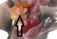
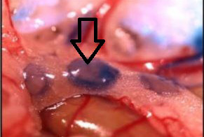
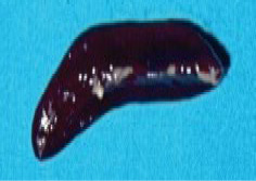
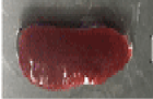
Macroscopic appearance: There are enlargement of lymph node structure with local hyperemia in some of them Figures 1 and 2 lymph nodes are may soft surface, when incised the paranchyma may bulge and the surface are wet with blood or lymph.
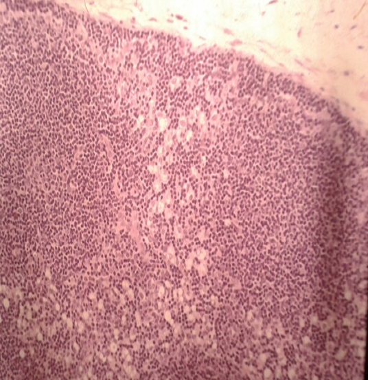
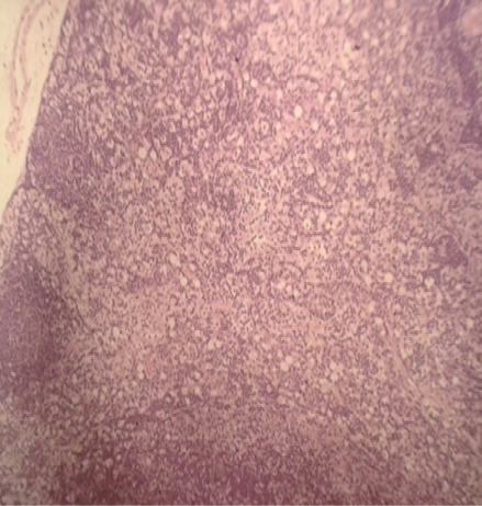
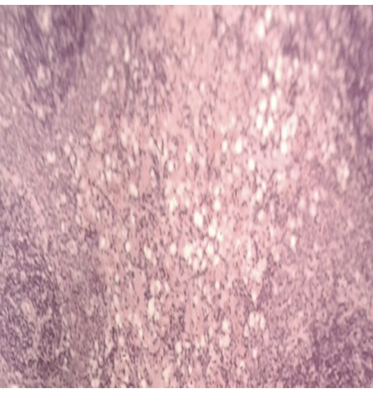
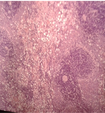
Microscopic appearance: The lymph node is hypermic and slight number neutrophils with erythrocytes are present in the sinuses and high numbers of vacoulated macrophages as foaming which are distended with lymph in other regional lymph nodes Figures 3 and 4.
The increased size of lymph node objectively reflected its response to peripheral inflammation, a set of inflammatory cytokines which is one of the most important functions of lymph node . Phagocytosis plays a main role in the scavenge of infectious agents or microbial cells and is major to regulating immune responses, inflammation, and tissues remodelling . Phagocytosis also plays a role in clearing inorganic particulate material from body such as inhaled carbon or mineral particles . Phagocytes Macrophages play a joint role with streptomycin in killing gram-positive bacteria, which helps in faster recovery than with other antibiotics . Macrophages play a joint role with streptomycin in killing gram-positive bacteria, which helps in faster recovery than with other antibiotics .
Spleen
Macroscopic appearance: The spleen tissue is so large that it exceeds it is capacity to enlargement and may colored by brown or black so it is present grossly enlarged or congested that is mean splenomegaly Figures 5 and 6.
Microscopic appearance: The response of spleen to injury are characterize by the red pulp spaces are atrophy, but in the white pulp are accumulated fibrinoide with hemorrehage and inflteration of macrophages with foam into the cytoplasm Figures 7 and 8.
The spleen filters blood in much the way that the lymph nodes filter lymph. Lymphocytes in the spleen react to pathogens in the blood and attempt to destroy them. Macrophages then engulf the resulting debris, the damaged cells, and the other large particles, our spleen also plays an important part in your immune system, which helps your body fight infection. Just as it detects faulty red blood cells, your spleen can pick out any unwelcome micro-organisms (like bacteria or viruses) in your blood, according many theories the macrophage role is carried out in the sinuses and cords of the red pulp and is a function of macrophages. Since these cells express IgG Fc receptors, red cells or platelets coated with IgG (auto)antibodies are avidly phagocytosed in the spleen .
Liver
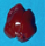
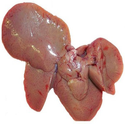
Macroscopic appearance: The grossly examination was rounded of egdes and swelling appearance with white spote in some area of surface lead to enlargement in their size Figure 9 but the high dose there are pale of color and enlargement of size Figure 10.
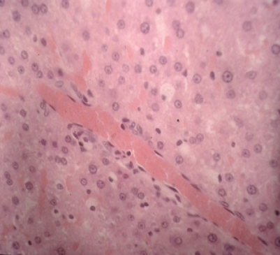
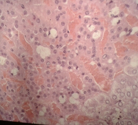
Microscopic appearance: Degeneration and even necrosis occurs in hepatocytes Figure 11 as well as hypermia in tissue Figure 12.
According to Durand et al. (1996), Sherlock and Dooley, (2002) and Stine and Lewis (2013), Free radicals induce hepatocyte damage via oxidative stress, which is the mechanism of aminoglycoside-induced hepatotoxicity. Histopathological changes increase hepatic damage because damaged hepatocytes leak their enzymes into the vascular compartment. Damage to the liver reduces its capacity to synthesize, which lowers serum levels of albumin and total protein . The obvious alterations in the liver in this investigation were pathological abnormalities in liver cells after injections of streptomycin and penicillin. This characteristic might be explained by the proposal that both of them documented histological alterations in liver cells as a result of free radical generation and free radicals may be disrupted, working on membrane phospholipids to increase cellular permeability and alter the signal transduction pathway, which finally causes serious damage to the liver tissue ; ; ; . As well as rats administered streptomycin exhibited changes in the histology of their livers and a statistically significant increase in the size of their organs in comparison to the control group. These results can indicate necrosis of the liver cells , degenerative alterations and hypofunction of the liver ; ; , and increased release of these enzymes into the bloodstream (Jaramillo-Jurez et al., 2008). According to Singh et al. (2005), Increased blood concentrations of these enzymes might be a sign of hepatocyte necrotic lesions brought on by drugs.
Kidneys
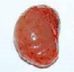
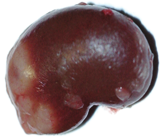
Macroscopic appearance: Grossly kidney of animals treated with 100 mg /kg are swelling, orange discoloration, and a rough surface Figure 13 and enlargement in size with brown colore and there are cyste formation in high dose 150 mg, Figure 14.
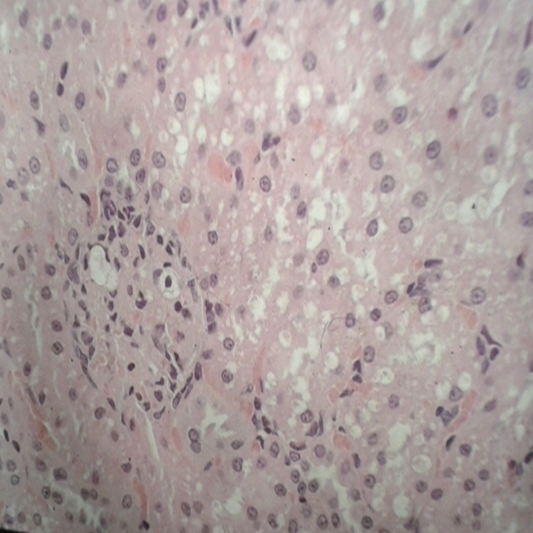
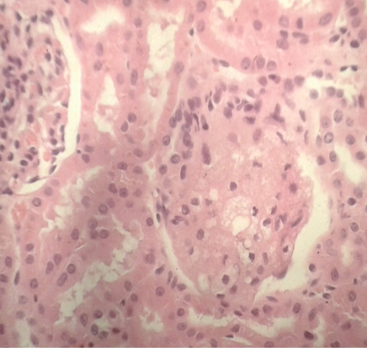
Microscopic appearance: Swelling the epithelium cells and foaming macrophages are present and hemorrhage (Figures 15 and 16).
Streptomycin is reabsorbed in the renal glands and concentrates in the proximal tubule cells. Renal injury is caused by a high trough value. Although the mechanism of renal injury is not as well understood, the contribution of free radical production and oxidative stress is questioned. Because of its ability to scavenge free radicals, vitamin C administered by CO may help avoid kidney damage . Histopathological analysis showed that the streptomycin treatment in this trial caused kidney injury. There was disarray in the renal anatomy, particularly in the proximal convoluted tubules. Significant degenerative alterations were noted.
Pyknotic nuclei were discovered to promote patchy necrosis in certain tubular cells. Because of their capacity to damage cells, free radicals have recently come under scrutiny for their role in a number of disorders (Fujita and Fujimoto., 1992; ; . Because of the unstable electron in their outer orbital ring, free radicals are extremely reactive and might potentially jeopardize the integrity of any cellular structure . Lipid peroxidation and cell damage are both caused by streptomycin. Streptomycin treatment for 30 days resulted in a significant decrease in all oxidative stress measures, including kidney damage marked by acute tubular necrosis following tobramycin 200 mg/kg/day. Boubred et al. (2006) examined the impact of increasing streptomycin concentrations on human proximal tubular cell culture.
Streptomycin’s toxicity is sufficiently low to support its use in treating severe or potentially dangerous infections. Conversely, after a few or more weeks of treatment, the rate of toxicity—particularly vestibular dysfunction—is high enough.
CONCLUSIONS AND RECOMMENDATIONS
We conclude that high-dose or prolonged streptomycin administration induces significant histopathological changes in rats, including vacuolation of macrophages in lymph nodes, splenomegaly, hepatocyte degeneration, and renal tubular damage. These findings highlight the potential risks of streptomycin overdose in clinical practice, particularly in veterinary settings where misuse may occur. Further studies should explore strategies to mitigate toxicity, such as adjunctive antioxidant therapy.
ACKNOWLEDGEMENTS
We want to thank head of department of surgery and obstetric, head of department of Patholgy and poultry disease, Dean of Veterinary Medicine and Basrah university’s president for their support in college laboratory.
NOVELTY STATEMENT
The novelty of our work entitled (Histopathological Effects of Streptomycin Treatment on Macrophages in Lymph Nodes, Spleen, Liver, and Kidneys of Rats) although there are many cases of drug poisoning, the effect of streptomycin drug adminsterition for long period still represents a challenge in veterinary medicine, and few researchers highlight the use of histopathological study after drugs adminsterition in rats.
AUTHOR’S CONTRIBUTIONS
Thaer A. Mohsin, Ibrahim M. H. Alrashid and Zainab W. Khudhair: Contributing to administering streptomycin doses, tissue sectioning, and Histopathological readings; Mohammed R. Abduljaleel, Alaa Jawad Radhi and Mosa F. Abbas: Analysis and interpretation of data, as well as writing and revision of the manuscript and journal communication.
Conflict of Interest
The authors have declared no conflict of interest.
Figures
Figure 1:.
Macroscopic picture of lymph node, was showed mesenteric lymph node after treated of 100 mg streptomycin, show enlargement with change in color.
Figure 2:.
Macroscopic picture of lymph node, was showed mesenteric lymph node after treated of 150 mg streptomycin.
Figure 3:.
Pathological section of lymph node after streptomycine 150mg, was showed macrophage with vacuolation induced by treatment with 100 mg streptomycin, lymph node notes Presence of vacuolated macrophage between lymph follicles(10x). H and E stain.
Figure 4:.
Pathological section of lymph node after streptomycine 150mg, was showed macrophage with vacuolation induced by treatment with 150 mg streptomycin, , lymph node notes Prescence of vacuolation macrophage between lymph follicles(10x). H and E stain.
Figure 5:.
Macroscopic picture of spleen with 100mg was showed splenomegaly after dose of streptomycin 100 mg.
Figure 6:.
Macroscopic picture of spleen with 150mg was showed splenomegaly after dose of streptomycin 150 mg.
Figure 7:.
Pathological section of spleen after streptomycine 100mg. Show areas of vacuolated macrophages in the red pulp (10x). H and E stain.
Figure 8:.
Pathological section of spleen after streptomycine 150mg. Show areas of vacuolated macrophages in the red pulp with fibrinoid (10x). H and E stain.
Figure 9:.
Macroscopic appearance of liver with 100mg , was showed eghed rounded, enlargement of liver lobes after toxic dose of streptomycin.
Figure 10:.
Macroscopic picture of liver with 150mg, was showed shrinking of liver lobes and pale colour after toxic dose of streptomycin.
Figure 11:.
Pathological section of liver after streptomycine 100mg, show vacuolated of hepatocytes and cells in sinusoid (10x) and H and E stain.
Figure 12:.
Pathological section of liver after streptomycine 150mg, vacuolated of hepatocytes and kuffer cells and hemorrhge of sinusoid (10x) and H and E stain.
Figure 13:.
Macroscopic picture of kidneys, was showed enlargement of kidneys after toxic dose of streptomycin with low dose100 mg/ kg.
Figure 14:.
Macroscopic picture of kidneys, was showed enlargement of kidneys after toxic dose of streptomycin with high dose 150mg showed cyst on surface.
Figure 15:.
Pathological section of kidney after streptomycine 100mg, vacuolated macrophages between cortical tubules (10x), H and E.
Figure 16:.
Pathological section of kidney after streptomycine 150mg, vacuolated macrophages between renal tubules, enlargement of glomerulus with vacolated. (10x)H and E staining.
References
Abdel-Wahhab MA, Aly SE (2005). Antioxidant property of Nigella sativa (black cumin) and Syzygium aromaticum (clove) in rats during aflatoxicosis. J. Appl. Toxicol., 25(3):218–23. https://doi.org/10.1002/jat.1057[CrossRef]
Abduljaleel MR (2024). Xylazine-Ketamine Outperforms Diazepam-Ketamine in Rabbit Anesthesia. Academia Open, 9(2): 9969-10.21070. https://doi.org/10.21070/acopen.9.2024.9969[CrossRef]
Adebajo AC, Iwalewa EO, Obuotor EM, Ibikunle GF, Omisore NO, Adewunmi CO, Obaparusi OO, Klaes M, Adetogun ,GE, Schmidt TJ, Verspohl EJ (2009). Pharmacological properties of the extract and some isolated compounds of Clausena lansium stem bark: anti-trichomonal, antidiabetic, anti-inflammatory, hepatoprotective and antioxidant effects. J. Ethnopharmacol., 122(1): 10-19. https://doi.org/10.1016/j.jep.2008.11.015[CrossRef]
Aderem A, Underhill DM (1999). Mechanisms of phagocytosis in macrophages. Ann. Rev. Immunol., 17(1): 593-623. https://doi.org/10.1146/annurev.immunol.17.1.593[CrossRef]
Al-Awara MS, AL-Shaibanib EA, Salihc EM, Al-Eryania MA (2013). The protective effect of nabk honey against pathological effects of penicillin and streptomycin: Histological structure and functions of Guinea pigs liver. J. Appl. Pharm. Sci., 3(4): S1-S6.
Alrafas HR, Alahmed JAS, Essa IM, Kadhim SZ, Al-Tameemi HM, Abduljaleel MR, Zameer F, Al-Hejjaj MY (2023). Role of anti-inflammatory interleukin 10 in asymptomatic heartworm infection (Dirofilariasis) in dogs. Adv. Life Sci., 10(3): 412-417.
Amara IB, Soudani N, Troudi A, Bouaziz H, Boudawara T, Zeghal N (2011). Antioxidant effect of vitamin E and selenium on hepatotoxicity induced by dimethoate in female adult rats. Ecotoxicol. Environ. Saf., 74(4): 811-819. https://doi.org/10.1016/j.ecoenv.2010.11.007[CrossRef]
Arsène MMJ, Davares AKL, Viktorovna PI, Andreevna SL, Sarra S, Khelifi I, Sergueïevna DM (2022). The public health issue of antibiotic residues in food and feed: Causes, consequences, and potential solutions. Vet. World, 15(3): 662. https://doi.org/10.14202/vetworld.2022.662-671[CrossRef]
Bacha WJ, Bacha LM (2012). Color Atlas of Veterinary Histology. 3rd edition.John Wiley Sons, Ltd. UK, USA, ISBN-13: 978-0-470-95851-3.
Biswas SK, Mantovani A (2010). Macrophage plasticity and interaction with lymphocyte subsets: cancer as a paradigm. Nat. Immunol., 11(10): 889-896. https://doi.org/10.1038/ni.1937[CrossRef]
Boubred F, Vendemmia M, Garcia-Meric P, Buffat C, Millet V, Simeoni U (2006). Effects of maternally administered drugs on the fetal and neonatal kidney. Drug Saf., 29: 397-419. https://doi.org/10.2165/00002018-200629050-00004[CrossRef]
Brown K, Mugoh M, Call DR, Omulo S (2020). Antibiotic residues and antibiotic-resistant bacteria detected in milk marketed for human consumption in Kibera, Nairobi. Plos One, 15(5): e0233413. https://doi.org/10.1371/journal.pone.0233413[CrossRef]
Burke B, Lewis CE (2002). The Macrophage Second edition (New York, NY, 2002; online edn, Oxford Academic, 31 Oct. 2023. https://doi.org/10.1093/oso/9780192631978.001.0001[CrossRef]
Cherubini A, Vigna GB, Zuliani G, Ruggiero C, Senin U, Fellin R (2005). Role of anti-oxidants in atherosclerosis: epidemiological and clinical update. Curr. Pharm. Des., 11(16): 2017-2032. https://doi.org/10.2174/1381612054065783[CrossRef]
Crowle AJ, Sbarbaro JA, Judson FN, Douvas GS, May MH (1984). Inhibition by streptomycin of tubercle bacilli within cultured human macrophages. Am. Rev. Respir. Dis., 130(5): 839-844.
Daniel TM (2005). Selman Abraham Waksman and the discovery of streptomycin, Founders of Our Knowledge. Int. J. Tuberc. Lung Dise., 9(2): 120-122.
Durand F, Jebrak G, Pessayre D, Fournier M, Bernuau J (1996). Hepatotoxicity of antitubercular treatments: rationale for monitoring liver status. Drug Saf., 15: 39. https://doi.org/10.2165/00002018-199615060-00004[CrossRef]
Fujita T, Fujimoto Y (1992). Formation and removal of active oxygen species and lipid peroxides in biological systems. Nihon Yakurigaku zasshi. Folia Pharmacol. Jpn., 99(6): 381-389. https://doi.org/10.1254/fpj.99.381[CrossRef]
Germovsek, E, Barker CI, Sharland M (2017). What do I need to know about aminoglycoside antibiotics?. Arch. Dis. Child. Educ. Pract., 102(2): 89-93. https://doi.org/10.1136/archdischild-2015-309069[CrossRef]
Jaramillo-Juárez F, Rodríguez-Vázquez ML, Rincón-Sánchez AR, Martínez MC, Ortiz GG, Llamas J, Posadas FA, Reyes JL (2008). Acute renal failure induced by carbon tetrachloride in rats with hepatic cirrhosis. Ann. Hepatol., 7(4): 331-338. https://doi.org/10.1016/S1665-2681(19)31833-2[CrossRef]
Jasim MM, Naeem RM, Abduljaleel MR, Sanad NH, Ibrahim AA, Alrafas HR (2025). Efficacy of autogenic, allogenic and heterogenic platelet rich plasma (PRP) on Avulsion skin wounds in rabbit model. Adv. Life Sci., 12(1): 91-97. https://doi.org/10.62940/als.v12i1.2907[CrossRef]
Jassim MM, Abduljaleel MR, Abdulkareem ZB, Sanad NH, Alrashid IMH (2023). Study the effect of the magnetic field on the healing of bone fracture after implant avian bone in femoral bone in rabbits. Adv. Anim. Vet. Sci., 11(11):1779-1784. https://doi.org/10.17582/journal.aavs/2023/11.11.1779.1784[Full text][CrossRef]
Jones AL, Schnabel EL (2000). The development of streptomycin resistant strains of Erwinia amylovora. In Fire blight: the disease and its causative agent, Erwinia amylovora, Ed. Vanneste, J.L, 235-251. https://doi.org/10.1079/9780851992945.0235[CrossRef]
Kaplan MM, Gershwin ME (2005). Primary biliary cirrhosis. N. Engl. J. Med., 353(12): 1261-1273. https://doi.org/10.1056/NEJMra043898[CrossRef]
Leonard I, Zanen J, Nonclercq D, Toubeau G, Heuson-Stiennon JA, Beckers JF, Laurent G (1994). Modification of immunoreactive EGF and EGF receptor after acute tubular necrosis induced by tobramycin or cisplatin. Ren. Fail., 16(5): 583-608. https://doi.org/10.3109/08860229409044887[CrossRef]
Liu Z, Huang Y, Wang X, Jia-Yi, Li JY, Zhang C, Yang Y, Jing Zhang J (2023).The cervical lymph node contributes to peripheral inflammation related to Parkinson’s disease. J. Neuro inflamm., 20:93. https://doi.org/10.1186/s12974-023-02770-5[CrossRef]
Luna LG (1968). Manual of histologic staining methods of the Armed Forces Institute of Pathology. 3rd Edition, McGraw-Hill, New York.
Mantovani A, Sica A, Sozzani S, Allavena P, Vecchi A, Locati M (2004). The chemokine system in diverse forms of macrophage activation and polarization. Trends Immunol., 25(12): 677-686. https://doi.org/10.1016/j.it.2004.09.015[CrossRef]
Martinez FO, Helming L, Gordon S (2009). Alternative activation of macrophages: an immunologic functional perspective. Annu. Rev. Immunol., 27(1): 451-483. https://doi.org/10.1146/annurev.immunol.021908.132532[CrossRef]
Mohammad A, Molavi F, Dolatabadi S (2022). Synergistic effect of silver nanoparticles and streptomycin antibiotic on the MexX gene expression of pump efflux system in drug-resistant Pseudomonas aeruginosa strains. J. Ilam Univ. Med. Sci., 30(2): 41-50. https://doi.org/10.52547/sjimu.30.2.41[CrossRef]
Mosser DM, Edwards JP (2008). Exploring the full spectrum of macrophage activation. Nat. Rev. Immunol., 8(12): 958-969. https://doi.org/10.1038/nri2448[CrossRef]
Robards K, Prenzler PD, Tucker G, Swatsitang P, Glover W (1999). Phenolic compounds and their role in oxidative processes in fruits. Food Chem., 66(4): 401-436. https://doi.org/10.1016/S0308-8146(99)00093-X[CrossRef]
Sahnoun Z, Jamoussi K, Zeghal KM (1997). Free radicals and antioxidants: human physiology, pathology and therapeutic aspects. Therapie, 52(4): 251-270.
Sherlock S, Dooley J (2002). Drugs and Liver. In: Diseases of the Liver and Biliary System, 11th Edition. Blackwell Science: Oxford, UK; Malden, MA. 335-63.
Singh VK, Dixit P, Saxena PN (2005). Cybil induced hepatobiochemical changes in wistar rats. J. Environ. Boil., 26(4): 725-727.
Sisein EA (2014). Biochemistry of free radicals and antioxidants. Scholars Acad. J. Biosci., 2(2): 110-118.
Stecher B, Chaffron S, Käppeli R, Hapfelmeier S, Freedrich S, Weber T, Hardt WD (2010). Like will to like: abundances of closely related species can predict susceptibility to intestinal colonization by pathogenic and commensal bacteria. PLoS Pathog., 6(1): e1000711. https://doi.org/10.1371/journal.ppat.1000711[CrossRef]
Stecher B, Robbiani R, Walker AW, Westendorf AM, Barthel M, Kremer M, Chaffron S, Macpherson AJ, Buer J, Parkhill J, Dougan G, von Mering C, Hardt WD (2007). Salmonella enterica serovar Typhimurium exploits inflammation to compete with the intestinal microbiota. PLoS Biol., 5:2177–2189. https://doi.org/10.1371/journal.pbio.0050244[CrossRef]
Stine JG, Lewis JH (2013). Hepatotoxicity of antibiotics: a review and update for the clinician. Clin. Liver Dis., 17(4): 609-642. https://doi.org/10.1016/j.cld.2013.07.008[CrossRef]
Underhill DM, Goodridge HS (2012). Information processing during phagocytosis. Nat. Rev. Immunol., 12(7): 492-502. https://doi.org/10.1038/nri3244[CrossRef]
Vianna JF, Bezerra KS, Oliveira JI, Albuquerque EL, Fulco UL (2019). Binding energies of the drugs capreomycin and streptomycin in complex with tuberculosis bacterial ribosome subunits. Phys. Chem. Chem. Phy., 21(35): 19192-19200. https://doi.org/10.1039/C9CP03631H[CrossRef]
Walker PD, Barri Y, Shah SV (1999). Oxidant mechanisms in gentamicin nephrotoxicity. Ren. Fail., 21(3-4): 433-442. https://doi.org/10.3109/08860229909085109[CrossRef]
Williams, Wilkins (2014). Briggs GGFR. Drugs in pregnancy and lactation. Baltimore, A Review of Antibiotic Use in Pregnancy. MD. Clin. Perinatol., 4:701–33.
Zhao W, Hu B, Zhang X, Wang P (2020). Pen-strep influence macrophage mechanical property and mechano-response to microenvironment. BioRxiv, 2020-04. https://doi.org/10.1101/2020.04.09.034884[CrossRef]
Zhu M, Burman WJ, Jaresko GS, Berning SE, Jelliffe RW, Peloquin CA (2001). Population pharmacokinetics of intravenous and intramuscular streptomycin in patients with tuberculosis. Pharmacotherapy: J. Hum. Pharmacol. Drug Ther., 21(9): 1037-1045. https://doi.org/10.1592/phco.21.13.1037.34625[CrossRef]