Research Article · Volume 14, Issue 3, 12 July 2026 · Open Access
Histopathological Effects of Aflatoxins in Broiler Chickens Produced by Aspergillus flavus and the Ameliorative Effects of Clove (Syzygium aromaticum) Powder
· · · ·
Affiliations & Notes ⌄
1Department of Animal Production, Faculty of Agriculture, University of Kerbala, Iraq
2Department of Forensic Science, College of Science, Al-Karkh University of Science, Baghdad, 10001, Iraq
3Department of Food Sciences, Faculty of Agriculture, University of Kerbala, Iraq. *Correspondence | Zeinab A.M. Al-Tememe, Department of Animal Production, Faculty of Agriculture, University of Kerbala, Iraq
This methodological precision strengthens the core findings of the study, demonstrating that pellet feed is a highly susceptible substrate for the proliferation of A. flavus and subsequent aflatoxin contamination.
The observed systemic histopathological degradation in liver tissues was significantly mitigated by the dietary inclusion of clove powder.
Consequently, these results validated by standardized experimental conditions confirm that cloves represent a potent, safe, and cost-effective natural alternative to synthetic antifungals, providing superior bio-protection against Aspergillus-induced pathogenesis in poultry production.
Based on the potent protective efficacy demonstrated by clove powder against A. flavus toxicosis, we recommend the dietary inclusion of clove powder at a concentration of 5 g/kg as a standard feed additive in poultry production particularly in high-humidity environments.
Selected from the article's own text.
Abstract
This study investigated the presence of Aspergillus flavus in commercial poultry pellet feed in Karbala province, its aflatoxin-producing capacity, and the potential ameliorative effects of Nystatin and clove powder on aflatoxin-induced hepatic injury in broilers. A. flavus was isolated from feed samples and confirmed via morphological and molecular techniques. Broilers were exposed to isolated aflatoxins and subsequently treated with nystatin (1 mL/L) and clove powder (5 g/kg), either individually or in combination. Histopathological analysis of aflatoxin-treated groups revealed severe hepatic architectural disruption, characterized by extensive hepatocyte degeneration, tissue necrosis, and central vein destruction with proteinaceous deposition. In contrast, the control group exhibited normal radial uniformity of hepatocytes with distinct cytoplasmic granules. Treatment with nystatin alone significantly mitigated inflammatory infiltrates and restored typical liver architecture with minimal lymphocytic presence. Most notably, the synergistic application of nystatin and clove powder effectively counteracted aflatoxin toxicity, resulting in a crowded uniform pattern of hepatocytes and a marked reduction in pathological secretory granules, indicating a potent hepatoprotective effect. The findings suggest that while aflatoxins cause devastating hepatic damage, the inclusion of clove powder as a feed additive alongside nystatin provides a robust defense mechanism against mycotoxicosis in poultry.
Keywords | A. flavus, Pellet feed, Poultry, Nystatin, Aflatoxicosis
INTRODUCTION
Aflatoxicosis remains a paramount challenge within the global poultry industry, primarily due to its profound impact on bird health and the subsequent economic repercussions. These potent mycotoxins, secondary metabolites produced predominantly by Aspergillus flavus, contaminate a wide spectrum of feed ingredients, especially those rich in protein and energy ; . The proliferation of these fungi is significantly exacerbated by tropical and sub-tropical environmental conditions, where high temperature and humidity provide an optimal niche for fungal metabolism (Aljebory and Naji, 2021a, b; . Beyond feed degradation and the emission of foul odors which reduce palatability , aflatoxins pose a severe clinico-pathological threat. Upon ingestion, even in sub-lethal concentrations, they induce multi-organ dysfunction, with the liver being the primary target organ. The hepatotoxic effects manifest as acute poisoning, impaired metabolic functions, and chronic immunosuppression, which drastically reduce growth rates and increase mortality in broiler populations ; . Furthermore, the bioaccumulation of these toxins in poultry tissues poses a significant public health risk due to their carry-over into the human food chain . Despite rigorous management protocols, traditional antifungal treatments often face limitations regarding cost and chemical residues. Consequently, there is an urgent need to explore sustainable, bio-active alternatives. Syzygium aromaticum (clove) has emerged as a promising candidate due to its rich content of phenolic compounds, particularly eugenol, which exhibits potent antifungal and antioxidant properties thereby could be used as a potential candidate in poultry to improve health and production ; . This study aimed to investigate the histopathological alterations induced by A. flavus aflatoxins in the liver tissues of broiler chickens and to evaluate the potential ameliorative efficacy of clove powder as a feed additive.
MATERIALS AND METHODS
Fungal isolation and identification
Sampling procedure
Random samples of pellet feed (200 g each) were collected from local commercial poultry feed stores in Karbala Governorate. To ensure representative analysis, samples of the same feed type were thoroughly homogenized before processing a sub-sample for fungal isolation. The culture medium was prepared by dissolving 42 g of medium in one liter of distilled water. It consisted of potato extract agar (PDA) supplemented with the antibiotic chloramphenicol at a rate of 250 mg L⁻¹. The medium was sterilized in an autoclave at 121 °C and 15 psi for 20 minutes, and the antibiotic was added before solidification.
Isolation technique
The isolation was performed using the direct plate method on Potato Dextrose Agar (PDA). The medium was prepared by dissolving 42 g of PDA in 1 L of distilled water, followed by sterilization via autoclaving at 121ºC and 15 psi for 20 minutes. To inhibit bacterial proliferation, the medium was supplemented with chloramphenicol (250 mg/L) prior to solidification. Approximately 0.5 g of the feed sample was uniformly distributed onto the PDA plates (9 cm diameter). The plates were then incubated in a BOD (biochemical oxygen demand incubator) incubator at 25 ºC for 5–7 days.
Purification and morphological identification
Emerging fungal colonies were purified using the hyphal tip technique to ensure monocultural isolates. The purified isolates were maintained on PDA slants at 4 ºC for subsequent experimental phases. Preliminary identification was conducted based on macromorphological characteristics (colony color, texture, and growth rate). Final taxonomic identification was confirmed through micromorphological examination of conidial heads, spores, and reproductive structures using the lactophenol cotton blue staining technique. The identification process followed standard mycological taxonomic keys .
Molecular identification of fungal isolates
DNA extraction and PCR amplification
The identity of the most prevalent fungal isolate was confirmed using molecular techniques. Genomic DNA was extracted from pure fungal colonies using the DNeasy Plant Mini Kit (QIAGEN, Hilden, Germany), following the manufacturer’s instructions, as described by White et al. (1990). The Internal Transcribed Spacer (ITS) region of rDNA was amplified using universal primers: ITS1 (5`-TCCGTAGGTGAACCTGCGG-3`) and ITS4 (5`-TCCTCCGCTTATTGATATGC-3`). PCR reactions were carried out in a total volume of 25 µl using Ready-To-Go PCR beads (GE Healthcare, IL, USA), with 1 µl of each primer (5 pmol) and 2 µl of template DNA (50–100 ng).
Sequencing and phylogenetic analysis
The amplified PCR products were purified and dispatched to Macrogen, Inc. (Seoul, South Korea) for bidirectional sequencing. The obtained sequences were analyzed using the Basic Local Alignment Search Tool (BLAST) against the NCBI GenBank database. Phylogenetic relationships were constructed using MEGA6 software, employing the Neighbor-Joining method with 1,000 bootstrap replicates to ensure taxonomic accuracy . The diagnosis of one of the most common fungal species was confirmed by PCR and nucleotide sequencing at the Asco learning center, Baghdad.
Production and preparation of aflatoxins
Fungal inoculation and cultivation
Aflatoxin production was induced using Potato Dextrose Broth (PDB) supplemented with 250 mg/L of chloramphenicol to prevent bacterial contamination. The medium was dispensed into 250 ml Erlenmeyer flasks and sterilized. Each flask was inoculated with a 5 mm diameter mycelial disc from a 7-day-old pure culture of Aspergillus flavus. The cultures were incubated in the dark at 25±2 ºC for 14 days to allow for secondary metabolite biosynthesis.
Extraction and filtration of mycotoxins
Following incubation, the fungal cultures (mycelia and broth) were homogenized using a high-speed electric blender for 3 minutes. The crude extract was initially filtered through a Buchner funnel under vacuum pressure. To ensure a cell-free filtrate, the extract was subsequently passed through a 0.45 µm syringe filter. The final purified extract was stored in sterile amber glass bottles at 4 ºC and utilized within 24 hours to maintain toxin stability.
Animal ethics and husbandry
All animal procedures were approved by the Animal Ethics Committee, College of Agriculture, University of Kerbala (Ethical approval code: UOK. Agri. No. ٨.2026). One-day-old broiler chicks (ROSS 308) were obtained from a commercial hatchery in Karbala. The birds were housed in a controlled environment with standardized ventilation and lighting. Temperature was maintained at 33 ºC for the first week, then gradually decreased by 2 ºC weekly. All procedures followed the institutional ethical standards for animal care.
Experimental groups and treatments
The chicks were randomly divided into five experimental groups (3 replicates per group) to evaluate the protective role of clove powder and nystatin against aflatoxicosis. The experimental period lasted 45 days. The treatment groups were structured as follows:
G1 (Control): Received a basal diet and sterile distilled water (placebo).
G2 (Aflatoxin - Positive Control): Received a basal diet and were challenged with A. flavus aflatoxins via oral gavage (1 ml/50 g BW).
G3 (Nystatin Intervention): Received aflatoxin (as in G2) + Nystatin in drinking water (1 ml/L).
G4 (Clove Intervention): Received aflatoxin (as in G2) + basal diet supplemented with clove powder (5 g/kg).
G5 (Synergistic/Combination): Received aflatoxin (as in G2) + clove-supplemented diet (5 g/kg) + Nystatin in drinking water (1 ml/L).
Histopathological processing
At the conclusion of the 45-day trial, liver samples were collected from each group for comparative histopathological analysis. The tissues were immediately fixed in 10% neutral buffered formalin for 48 hours, then processed by dehydration through ascending grades of ethyl alcohol, clearing in xylene, and embedding in paraffin wax. Tissue sections were cut at a thickness of 5 µm using a rotary microtome and subsequently stained with hematoxylin and eosin (H & E) to visualize structural alterations. The prepared slides were examined under a light microscope to identify and compare pathological features, including fatty changes, necrosis, and inflammatory cell infiltration, across all groups.
RESULTS AND DISCUSSION
Morphological and microscopic identification of A. flavus
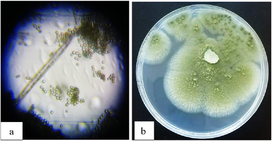
The mycological analysis of the pellet feed samples confirmed the presence of A. flavus. Macroscopically, the isolated colonies on Potato Dextrose Agar (PDA) exhibited rapid growth, initially appearing as white cottony mycelia before transforming into a characteristic yellowish-green granular texture due to profuse sporulation (Figure 1). The reverse side of the colonies showed a pale yellow to gold coloration, often accompanied by radial wrinkling. Microscopic examination (40x and 100x) revealed typical diagnostic features of A. flavus. The conidiophores were long, hyaline, and characterized by a distinct rough-walled texture, arising from well-defined foot cells. These structures terminated in globose to sub-globose vesicles. The seriation was observed to be predominantly biseriate (possessing both metulae and phialides) covering the entire surface of the vesicle. The conidia were globose, finely roughened, and arranged in divergent chains. These morphological characteristics are consistent with the standard taxonomic descriptions provided by Layton (1977) and Klich (2002).
Molecular diagnosis of A. flavus
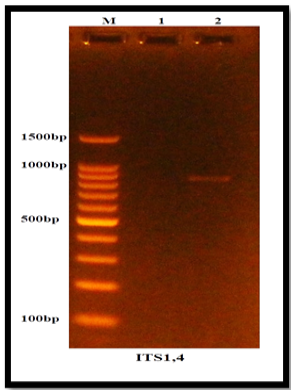
The results of DNA extraction from the fungus under study, after treatment with polymerase chain reaction (PCR) technique, were almost successful in amplifying the DNA. This technique demonstrated the ability to amplify DNA products with an estimated size of approximately 1,500 base pairs (Figure 2).
DNA analysis revealed that the fungal isolate under study is A. flavus (isolate No. 2). Traditional techniques for diagnosing fungi are currently insufficient, as the characteristics of some species overlap, making differentiation difficult. For this reason, previous experiments have been adopted on the polymerase chain reaction (PCR). This technique is known for its high accuracy in identifying organisms, including fungi, ensuring reliable and accurate diagnoses . While traditional methods rely on microscopic examination and phenotypic description, which can be slow and inaccurate, PCR technology allows for accurate and rapid identification of fungi by examining their DNA. According to Al-Tememe et al. (2019), nucleotide sequencing enables the identification of several fungal species, including Aspergillus versicolor, which has been isolated from forests, plants (Al-Hakim et al., 2023), and fish .
Histopathological evaluation of liver tissue
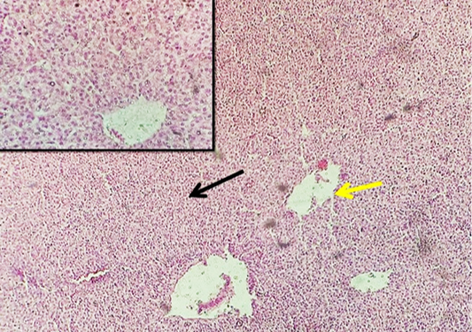
Group 1 (Control): Microscopic examination of the liver sections revealed a normal histological architecture. The hepatocytes exhibited uniform cord-like arrangements around the central vein, with distinct nuclei and normal granular cytoplasmic consistency. No evidence of inflammatory infiltration or vascular congestion was observed (Figure 3).
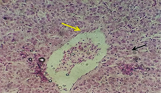
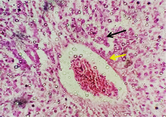
Group 2 (Aflatoxin-treated): Exposure to aflatoxins induced severe pathological alterations. Sections showed widespread hydropic degeneration and focal areas of coagulative necrosis in hepatocytes. The central veins exhibited structural disruption with perivascular proteinaceous deposition. Additionally, marked inflammatory cell infiltration and loss of the typical hepatic cord pattern were prominent features (Figure ٤).
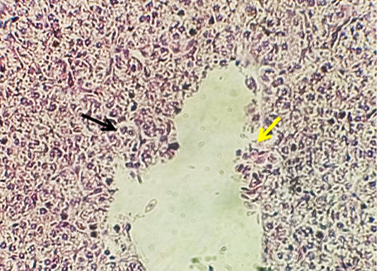
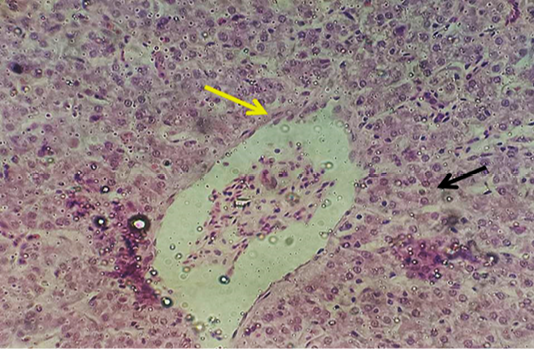
Group 3 (Aflatoxin + Nystatin): Treatment with nystatin (1 ml/L) showed a moderate palliative effect. The hepatic tissue maintained its general structural integrity with a noticeable reduction in inflammatory infiltrates compared to the aflatoxin-only group. Only scattered lymphocytic aggregates were observed, and the endothelial lining of the central veins appeared relatively intact (Figure 5).
Group 4 (Aflatoxin + Clove Powder): The liver sections in this group demonstrated a significant regenerative response. Hepatocytes appeared more active with intact nuclei and normal cytoplasmic volume. The tissue showed a high degree of cellular uniformity in size and form, indicating the protective role of clove powder against aflatoxin-induced cytotoxicity (Figure 6).
Group 5 (Aflatoxin + Nystatin + Clove): The combined treatment resulted in the most pronounced ameliorative effect. The liver parenchyma showed a crowded, uniform pattern of hepatocytes closely resembling the control group. A significant regression of pathological lesions was noted, with only minimal cytoplasmic secretory granules observed, reflecting a near-complete restoration of hepatic function and structure (Figure 7).
The histopathological findings of the current study demonstrate that aflatoxin exposure induced significant hepatic architecture disruption in broiler chicks. These alterations were characterized by vacuolar degeneration and severe necrosis, which are indicative of acute hepatotoxicity. The observed liver damage directly impairs the synthesis of essential proteins, particularly apoproteins. This deficiency hinders the formation of lipoprotein complexes required for lipid transport, thereby triggering lipid accumulation within hepatocytes and a subsequent rise in serum lipid levels a mechanism consistent with the findings of Basmacioglu et al. (2005). Our results align with Bakeer et al. (2013) and Ibrahim (2013), who documented similar pathological changes in broiler chicks and quails exposed to fungal toxins.
The ameliorative effect of clove powder observed in this study can be attributed to its high concentration of eugenol. This active compound exerts potent antifungal and anti-aflatoxigenic activities by disrupting fungal cell wall integrity and plasma membrane stability, leading to the leakage of intracellular contents and fungal cell death . Beyond its fungicidal role, eugenol effectively interferes with the biosynthetic pathways of aflatoxin B1, even at sub-inhibitory concentrations, thus mitigating the toxic load on the liver. This explains the marked improvement in the hepatic tissue integrity in the clove-supplemented groups (group 4 and 5).
Furthermore, the inclusion of nystatin provided a complementary protective layer. Nystatin’s antifungal efficacy is attributed to its high affinity for ergosterol, a key component of the fungal cell membrane. By binding to ergosterol and forming transmembrane pores, nystatin disrupts membrane integrity and induces lethal ionic imbalance within fungal cells . A key advantage of nystatin in this poultry model is its minimal systemic absorption; its localized action within the gastrointestinal tract effectively neutralizes A. flavus before systemic toxin absorption occurs, thereby reducing the histopathological burden on the liver. The synergistic or additive effects observed in the combined treatment (group 5) underscore the potential of integrating natural antioxidants with conventional antifungals to combat the pervasive threat of Aspergillus contamination in poultry feed .
Conclusions and Recommendations
To ensure maximum reproducibility and scientific rigor the experimental design utilized a robust sample size across five treatment groups with triple replicates, maintaining strictly controlled environmental parameters. This methodological precision strengthens the core findings of the study, demonstrating that pellet feed is a highly susceptible substrate for the proliferation of A. flavus and subsequent aflatoxin contamination. The observed systemic histopathological degradation in liver tissues was significantly mitigated by the dietary inclusion of clove powder. Consequently, these results validated by standardized experimental conditions confirm that cloves represent a potent, safe, and cost-effective natural alternative to synthetic antifungals, providing superior bio-protection against Aspergillus-induced pathogenesis in poultry production.
Based on the potent protective efficacy demonstrated by clove powder against A. flavus toxicosis, we recommend the dietary inclusion of clove powder at a concentration of 5 g/kg as a standard feed additive in poultry production particularly in high-humidity environments. Furthermore, this natural alternative should be integrated into biosecurity protocols as a safe and cost-effective substitute for synthetic antifungals. We also emphasize the necessity of stringent monitoring for pellet feed storage conditions to prevent initial fungal proliferation and subsequent tissue degradation.
Acknowledgments
We express our sincere thanks and gratitude to all those who played a role in the successful completion of this research.
Novelty Statement
This study demonstrates for the first time the potent synergistic hepatoprotective efficacy of combining Nystatin with clove powder (5 g/kg) as a novel feed additive strategy to completely counteract Aspergillus flavus-induced severe hepatic injury in broilers.
Author’s Contribution
All Authors contributed equally to the mansucript.
Generative AI and AI assisted technology statement
The authors declare that no generative AI and AI assisted technology was used in the creation of this manuscript.
Conflict of interest
The authors have declared no conflict of interest.
Figures
Figure 1:.
A. flavus (a): An undivided, unbranched conidiophore ending in a head that bears conidia in chains. (b): colony appearance on PDA
Figure 2:.
Molecular identification of Aspergillus flavus. The amplified DNA bands obtained by gel electrophoresis are aligned against a DNA ladder (marker) on the left side, and their fragment sizes are estimated in base pairs (bp).
Figure 3:.
Photomicrograph of liver tissue showing densely packed, uniform hepatocytes with clearly visible cytoplasmic granules (control group).
Figure 4:.
Photomicrograph of liver tissue showing severe hepatocellular degeneration and necrosis with inflammatory cell infiltration (black arrow), along with destruction of the central vein (aflatoxin-treated group only).
Figure 5:.
Photomicrograph of liver tissue showing reduced inflammatory cell infiltration with preserved hepatic architecture and a small number of lymphocytes (black arrow). The central vein exhibits a normal endothelial lining (yellow arrow) (Nystatin 1 mL/L + aflatoxin group).
Figure 6:.
Photomicrograph of liver tissue showing increased hepatocellular activity, with hepatocytes exhibiting intact nuclei, normal cytoplasmic consistency, and uniform size and shape (Clove powder + aflatoxin group).
Figure 7:.
Photomicrograph of liver tissue showing densely packed, uniform hepatocytes with few cytoplasmic secretory granules (Nystatin 1 mL/L of water + clove + aflatoxin group).
References
Adelusi, O.A., Gbashi, S., Adebo, J.A., Aasa,A.O., Oladeji, O.M., Kah, G., Adebo, O.A., Changwa, R., Njobeh, P.B., (2023). Seasonal andgeographical impact on the mycotoxigenicity of Aspergillus and Fusarium species isolated fromree State and Limpopo provinces of SouthAfrica. Toxins 15, 128. https://doi.org/10.3390/TOXINS15020128/S1[CrossRef]
Adelusi, O.A.; Njobeh, P.B. (2024). Insights from modelling sixteen years of climatic and fumonisin patterns in maize in South Africa. Sci. Rep. 14, 1–12. https:// doi.org/10.1038/s41598-024-60904-y.[CrossRef]
Al-Asadi SHAA (2006). The effect of some fungal filtrates isolated from poultry feed on the growth of chicks in some areas of Karbala. Master’s thesis, College of Education for Pure Sciences, University of Kerbala, Iraq.
Al-Hakeem AM, Al-Tememe ZA, AbdAlssirag M (2023). Morphological and molecular identification of fungi associated with Zuri fish (Pilchard sp.) in Karbala/Iraq. In: IOP Conference Series: Earth and Environmental Science. IOP Publishing. 1158(5): 052006. https://doi.org/10.1088/1755-1315/1158/5/052006[CrossRef]
Al-Jebory HH, Naji SAH (2021b). Effect of Pelleted fermented feed in production performance of laying hens. Fourth international conference for agricultural and sustainability sciences IOP Conf. Series: Earth and Environmental Science, IOP Publishing. 910(2021): 012007. https://doi.org/10.1088/1755-1315/910/1/012007[CrossRef]
Al-tememe ZA, Abdalmoohsin RG, Mohammadali MT, Al-Mosawy MM, Al-Masoudi ZM (2019). Molecular diagnosis of the fungus Sclerotinia sclerotiorum: A causal agent of white rot disease in Solanum melongena L. and its control using zinc oxide nanoparticles. Biopestic. Int., 15(1): 51–56.
Al-Tememe ZA, Lahuf AA, Kareem AA, Kadhim AA, Al-Mosawy MM (2019). A survey and molecular identification of Aspergillus versicolor causing brown rot on imported spruce (Picea canadensis) wood in Karbala province, Iraq and control it using copper boron chromate. In: IOP Conf. Ser. Earth Environ. Sci., IOP Publishing. 388(1): 012010. https://doi.org/10.1088/1755-1315/388/1/012010[CrossRef]
Al-Tememe ZAM, Hamid MH, Al-Musodi MFH (2024). The effect of the mycotoxins of some fungi isolated from poultry feeds on the relative weight of the liver and gizzard of broiler chicks in different regions of Karbala, Iraq. Open Vet. J., 15(2): 1050–1055.
Ashry A, Taha N M, Lebda MA, Fadl SE, Elkamshishi M M (2022). Effect of aflatoxin on kidney pathology in broilers. Matrouh J. Vet. Med., 2(1): https://doi.org/10.21608/mjvm.2022.122711.1009[CrossRef]
Bakeer AM, Farid AS, Gad El-Karim MF (2013). The hepatotoxic and nephrotoxic effects of mycotoxin in broiler chickens. Benha Vet. Med. J., 25(1): 29-45.
Basmacioglu H, Oguz H, Ergul M, Çöl R, Birdane YO (2005). Effect of dietary esterified glucomannan on performance, serum biochemistry and haematology in broilers exposed to aflatoxin. Czech J. Anim. Sci., 50(1): 31-39. https://doi.org/10.17221/3992-CJAS[CrossRef]
Biswas A, Ahmed M, Bharti VK, Singh SB (2011). Effect of antioxidants on physio-biochemical and hematological parameters in broiler chicken. at high altitude. Asian-Austrlras J. Anim. Sci., 24: 246-249. https://doi.org/10.5713/ajas.2011.10060[CrossRef]
Do JH, Choi DK (2007). Aflatoxins: Detection, toxicity, and biosynthesis. Biotechnol. Bioprocess. Eng., 12: 585-593. https://doi.org/10.1007/BF02931073[CrossRef]
Dutta AK, Singh SK, Upadhyay AK (2010). Identification of antifungal compound in clove that inhibits Aspergillus spp. colonizing rice grains. Int. J. Food Sci. Nutr., 61(6): 614-622.
Gbashi, S., Adelusi, O.A.; Njobeh, P.B. (2024). Insights from modelling sixteen years of climatic and fumonisin patterns in maize in South Africa. Sci. Rep. 14: 1–12. https://doi.org/10.1038/s41598-024-60904-y[CrossRef]
Ibrahim QQ (2013). Histopathological study of quails liver experimentally induced by aflatoxin. Bas. J. Vet. Res., 12(1): 116-127. https://doi.org/10.33762/bvetr.2013.76194[CrossRef]
Khalil II, Al-Jabouri KMT (1998). Mycotoxins. Second Edition. Ibaa Center for Agricultural Research.
Klich MA ((٢٠٠٢). Identification of common Aspergillus Species; Centraalbureau voor Schimmelcultures: Utrecht, The Netherlands, 2002; pp. 426–432.
Layton YM (1977). Medically important fungi: A guide to identification. Proc. R. Soc. Med., 70: 359. https://doi.org/10.1177/003591577707000519[CrossRef]
Mlaghee SM, Abbas M, Alwan MA, Fidhe MA, Tauma JA (2025). Antibacterial impact of Syzygium aromaticum extract on clinical and sub-clinical methicillin-resistant Staphylococcus aureus. J. Anim. Health Prod., 13(3): 649-653. https://doi.org/10.17582/journal.jahp/2025/13.3.649.653[Full text][CrossRef]
Mutter ZA, Alallawee MHA, Gheni QJ, Adlan RH (2026). Effect of dietary supplementation of Curcuma longa and Syzygium aromaticum on growth performance, antioxidant enzymes, and chemical composition of broiler meat (Ross 308). J. Anim. Health Prod., 14(2): 442-448. https://doi.org/10.17582/journal.jahp/2026/14.2.442.448[Full text][CrossRef]
Oladele OO, Ameji NO, Odey M, Gurumyen GY, Adanu AW, Bitrus AA, Durkwa HU, Anifowose OR, Unanam ES (2022). Management of acute Aspergillus in newly hatched chicks: A case report. Niger. Vet. J., 43(4): 64-73. https://doi.org/10.4314/nvj.v43i4.6[CrossRef]
Pinto E, Pina-Vaz C, Salgueiro L, Gonçalves MJ, Costa-de-Oliveira S, Cavaleiro C, Rodrigues RP (2009). Antifungal activity of clove oil and its main component eugenol against Candida, Aspergillus, and Dermatophytes fungi. J. Med. Microbiol., 58(10): 1368-1375.
Pitt JI, Hocking AD (1997). Fungi and food spoilage (2nd ed.). Springer Science & Business Media. https://doi.org/10.1007/978-1-4615-6391-4[CrossRef]
Rashed, A. H. and El-Nady, E. S. (2023). Mycotoxin contamin ation in livestock feed: A comprehensive review of recent trends, health effects, and control measures. Journal of Applied Animal Research, 51(1), 1-15.
Saleem F, Saleem MA, Khan AZ, Khan MS (2011). Impact of aflatoxins on poultry health and production: A review. J. Anim. Plant Sci., 21(2): 224-232.
Samson RA, Pitt JI (1994). Modern concepts in Penicillium and Aspergillus classification. Plenum Press. New York, NY.
Tamura K, Stecher G, Peterson D, Filipski A, Kumar S (2013). MEGA6: Molecular evolutionary genetics analysis version 6.0. Mol. Biol. Evol., 30: 2725–2729. https://doi.org/10.1093/molbev/mst197[CrossRef]
White TJ, Bruns T, Lee SJWT, Taylor J (1990). Amplification and direct sequencing of fungal ribosomal RNA genes for phylogenetics. PCR protocols: A guide to methods and applications, 18(1): 315-322. https://doi.org/10.1016/B978-0-12-372180-8.50042-1[CrossRef]
Histopathological Effects of Aflatoxins in Broiler Chickens Produced by Aspergillus flavus and the Ameliorative Effects of Clove (Syzygium aromaticum) Powder · ResearchersLinks