Syrinx of black francolin was tracheobronchial in type; it consists from three different groups: Tympanum, Tracho-Syringeal part, Brancho-Syringeal part (Figure 1 and 2). In addition, there were branchidesmus (interbronchial ligament), lateral and medial vibrating membranes, pessulus and triangular voice. Similar to this syringeal division was mentioned in turkeys (Khaksar et al., 2012). Meanwhile, in the other song birds, the syrinx is consisted from either the tracheal or bronchial part or both (Seller, 2002).
Three incomplete rings were formed tympanum of francolin syrinx. The last rings of tympanum bifurcate into two modified rings to form the tracho-Syringeal part (three rings), this followed by last part of syrinx (Brancho-Syringeal) which consisted from four incomplete rings (Figure 3). Comparing with sea gulls, the tympanum is consisted from five rings (İnce et al., 2012), and in long-legged buzzard are two rings (Kabak et al., 2007).
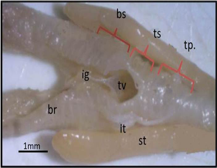
Figure 1: Macrograph of black francolin syrinx (ventral view): showing the parts of syrinx that formed by: tympanum (tp); trachea-syringeal part (ts); broncho-Syringeal part (bs); triangular voice (tv); ligament (ig); sternotrachilis (st); lateral trachealis (lt); primary bronchi (br).
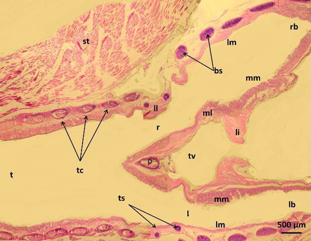
Figure 2: Micrograph of syrinx of male black francolin showing: trachea (t); tympanum cartilages (tc); trachea-syringeal cartilages (ts); pessulus (p); right passage (r); left passage (l); lateral labium (ll); medial labium (ml); lateral vibrating membrane (lm); medial vibrating membrane (mm); triangular voice (tv); sternotrachealis (st): ligament (li). H&E.
In francolin, the tympanum was lined by the pseudostratified ciliated columnar epithelia with goblet cells, the later however tended to aggregates in middle and lower parts of syrinx to form intra epithelia mucous glands which appeared in different numbers and sizes (Figure 4). These glands or the goblet cells showed positive reaction with PAS stain and Alcian blue stain (Figure 5). However, these glands appeared magenta due to its contains of sulphated mucin (Figure 6). In Bursa roller pigeon this mucosa is compose of pseudostratified prismatic epithelium or bistratified squamous, columnar (Yıldız et al., 2005; Demir kan et al., 2006) had recorded that the gland and goblet cells increased toward the broncheo-syringeal part in bursa roller pigeon. In birds, (Bacha and Bacha, 2000) and goose, (Jeffery, 2004) mentioned that plentiful goblet cells constituted the intraepithelial glands which are contain mucin with a predominance of sulphate ester salt for protection.
In the current study, the lamina propria-submucosa was consisted from loose connective tissue with high amount of elastic fibers which showed continuity with the perichondrium that surround the hyaline cartilage. The chondrocytes of the later located inside the lacunae and surrounded by homogenous matrix. These hyaline cartilages were mostly ossified at its center (Figure 7). Similar results were reported by (Aughey and Frye, 2001) however, there was partly ossified perichondrium of the syringeal hyaline cartilages in avian.
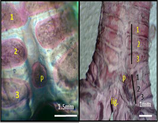
Figure 3: Macrograph of syrinx (dorsal view) of black francolin: showing tympanum which formed by (1, 2, 3, yellow color) C shaped cartilages; trachea-syringeal part which formed by (1, 2, 3, black color) C shaped cartilages; pessulus (p); ligament (lig.) Alcian blue-Alizarin red technique.
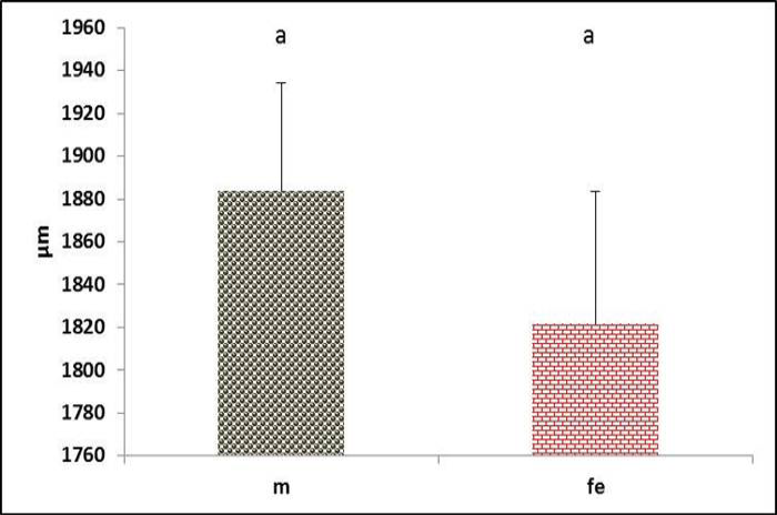
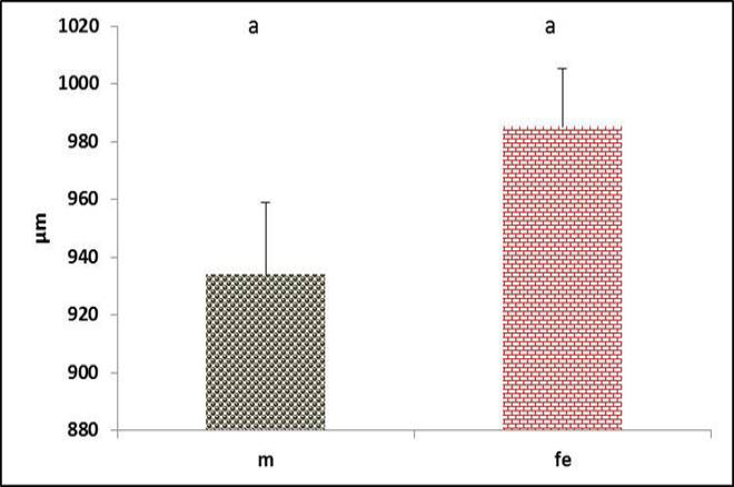
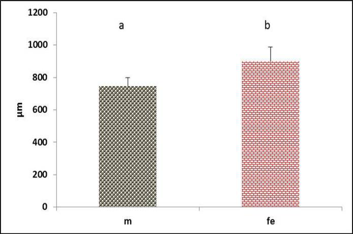
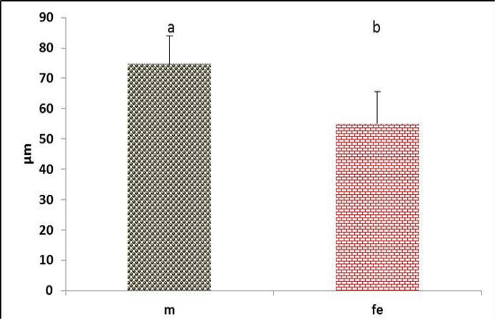
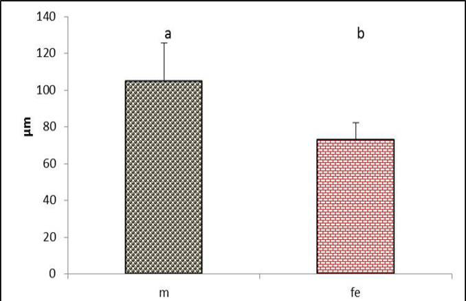
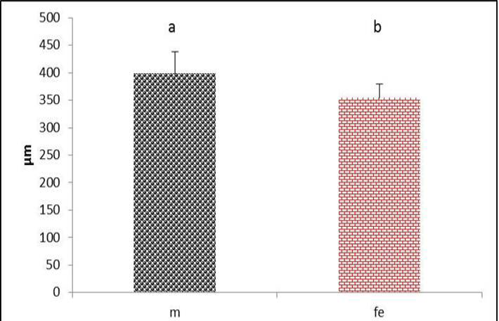
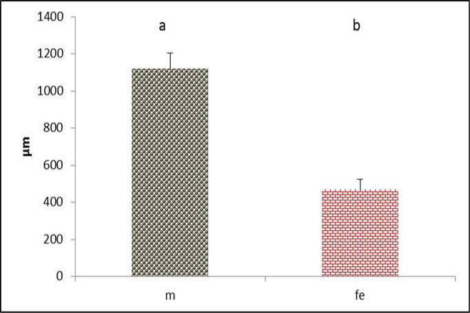
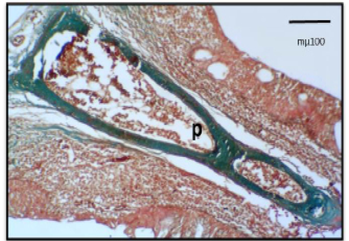
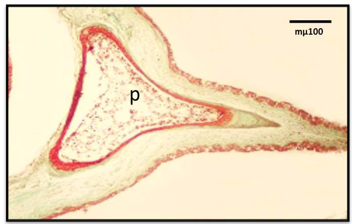
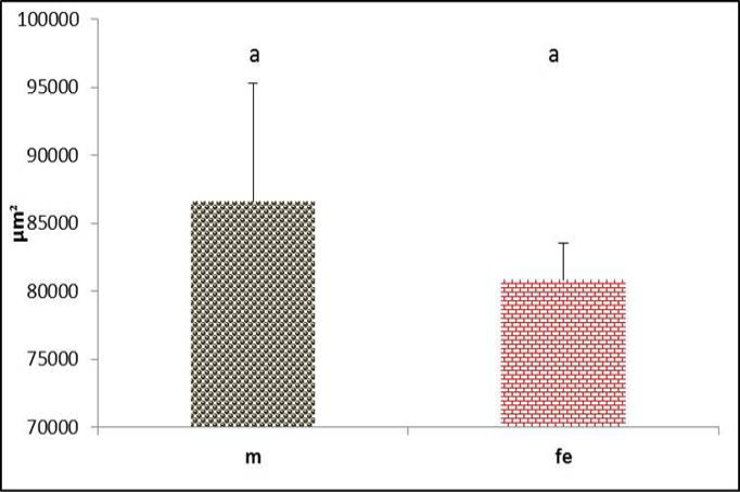
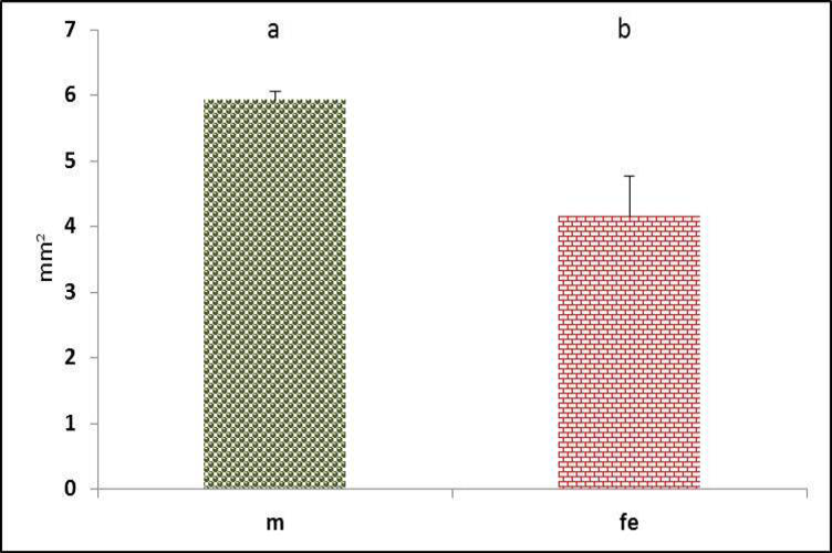
The tympanum diameter showed no difference between genders (Figure 8). Meanwhile, the diameter of right passage appeared significantly (P<0.05) narrower in male (Figure 9). In contrast, the left passage showed no difference between genders (Figure 10). These data conceded with (Suthers and Zollinger, 2004) report that in songbird, the left passage is source sound produces lower than the right side.
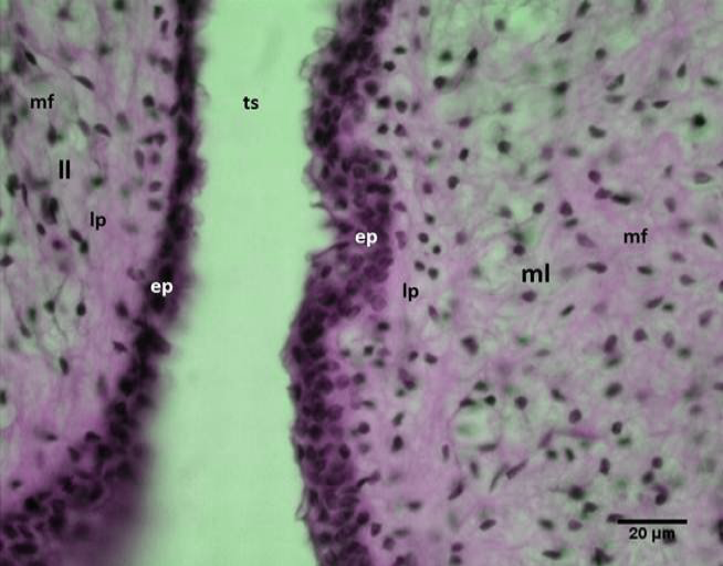
In the present study, the medial and lateral labia were thickening of the mucous membrane that protruded into the syringeal lumen between tympanum and the broncheo-syringeal parts of syrinx. The medial labia attached with the pesselus cranially and continue with thin medial tympanic (vibrating) membranes caudally, in which the later has attached with the bronchial wall medially. Meanwhile, the syringeal muscles attached directly with medial labia (Figure 11).
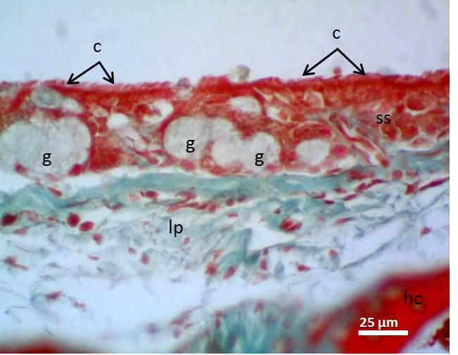
Figure 4: Micrograph of syrinx epithelium of black francolin. Whereas, cilia (c); pseudostratified ciliated columnar epithelia (ss); aggregation of goblet cells (intra epithelia mucous glands) (g); lamina propria (lp); hyaline cartilage (hc). Massson’s trichrom stain.
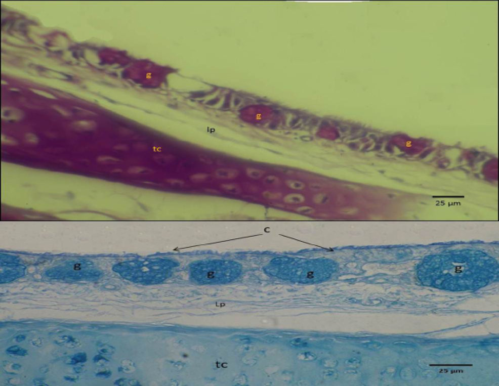
Figure 5: Micrograph of syrinx epithelium of black francolin. Showing mucin reaction of the mucous epithelia (upper with PAS) and (lower with Alcian blue stan), whearas, trachea-syringeal part (ts); aggregation of goblet cells (intra epithelia mucous glands) (g); lamina propria (lp).
The epithelium lining these labia had characteristic features, it not appeared as typical stratified columnar cells, and these cells appeared lower in height and more compact. This may be due to continues direct effects of the air flow on this part of mucosa. The extra cellular matrix of these labia was consisted mainly from collagen and smooth muscle fibers. These fibers were oriented randomly particularly in the superficial layers (Figure 12).
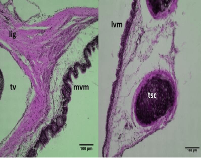
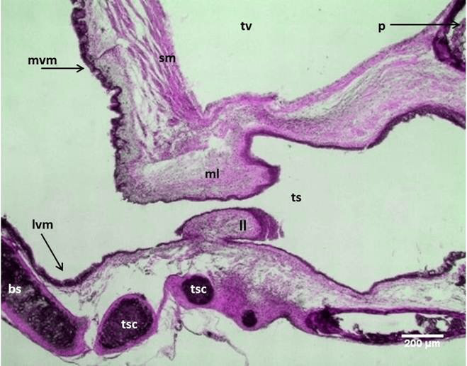
Figure 6 Micrograph: Medial vibrating membranes (mvm) notice the folded; lateral vibrating membranes (lvm); tracheo-syringeal cartilages (tsc); triangular voice (tv); sternotrachealis ligament (lig). (Combined Aldehyde Fuchsin-Alcian blue method).
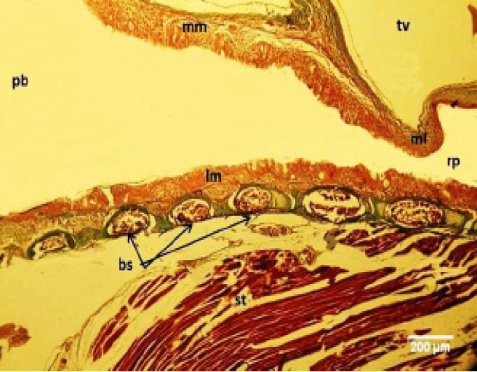
Figure 7: Micrograph of male black francolin syrinx. Showing ossification of the syringeal cartilages (bc); pseudostratified ciliated columnar epithelia lining the lateral vibrating membrane (lm) and medial vibrating membrane (mm); right syringeal passage (rp); primary bronchi (pb); triangular voice (tv); sternotrachealis (st). Massson’s trichrom stain.