Research Article · Volume 10, Issue 2, 15 June 2022 · Open Access
Hepato-Nephrotoxic Effects of Induced Fluorosis in Rabbits and Broilers
· · · · ·
Affiliations & Notes ⌄
1Department of Veterinary Pathology, Sindh Agriculture University Tandojam-70060, Pakistan
2Department of Poultry Husbandry, Sindh Agriculture University Tandojam-70060, Pakistan
3Department of Pharmacology & Toxicology, College of Veterinary Medicine, China Agriculture University, Beijing, China. *Correspondence | Zaheer Ahmed Nizamani, Department of Veterinary Pathology, Sindh Agriculture University Tandojam-70060, Pakistan
Fluorosis in humans, animals and birds is often caused by drinking of fluoride contaminated groundwater. The study was designed to evaluate nephro-hepatotoxic effects of fluorosis on liver and kidneys in rabbits and broilers. Sixteen rabbits and sixteen broilers of four weeks age were divided into four subgroups each were given 0mg (control),50 mg, 100 mg and 200 mg Sodium Fluoride /liter in water daily for 18 days. The clinical signs and mortality were noted. Blood was collected at days 0, 5, 10, 15 and 18 of experiment, for evaluation of liver and kidney functions. Dose and time dependent significant (p<0.05) increase in serum Alanine aminotransferase (GPT), Alkaline phosphatase (ALP), Gamma glutamyl transferase (γGT), uric acid and creatinine levels and significant (p<0.05) decrease of serum calcium levels were noted in all treatment groups of both species as compared to control. In rabbits and broilers, necropsy findings included mild inflammation and discoloration of liver along with nephritis. While in broilers, toxic lesions were observed on mucus membranes of duodenum and proventriculus along with nephritis. Histological lesions observed in livers of both rabbits and broilers included dilation of central vein and sinusoids and fatty degeneration of hepatocytes. Kidney tissues of both rabbits and broilers revealed marked shrinkage of glomeruli with widened bowman’s spaces along with inflammatory cellular infiltration. It is concluded that high dose (200mg/l) of Sodium Fluoride causes liver and kidney dysfunction in both species along with lesions in digestive system of broilers.
Fluoride is a micronutrient naturally found in ground water, soil and plants in variable concentrations but is also sometimes added in small amounts in drinking water to maintain desired level . In the last few decades, due to water shortages the use of poor-quality groundwater having high fluoride contamination has become a serious threat to public health, wildlife, livestock and poultry. The World Health Organization (WHO) recommended Fluoride level in drinking water is 1.5 mg/l. Li et al. (2021), in their studies showed that more than recommended level causes health problems in animals. Fluoride contaminated groundwater has been reported in various areas of Pakistan and India with concentration ranging from 0.93 to 49.3 mg/L ; ; ; ; .
Fluoride is good for dental and bone but excess amount of (>1.5 mg/L) in long term causes dental and skeletal fluorosis; renal and neuronal disorders along with myopathy . Humans, animals and birds are severely affected in fluoride contaminated areas. The fluoride concentration of up to 1.5 ppm in water causes chronic fluorosis in animals . Several studies have been conducted on fluorosis in birds, cattle, rabbits and sheep while outbreaks of fluorosis in pigs have also been reported in china ; ; .Excess fluoride intake in drinking water results decrease in milk production of cows and an increase in calving interval .
In Pakistan, the groundwater quality is poor in many areas due to high fluoride levels. Most affected areas are Indus deltaic plain and Thar Desert in Sindh province ; . As a result, people and animals are suffering from fluorosis in these areas . Keeping in view the above facts; the study was designed to evaluate the hepato-nephrotoxicity produced by induced fluorosis in mammalian (rabbit) and avian (chicken) models.
MATERIALS AND METHODS
Animal management and experimental protocol
The animals and birds were kept at the Animal House of Department of Veterinary Parasitology, Sindh Agriculture University Tandojam. A total of 24 rabbits (~ 1.5 kg BW) and twenty-four broilers (04 weeks age) were purchased from local market. An approval for the study was granted by the ethics committee, Faculty of Animal Husbandry and Veterinary Sciences, Sindh Agriculture University Tandojam. Rabbits and broilers were divided into four subgroups, each having six animals, viz; A1, A2, A3, A4 and B1, B2, B3, B4. The animals in group A1 and B1 were kept as control and given normal water. The Groups A2-B2, A3-B3 and A4-B4 were given 50, 100 and 200 mg Sodium Fluoride/litter in drinking water respectively for 18 days.
Serum Biochemistry
The 3ml blood was collected on days 0, 5, 10, 15 and 18 of experiment. For evaluation of various biochemical parameters relating to liver and kidney functions, serum levels of Alanine aminotransferase (GPT), Alkaline phosphatase (ALP), Gamma glutamyl transferase (γGT), Creatinine, Uric acid along with calcium were analyzed spectrophotometrically by using commercially available kits (HUMAN Diagnostic Co., Wiesbaden, Germany) through kinetic method as recommended by IFCC (International Federation of Clinical Chemistry) (Ceriotti et al., 2008).
Recording morbidity and mortality
All animals and birds were observed daily for development of clinical findings, morbidity and mortality.
Gross and Histopathological Examination
For studying lesions on various visceral organs necropsies were performed on dead while surviving broilers and rabbits were euthanized and dissected. For histopathological examination, the kidney and liver samples were cut into small pieces through sharp scalpel blade and were preserved in 10% formalin. Tissue samples were dehydrated in ethanol, cleared in Xylene, infiltrated / embedded in hard paraffin wax, sectioned and stained with Hematoxylin and Eosin stains. The histological lesions were recorded at 10 and 40X.
Statistical Analysis
Data was analyzed using two-way analysis of variance (ANOVA) to observe the difference between control group and experimental groups at different time intervals. Statistical differences among different treatment groups were determined by Duncan’s Multiple Range test (DMR) at 5% level of significance .
RESULTS
Serum biochemical profile
Serum biochemical profile of rabbits and broilers treated with various concentrations of Sodium fluoride in drinking water and showing serum Alanine aminotransferase (GPT), Alkaline phosphatase (ALP), Gamma glutamyl transferase (γGT), Serum creatinine, uric acid and calcium levels are shown in Table 1-3. Serum levels of Alanine aminotransferase (GPT), Alkaline phosphatase (ALP), Gamma glutamyl transferase (γGT),in all treatment groups of both rabbits and broilers significantly (p<0.05) increased in time and dose dependent manner as compared to control (Table 1). Serum creatinine and uric acid levels were significantly higher (p<0.05), in all treatment groups of both rabbits and broilers, as compared to control group (Table 2). The increases were dose dependent and time dependent. Moreover, time and dose dependent significant (p<0.05) decrease was seen in serum calcium levels in the groups treated with Sodium Fluoride as compared to control group (Table 3).
Clinical findings
There were no clinical signs observed in Rabbits during experiment, while in broiler chickens, paralysis of legs in few birds, decreased feed intake, increased water intake and intermittent diarrhea were observed in treatment groups (B2, B3, and B4) with signs being more pronounced with higher dose. There were only three deaths, one in each treatment group of broilers.
Table 1: Serum ALP, GPT and γGT levels of rabbits and broiler chicken given different concentrations of Sodium Fluoride in drinking water.
Test
Group
Day 0
Day 5
Day 10
Day 15
Day 18
ALP
(IU/L)
A1 (0)
70.67±0.0aA
70.91±0.01dA
70.9±0.02dA
70.9 ±0.04dA
70.9 ±0.03dA
A2(50)
70.8±0.03aD
72.5±0.14cCD
73.3±0.05cC
78.6 ±0.14cB
83.3 ±0.15cA
A3 (100)
70.9±0.0aE
77.51±0.23bD
80.5±0.07bC
83.3±0.29bB
86.3±0.23bA
A4 (200)
70.9±0.0aE
79.45±0.06aD
86.6±0.13aC
90.5 ±0.12aB
94.24±0.84aA
B1 (0)
76.2±0.08aA
76.10±0.02dA
76.2±0.03dA
76.1±0.07dA
76.2±0.06dA
B2(50)
75.4±0.54abE
77.4±0.04cD
79.6±0.14cC
82.38±0.04cB
85.05±0.15cA
B3 (100)
74.5±1.03bE
78.2±0.08bD
80.3±0.12bC
84.5±0.15bB
88.17±0.17bA
B4 (200)
75.4±0.18abE
83.3±0.1aD
85.1±0.03aC
88.62±0.19aB
92.42 ±0.15aA
GPT
(IU/L)
A1 (0)
25.8±0.01aA
25.8±0.01dA
25.8±0.01dA
25.8±0.01dA
25.8±0.01dA
A2(50)
25.7±0.03aE
28.3±0.02cD
30.8±0.02cC
33.8±0.06cB
35.2±0.07cA
A3 (100)
25.6±0.09aE
31.7±0.01bD
34.3±0.13bC
36.62±0.05bB
38.6±0.06bA
A4 (200)
25.6±0.14aE
35.6±0.05aD
37.4±0.13aC
42.33±0.08aB
45.58±0.12aA
B1 (0)
37.5±0.13aA
37.6±0.11dA
37.6±0.14dA
37.7±0.06dA
37.7±0.01dA
B2(50)
37.4±0.16aE
39.1±0.02cD
41.7±0.77cC
43.2±0.11cB
46.7±0.08cA
B3 (100)
37.8±0.08aE
42.5±0.05bD
44.4±0.16bC
46.6±0.12bB
48.5±0.15bA
B4 (200)
37.6±0.07aE
45.7±0.07aD
47.3±0.08aC
49.3±0.2aB
49.3±0.24aA
γGT
(IU/L)
A1 (0)
6.2±0.02aA
6.1±0.01dA
6.2±0.02dA
6.2±0.14dA
6.3±0.1dA
A2(50)
6.2±0.05aE
10.3±0.16cD
13.8±0.29cC
16.5±0.08cB
18.4±0.1cA
A3 (100)
6.1±0.05aE
15.3±0.05bD
18.3±0.05bC
19.6±0.14bB
21.7±0.13bA
A4 (200)
6.1±0.03aE
20.1±0.04aD
20.7±0.01aC
21.2±1.33aB
24.3±0.95aA
B1 (0)
17.5±0.12aA
17.5±0.12dA
17.5±0.13dA
17.5±0.13dA
17.5±0.13dA
B2(50)
17.3±0.09aE
18.5±0.16cD
21.6±0.17cC
24.5±0.06cB
26.5±0.05cA
B3 (100)
17.4±0.2aE
20.4±0.02bD
24.3±0.16bC
28.6±0.12bB
31.1±0.33bA
B4 (200)
17.6±0.07aE
45.7±0.07aD
47.3±0.08aC
49.3±0.2aB
49.3±0.24aA
Means±SD with different lowercase superscripts, for each species, show significant differences among the treatments for each time point (comparison per column), whereas different capital letters show significant differences among the time points for each treatment (comparison per line) (p<0.05).
Table 2: Serum creatinine and uric acid levels of rabbits and broiler chicken given different concentrations of Sodium Fluoride in drinking water.
Test
Group
Day 0
Day 5
Day 10
Day 15
Day 18
Creatinine
(mg/dl)
A1 (0)
1.9±0.05aA
1.9±0.11dA
1.9±0.52dA
1.9±0.17dA
1.9±0.31dA
A2(50)
1.9±0.0aD
3.3±0.1cC
4.4±0.17cCB
5.4±0.24cB
7.6±0.13cA
A3 (100)
1.9±0.0aD
4.8±0.02bC
5.4±0.1bCB
6.6±0.16bB
8.9±0.02bA
A4 (200)
1.9±0.0aE
8.6±0.1aD
10.2±0.08aC
13.9±0.09aB
17.4±0.42aA
B1 (0)
1.9±0.02aA
1.9±0.04dA
1.9±0.34dA
1. 9±0.13dA
1.9±0.27dA
B2(50)
1.8±0.04aE
2.8±0.03cD
3.3±0.11cC
3.6±0.12cB
5.2±0.15cA
B3 (100)
1.7±0.04aE
4.5±0.09bD
5.9±0.28bC
8.4±0.14bB
9.5±0.09bA
B4 (200)
1.8±0.08aE
5.4±0.08aD
7.2±1.12aC
9.2±0.19aB
14.7±1.54aA
Uric Acid (mg/dl)
A1 (0)
1.6±0.02aA
1.6±0.04dA
1.6±0.01dA
1.6±0.01dA
1.6±0.16dA
A2(50)
1.6±0.04aE
3.5±0.25cD
4.6±0.04cC
6.3±0.15cB
7.6±0.1cA
A3 (100)
1.6±0.01aE
4.1±0.04bD
7.1±0.22bC
8.4±0.15bB
10.3±0.25bA
A4 (200)
1.7±0.06aE
9.4±0.09aD
14.5±0.18aC
18.4±0.19aB
19.7±0.04aA
B1 (0)
12.1±0.05aA
12.2±0.13dA
12.1±0.1dA
12.1±0.03dA
11.9±0.18dA
B2(50)
12.1±0.08aE
14.1±0.22cD
23.3±1.15cC
28.1±0.64cB
29.9±0.49cA
B3 (100)
12.1±0.04aE
16.7±0.10bD
26.7±0.1bC
36.4±2.16bB
35.1±0.57bA
B4 (200)
12.1±0.05aE
18.9±0.33aD
39.1±2.53aC
58.5±2.4aB
68.1±4.57aA
Means±SD with different lowercase superscripts, for each species, show significant differences among the treatments for each time point (comparison per column), whereas different capital letters show significant differences among the time points for each treatment (comparison per line) (p<0.05).
Table 3: Serum calcium levels of rabbits and broiler chicken given different concentrations of Sodium Fluoride in drinking water.
Test
Group
Day 0
Day 5
Day 10
Day 15
Day 18
Calcium (mg/dl)
A1 (0)
21.4±0.03aA
21.4±0.33aA
21.4±0.46aA
21.4±0.07aA
21.4±0.61aA
A2(50)
21.5±0.01aA
21.2±0.05aA
20.4±0.1abB
19.2±0.04bC
18.8±0.01bD
A3 (100)
21.5±0.01aA
20±0.11abB
19.4±0.09bC
18.6±0.06cbD
16.4±0.17cE
A4 (200)
21.5±0.01aA
19.8±0.02bB
16.3±0.12cC
15.61±0.02cD
11.4±0.14dE
B1 (0)
13.4±0.07aA
13.5±0.01aA
13.3±0.18aA
13.3±0.07aA
13.5±0.01aA
B2(50)
13.6±0.06aA
12.4±0.01bB
10.5±0.06bC
8.5±0.03bD
6.9±0.22bE
B3 (100)
13.6±0.07aA
8.6±0.03cB
8.3±0.04cC
6.7±0.06cD
5.5±0.09cE
B4 (200)
13.6±0.06aA
6.5±0.05dB
5.5±0.12dC
4.3±0.06dD
3.5±0.18dE
Means±SD with different lowercase superscripts, for each species, show significant differences among the treatments for each time point (comparison per column), whereas different capital letters show significant differences among the time points for each treatment (comparison per line) (p<0.05).
Gross and Histopathological Examination
In rabbits, postmortem lesions observed in kidney (Figure 1) included enlarged and inflamed kidneys, while in liver (Figure 2) there were inflammation and discoloration. The most common postmortem lesions observed in broilers were the toxic lesions on mucus membranes of proventriculus, swollen and congested spleen and enlarged gallbladder. The kidneys and liver were discolored and enlarged.
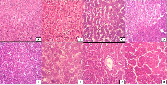
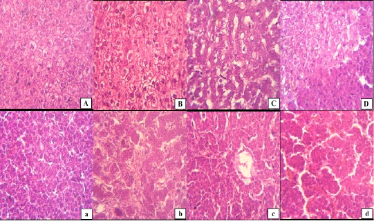
The kidney tissues, in both species had mild inflammatory cellular infiltration, hyperemia in intertubular space, shrinkage of glomeruli with widened bowman’s spaces (Figure 1). Sections of liver tissue of rabbit showed dilation of central vein and sinusoids and fatty degeneration of hepatocytes (Figure 2). On other hand, group of broilers showed hepatocytes surrounding the central vein were observed to possess darkly staining condensed nuclei and necrosis of the hepatocytes around the peri-portal and central vein area (Figure 2).
DISCUSSION
The fluorosis affects multiple organ systems of body including teeth, bones, gastrointestinal tract, nervous and muscular tissues. Among various modes of injury, reducing blood calcium levels, involvement of free radicals, increased generation of oxygen radicals and enhanced lipid peroxidation have been shown to play an important role in fluorosis ; ; . Most nutrients and toxins upon absorption, first pass through and are metabolized by liver and are excreted by kidneys. Therefore, these two organs are commonly affected in most toxicosis. While subtle damage is only reflected in changes of serum biochemical parameters, more severe damage is seen in on gross and microscopic levels. The present study was, therefore, designed to evaluate the effect of oral administration of high dose Sodium Fluoride in drinking water on liver and kidney structure and function.
In current study, both species belonging to two different taxonomic classes, showed structural and functional disturbance of liver and kidneys. The clinical signs of fluorosis and mortalities occurred only chicken, indicating greater susceptibility to fluoride toxicity. One probable reason could be the much higher growth and metabolic rate along with greater feed and water requirement in chickens.
Liver plays a major role in detoxification of various compounds. Serum ALP, γGT, GPT levels are important markers of liver function. In present study, increased serum levels of ALP, γGT and GPT show that liver is significantly affected in fluorosis. Similar results showed in rats and rabbits ; . It is in line with various other studies . Similarly, Faccini and Teotia (1974) have also reported the high plasma alkaline phosphatase in the patients affected with fluorosis. Livers in both rabbits and broiler chicken tissues of rabbit showed dilation of central vein and sinusoids and fatty degeneration of hepatocytes while in broiler showed hepatocytes surrounding the central vein having degenerative changes and necrosis in some areas (Figure 2). Similar lesions observed in liver of rats, mouse and ducks ; ;. Shashi et al. (2001) have also reported degenerative changes in liver of rabbits but changes were much more severe along with necrosis as duration of fluoride administration was 15 weeks. Similar findings have also been reported in fluoride exposed mice and rats ; ; ; .
The kidney is the major route for removal of fluoride from the body, and consequently, this organ is sensitive to damage because of excessive fluoride exposure. In our study, serum levels of uric acid and creatinine in both rabbits and broilers increased with dose as well time duration. This study is also in agreement with Al-safei & Al-Mashhadane, (2021) and Dembińska-Kieć et al. (2017) who reported that in fluorosis produced significant increase in the concentration of creatinine in the blood due to impaired renal function. Similarly, Appleton (1995) also reported that high doses of sodium fluoride injection into rats resulted in increases in the concentration of urea, and creatinine in the plasma. In rat, kidney is exposed to concentrations of fluoride about five times higher than in other organs, as the tissue/plasma ratio for the kidney is approximately 5 to 1. In current study, mild inflammatory cellular infiltration, hyperemia in intertubular space, shrinkage of glomeruli with widened bowman’s spaces were observed. Other researchers have found more or less severe gross and histopathological changes in kidneys as fluoride doses, routes of administration, duration of treatment and species vary. Basha and Rao (2014) found necrosis in glomerulus, degenerative changes in Bowman’s capsule and alterations in glomerulus’s tubular region in fluoride-treated albino mice. Similarly, Shashi et al. (2002) also reported same investigation necrotic and degenerative changes in kidney on rabbit however duration of treatment was fifteen weeks. Xu et al. (2006) reported similar the hypertrophy and hyperplasia in the renal tubules and damage kidney structure in fluoride-treated rats.
In the present study, rabbits and broilers were tested for serum calcium levels. The serum concentration of calcium in both rabbits and broilers decreased with fluoride dose as well time duration of treatment. Similar decrease has also been reported by various other researchers as well ; ; ; . Similar observations were also reported by Faccini and Teotia (1974) who reported that increased urinary fluoride, a low urinary calcium excretion along with increased Ca and F in bone in the patients affected with fluorosis. Similarly, Appelton (1995) reported calcium decreased in blood due to renal insufficiency and cause in hypocalcemia in fluoride- treated rats. The decrease in serum calcium is related to a decrease of intestinal absorption of calcium by fluorine .
CONCLUSION
It is concluded that, there are dose and time dependent hepato-nephrotoxic effects of fluorosis in the rabbits and broilers as is reflected by gross and histopathological changes along with significant increase in serum ALP, γGT, GPT, creatinine, uric acid levels and decrease in calcium concentration.
acknowledgements
The authors thank the department of Veterinary Parasitology, Faculty of Animal Husbandry and Veterinary Sciences, Sindh Agricutlure University Tandojam for providing the facility of Animal House for experiments.
Conflict of Interest
The authors declare that there is no conflict of interests regarding the publication of this article.
novelty statement
Livestock and poultry farming industries in many parts of Pakistan have difficulties due to high fluoride content of groundwater. In the manuscript we have used a mammalian (rabbit) and avian (chicken) model to investigate the hepato-nephropathic effects of induced fluorosis. We report that dose-dependent subacute fluoride toxicity affects both function as well structure of liver and kidneys of rabbits and chicken.
Authors Contributions
Barirah Rehman Talpur conducted all experiments as part of MPhil research. Dr. Zaheer Ahmed Nizamani designed experiments and analyzed the data. Dr. Mansoor Tariq, Dr. Imdad Hussain Leghari and Aisha Rehman helped in data collection. Dr. Shahnawaz Kumbhar assisted in histopathological evaluation of slides.
Figures
Figure 1. Figure 1.
Photomicrographs of the kidneys of rabbits (Uppercase letters) and broilers (Lowercase letters) respectively, intoxicated with Sodium Fluoride. Sections from control group (A, a), groups given 50 mg/L (B, b), 100 mg/L (C, c) and 200 mg/L of NaF (D, d) in drinking water. (H&E) 400X.
Figure 2.
Photomicrographs of the Liver of rabbits (Uppercase letters) and broilers (Lowercase letters) respectively, intoxicated with Sodium Fluoride. Sections from control group (A, a), groups given 50 mg/L (B, b), 100 mg/L (C, c) and 200 mg/L of NaF (D, d) in drinking water. (H&E) 200X.
References
Akdogan M, G Eraslan, F Gultekin, F Sahindokuyucu, D Essiz (2004), D. Effects of fluoride on lipid peroxidation in rabbits. Fluoride. 37(3): 185-189.
Akinrinde, Akinleye SM, T Monsuru, Awodele A, Olusola AA., Oyagbemi A (2021). Fluoride-induced hepatotoxicity is prevented by L-Arginine supplementation via suppression of oxidative stress and stimulation of nitric oxide production in rats. Toxicol. Environ. Health Sci. 13(1): 57-64. https://doi.org/10.1007/s13530-020-00070-6[CrossRef]
Al-safei, Amina N, Al-Mashhadane, Faehaa A (2021). Effects of Sodium Fluoride on Liver and Kidney in Rabbits. Egyptian J. Chem. 64(10): 3-4.
Appleton J (1995). Changes in the plasma electrolytes and metabolites of the rat following acute exposure to sodium fluoride and strontium chloride. Archiv. Oral Biol. 40(4): 265-268. https://doi.org/10.1016/0003-9969(94)00186-F[CrossRef]
Ayoob S, Gupta AK (2006). Fluoride in drinking water: a review on the status and stress effects. Crit. Rev. Environ.Sci. Technol. 36(6): 433-487. https://doi.org/10.1080/10643380600678112[CrossRef]
Basha SK, Rao KJ (2014). Sodium fluoride induced histopathalogical changes in liver and kidney of albino mice. Acta Chim. Pharmaceut. Indica. 4(1): 58-62.
Brahman, Kapil D, Kazi, Tasneem G, Afridi H, I Naseem, S Arain, S Sadaf, Wadhwa, Sham K, Shah F (2013). Simultaneously evaluate the toxic levels of fluoride and arsenic species in underground water of Tharparkar and possible contaminant sources: a multivariate study. Ecotoxicol. Environ. Safety. 89: 95-107. https://doi.org/10.1016/j.ecoenv.2012.11.023[CrossRef]
Ceriotti, Ferruccio B, James C, Klein, Gerhard H, Joseph, Queralto J, Kairisto V (2008). Reference intervals for serum creatinine concentrations: assessment of available data for global application. Clin. Chem. 54(3): 559-566. https://doi.org/10.1373/clinchem.2007.099648[CrossRef]
Chandra S (1997). Fluoride: experimental toxicosis, toxicokinetics and immunomodulation in calves: Indian Veterinary Research Institute Izatnagar, India.
Chen L, Kuang P, Liu H, Wei Q, Cui Hengmin F, Jing, Zhao L (2019). Sodium Fluoride (NaF) Induces Inflammatory Responses Via Activating MAPKs/NF-κB Signaling Pathway and Reducing Anti-inflammatory Cytokine Expression in the Mouse Liver. Biolog. Trace Element Res. 189(1): 157-171. https://doi.org/10.1007/s12011-018-1458-z[CrossRef]
Chinoy NJ, Sharma M, Michael M (1993). Beneficial effects of ascorbic acid and calcium on reversal of fluoride toxicity in male rats. Fluoride. 26(1): 45-56.
Dembińska-Kieć, Aldona N, Jerzy W, Solnica B (2017). Diagnostyka laboratoryjna z elementami biochemii klinicznej: Edra Urban & Partner.
Farooqi Abida, M Harue, Firdous N (2007). Toxic fluoride and arsenic contaminated groundwater in the Lahore and Kasur districts, Punjab, Pakistan and possible contaminant sources. Environ. Pollutt. 145(3): 839-849. https://doi.org/10.1016/j.envpol.2006.05.007[CrossRef]
Guo, Xiao-ying S, Gui-fan, Sun Y (2003). Oxidative stress from fluoride-induced hepatotoxicity in rats. Fluoride. 36(1): 25-29.
Kazi Tasneem G, Brahman, Kapil D, Afridi HI, Shah F, Arain MB (2018). Effects of high fluoride content in livestock drinking water on milk samples of different cattle in endemic area of Pakistan: risk assessment for children. Environ. Sci. Pollut. Res. 25(13): 12909-12914. https://doi.org/10.1007/s11356-018-1563-8[CrossRef]
Kisku Ganesh C, Sahu P (2020). Fluoride contamination and health effects: an Indian scenario Environ. Concerns Sustain. Develop., 213-233. https://doi.org/10.1007/978-981-13-5889-0_11[CrossRef]
Li A, Wang Y, He Y, Liu B, Iqbal M, Mehmood K, Li Y (2021). Environmental fluoride exposure disrupts the intestinal structure and gut microbial composition in ducks. Chemosphere., 277: 130222. https://doi.org/10.1016/j.chemosphere.2021.130222[CrossRef]
Madan J, Puri JP, Singh JK (2009). Growth, feed efficiency and blood profile of buffalo calves consuming high levels of fluoride. Trop. Anim. Health Prod. 41(3): 295-298. https://doi.org/10.1007/s11250-008-9189-3[CrossRef]
Ouyang Z, Yang B, Yi J, Zhu S, Lu S, Liu Y (2021). Hussain, R. Exposure to Fluoride induces apoptosis in liver of ducks by regulating Cyt-C/Caspase 3/9 signaling pathway. Ecotoxicol. Environ. Safety. 224: 112662. https://doi.org/10.1016/j.ecoenv.2021.112662[CrossRef]
Park Y, Kim D, Ku H, Kang H (2021). Fluoride levels and biochemical assessments in cattle accidentally exposed to hydrofluoric acid in Korea. Toxicolog. Res. 1-6. https://doi.org/10.1007/s43188-020-00076-9[CrossRef]
Rafique T, Naseem S, Bhanger MI, Usmani TH (2008) Fluoride ion contamination in the groundwater of Mithi sub-district, the Thar Desert, Pakistan. Environ. Geol. 56: 317–326.
Rafique T, Naseem S, Ozsvath D, Hussain R, Bhanger MI, Usmani TH (2015). Geochemical controls of high fluoride groundwater in Umarkot sub-district, Thar Desert, Pakistan. Sci. Total Environ. 530: 271-278.
Ranjan R, Ranjan A (2015). Fluoride toxicity in animals: Springer. https://doi.org/10.1007/978-3-319-17512-6[CrossRef]
Rasool A, Farooqi A, Xiao T, Ali W, Noor S, Abiola O, Nasim W (2018). A review of global outlook on fluoride contamination in groundwater with prominence on the Pakistan current situation. Environ. Geochem. Health. 40(4): 1265-1281. https://doi.org/10.1007/s10653-017-0054-z[CrossRef]
Rzeuski R (1998). Interactions between fluoride and biological free radical reactions. Fluoride., 31: 43-45.
Shah Mohammad T, Danishwar S (2003). Potential fluoride contamination in the drinking water of Naranji area, northwest frontier province, Pakistan. Environ. Geochem. Health. 25(4): 475-481. https://doi.org/10.1023/B:EGAH.0000004579.54947.e7[CrossRef]
Shashi A, Singh JP, Thapar SP (2002). Toxic effects of fluoride on rabbit kidney. Fluoride., 35(1): 38-50.
Shashi A, Thapar SP (2001). Histopathology of fluoride-induced hepatotoxicity in rabbits. Fluoride. 34(1): 34-42.
Shupe James L, Olson Arland E, Sharma RP (1972). Fluoride toxicity in domestic and wild animals. Clin. Toxicol. 5(2): 195-213. https://doi.org/10.3109/15563657208990999[CrossRef]
Takizawa S (2008). Groundwater management in Asian cities: Technology and policy for sustainability (Vol. 2): Springer Science & Business Media. https://doi.org/10.1007/978-4-431-78399-2[CrossRef]
Tao X, Xu Z, Rong, Wang YZ (2006). Effects of dietary fluoride levels on growth, serum indexes and antioxidant systems in growing pigs. Turkish J. Vet. Anim. Sci. 30(1): 65-70.
Wang JD, Zhan CW, Chen YF, Li JX, Hong JP, Wang WF, Cai JP (1992). A study of damage to hard tissue of goats due to industrial fluoride pollution. Fluoride., 25(3): 123-130.
Xu H, Jin X, Jing L, Li (2006). Effect of sodium fluoride on the expression of Bcl-2 family and osteopontin in rat renal tubular cells. Biolog. Trace Element Res. 109(1): 55-60. https://doi.org/10.1385/BTER:109:1:055[CrossRef]