
The present study was carried out to investigate the effects of lead toxicity on Japanese quail and its alleviation with dietary selenium. Thirty healthy Japanese quails at the age of about 3 to 4 weeks were divided into six groups viz., A (control group), B (selenium 1.0 mg/L), C (lead acetate 200 mg/L), D (lead acetate 400 mg/L ), E (lead acetate 200 + selenium 1.0 mg/L) and F (lead acetate 400 + selenium 1.0 mg/L). Both selenium and lead acetate were given by adding in the drinking water for 20 days. At the end of experiment, four birds from each group were slaughtered and blood was collected for evaluation of biochemical tests like Gamma-glutamyltransferase (GGT), bilirubin, albumin, uric acid and creatinine. After necropsy examination, the liver and kidney samples were collected for gross and histopathological examination. The mild clinical signs like reduced feed and water intake, and weight loss in group C, D, E and F was noted; whereas diarrhea, muscular tremors, and lethargy were observed in group C and D. Significantly increased (p <0.05) GGT, bilirubin, creatinine and uric acid levels were recorded in C and D (lead toxicity groups), as compared to control group; whereas group E and F (treated with selenium) showed the protective effect by decreasing (p <0.05) GGT, creatinine, bilirubin and uric acid concentration as compare to C and D groups. Gross pathology observed in liver of C, D and F groups revealed inflammation, and discoloration, whereas kidneys were found swollen and congested with distended ureters. Major histopathological changes observed in kidneys of group C, D and F like shrinkage of glomeruli with widened bowman’s spaces, necrosis and inflammation. Whereas, liver shows severe dilation of central vein and sinusoids, congestion and necrosis. Group E showed no major changes in liver and kidney while group F has less severity than groups C and D. The present study concluded that selenium has a protective effect against toxicity produced by lead acetate as evident from reduced morbidity, amelioration of liver and kidney function with decrease in serum GGT, creatinine and uric acid and albumin increase; thus selenium might be adapted for strategies against the toxicological effects of lead in avian species.
Research Article · Volume 11, Issue 3, 25 October 2023 · Open Access
Effect of Lead Toxicity on Japanese Quail and its Alleviation with Dietary Selenium
· · · · ·
Affiliations & Notes ⌄
- 1Department of Veterinary Pathology, Faculty of Animal Husbandry and Veterinary Sciences, Sindh Agriculture University, Tandojam, 70060 Pakistan
- 2Department of Veterinary Pharmacology, Faculty of Animal Husbandry and Veterinary Sciences, Sindh Agriculture University, Tandojam, 70060 Pakistan
- 3Department of Veterinary Microbiology, Faculty of Animal Husbandry and Veterinary Sciences, Sindh Agriculture University, Tandojam, 70060 Pakistan *Correspondence | Nusrat Habiullah, Department of Veterinary Pathology, Faculty of Animal Husbandry and Veterinary Sciences, Sindh Agriculture University, Tandojam, 70060 Pakistan
- 4Email: nkkeerio@gmail.com
Article Info ⌄
- Journal:
- Journal of Animal Health and Production
- Published:
- 25 October 2023

From the article
- This present research study was undertaken to investigate the probable effect of lead acetate toxicity on biochemical and histopathological changes in quail and its protection via selenium.
- In our present experiment, different dosages of lead acetate increased creatinine and uric acid values, which were again reduced by selenium.
- Based on the present findings, it can be concluded that, lead acetate had acute toxicity in liver and kidney of Japanese quail.
- Our study suggests that the addition of 0.1 mg/L selenium can ameliorate the toxicological effects induced by lead up to 200 mg/L. These results have implications for the development of strategies to mitigate lead toxicity in avian species, such as Japanese quail, through dietary selenium supplementation.
Selected from the article's own text.
Abstract
INTRODUCTIOn
Lead is a heavy metal that can be found in a variety of forms in soil, water, and plants. It is a natural component of the Earth’s crust. Lead is well as -known heavy metal contaminant with significant toxicity and a long-term environmental persistence due to its non-biodegradable nature . Lead poisoning in birds is a serious problem (Yazdanparast et al., 2022), when it comes to birds, As a result of the exposure to lead in the liver, histopathology liver and kidney damage, as well as a reduction in bone mineralization . Lead poisoning can influence laying hens’ performance in reproduction by reducing fertility . Lead Poisoning has a variety of negative consequences in the body including problems with the brain; kidneys, liver, circulation, and immune system are all affected (Offor and Orisakwe, 2017). Furthermore, low-dose lead exposure can cause considerable apoptosis in the liver and kidneys, eventually impairing their function .
Lead poisoning has resulted in a large number of bird deaths, and it continues to be a serious hazard to both terrestrial and aquatic species . It is a major cause of death of many endangered species like the California condor (Gymnogyps californianus); while many other species are at high risk . Lethargy, anorexia, crop paralysis, vomiting, diarrhea, ataxia, anemia, osteoporosis, emaciation, were all symptoms of lead poisoning. Moreover it may cause deterioration of sperm quantity and quality in male birds. Additionally, lead poisoning can cause memory loss in young birds (Vallverdu et al., 2016;.
Selenium is a mineral that is required by microorganisms, animals, and humans to some extent. Due to its’ potent antioxidant activity it has been observed beneficial for growth performance, reproduction, health, and immunity of farm animals ;. Selenium is found in a variety of selenoproteins, including glutathione peroxidases (GPx), which are antioxidant enzymes found in both animals and humans . Protective effects of selenium against acute and chronic toxicity has been well established in the literature (Qiao et al., 2022; . It has been reported that like other poultry species, quails are extremely vulnerable to lead poisoning, altering the weight of the body and its organs as well as the blood, serum biochemistry and immunity . The goal of this research was to investigate the harmful effects of long-term exposure of lead acetate and its protection with selenium in quails.
MATERIALS AND METHODS
Experimental design
This animal experiment was conducted in line with Animal Ethical Standards and was approved by Sindh Agriculture University Tandojam. The current research was conducted during March and April 2021 to evaluate the effects of lead acetate and ameliorative efficacy of selenium on liver and kidney functions of Japanese quail. In this experiment, 3- week old female Japanese quails (n=36) were obtained from a commercial hatchery and brought to the Faculty of Animal Husbandry and Veterinary Sciences. All the Birds were weighed immediately upon arrival. Japanese quails were randomly assigned to each five treatments, with six birds per cage. Treatment groups includes viz., A (control group, without any treatment), B (selenium 1.0 mg/L), C (lead acetate 200 mg/L), D (lead acetate 400 mg/L ), E (lead acetate 200 + selenium 1.0 mg/L) and F (lead acetate 400 + selenium 1.0 mg/L). Both selenium (sodium selenite) and lead acetate were given by adding in the drinking water. The birds were allowed for water and feed ad libitum. Commercial feed as well as all environmental conditions were provided according to standard (NRC, 1994). The experiment lasted for 20 days.
Sample collection
All the birds were observed daily for behavioral changes, body weight and development of clinical signs like dullness, morbidity and mortality etc. At the end of trial, 4 birds were randomly selected and blood was collected from the wing vein aseptically using a 1 ml sterile needle and was placed into 1.5 ml tube (without anticoagulant) for the biochemical analysis. After the completion of blood collection all the birds were slaughtered by halal method. Liver and kidney samples were collected, washed with normal saline twice and preserved in formalin for histopathology.
Biochemical tests
Commercially available kits (HUMAN Diagnostic Co., Wiesbaden, Germany) were used to perform liver and kidney function tests in accordance with given protocol methods of manufacturer. The kits includes gama- glutamyl transferase (GGT), bilirubin, albumin, alkaline phosphate, uric acid and creatinine. Prescribed protocols of IFCC (International Federation of Clinical Chemistry) were adopted according to methods of Talpur et al. (2022).
Histopathological examination
For histopathological examination, the liver and kidney samples were dissected into small pieces using scalpel blade and were preserved in formalin. Tissue samples were dehydrated in ethanol, cleared in xylene then infiltrated / embedded in hard paraffin wax. It was cut into small sections (5 μm), and stained using HandE (Hematoxylin and Eosin) stains .
Statistical analysis
The data was statistically analyzed using Statistix 8.1 software. Groups were compared by analysis of variance (ANOVA) and LSD test to know level of significance (p<0.05).
RESULTS
Effect of selenium on weight gain in lead induced Japanese quail
The weight gain results are depicted in Figure 1. At the end of experiment, the weight was highest (132 g) in group A, while it was lowest in the lead induced group C (66.66g). Selenium treatments significantly increased weight when compared with positive control groups (group C, and D), but group E showed a better amelioration. The statistical analysis indicated significant (p<0.05) differences between all groups except group B and D.
Effect of selenium on GGT in lead induced Japanese quail
The findings of different dosages of lead and selenium are presented in Figure 2 for GGT level in quails. The results indicated that maximum GGT (3.36 U/L) was recorded in group D followed by group F (2.98 U/L), group C (2.81 U/L), and group E (2.68 U/L); while minimum GGT level was observed in group A (1.78 U/L) and B (1.82 U/L). Selenium treated group F showed a significant reduction (p<0.05) in GGT level as compared to its’ corresponding positive control (group D).
Effect of selenium on albumin in lead induced
Japanese quail
The effects of different dosages of lead and selenium on albumin level in quails was presented in Figure 3. The results indicated that maximum albumin (13.84 g/L) was recorded in group A followed by group B (12.94 g/L), group E (11.3 g/L), group C (9.55 g/L), group F (8.58 g/L) and group D (6.57 g/L). Albumin values differences between various groups were significant (p<0.05) except between group A and B. Albumin level was significantly improved (p < 0.05) in selenium treated groups ( group E and F) as compared to their corresponding positive controls (group C and D respectively).
Effect of selenium on bilirubin in lead induced Japanese quail
Mean serum bilirubin values obtained in quails at the end of trial were depicted in Figure 4. Significant difference (p < 0.05) in serum bilirubin level was recorded between various groups. Selenium treated groups (group E and F) exhibited significant reduction in the bilirubin level as compared to their corresponding positive control groups (group C and D respectively).
Effect of selenium on uric acid in lead induced quail
The impact of dietary lead and selenium on the blood uric acid level was illustrated in Figure 5. The uric acid concentrations were increased significantly (p < 0.05) in all the lead acetate supplemented groups. Selenium treated groups (group E and F) reduced the uric acid concentration (p < 0.05) as compared to both negative and their corresponding positive controls (group C and D respectively).
Effect of selenium on Creatinine in lead induced quail
The effects of selenium against dietary lead on the blood creatinine concentrations were illustrated in Figure 6. Dietary lead significantly increased (p < 0.05) creatinine level as compared to the control group. However, creatinine level was significantly reduced (p < 0.05) by the dietary selenium in both groups E and F when compared with their corresponding positive control groups.
Gross and Histopathological lesions
Gross pathology of liver of quail treated with lead acetate and selenium was shown in Figure 7. Livers of lead-treated groups exhibited high intensity of discoloration and inflammation, while selenium-treated groups showed recovery towards normal color and texture. Histopathology of slides shows collapsed tubules with pinkish proteinaceous and sloughed tubular cells from basement membrane were suggestive of chronic degenerative changes. Liver tissues in lead acetate groups showed that hepatocytes were severely swollen, and have granule denaturation and macrovesicular steatosis, however such deteriorative changes in liver were milder in selenium treated groups (Figure 8). Tubular necrosis, aggregates of mononuclear cells in parenchyma along with increased cellular infiltration around proximal convoluted tubules in kidneys of lead treated birds.
discussionS
Lead considered as the harmful or deleterious heavy metal which produce toxicity in humans, animals and birds (Rice and Silbergeld 2023). This present research study was undertaken to investigate the probable effect of lead acetate toxicity on biochemical and histopathological changes in quail and its protection via selenium.
Our results exhibited that body weight was decreased in quail received lead acetate @ 200 400 mg/L in contrast with control group. The selenium supplementation ameliorated the deleterious effects of lead which is evidenced by raise in body weight in E group. Our present findings corroborates with findings reported by Kou et al. (2020). Decrease in body weight in lead acetate supplemented groups could be due to decrease in the feed consumption or owing to metabolic disorders associated with lead toxicity production, such as inhibition of enzyme involved in the catabolism process or the oxidase reduction system resulting in loss of cellular functions and tissue damage .
There are many clinical examples where exposure to lead might result in diminished aminotransferase action when the enzyme pyridoxal-5-phosphate become lower under stress . It is reported that when the lead acetate contaminated feed was fed to the chickens, significant increase in apoptotic cell number in liver was recorded . It is explained that infiltration of provocative cells within the hepatic structure due to lead toxicity produce cellular disruptions that leads to disruption of the liver morphology . Similar results have seen in mice . In our current research, various doses of lead acetate (200 and 400 mg/L) raised the values of gamma-glutamyltransferase (GGT) in Japanese quails. Al-Wabel et al. (2007), reported that when 50 mg/kg lead acetate was available to rats, the serum concentration of GGT was gradually elevated from 24 IU/L to 38.3 IU/L. Gani et al. (2016) revealed the familiar research conclusion when exposed the feed of rats with lead acetate. The authors reported elevated serum values of glutamate pyruvate transaminase and recorded detrimental effects on the hepatic system.
Uric acid is a crystalline pure white organic compound which is barely soluble in water and insoluble in ether and alcohol. It is a waste product of body that results from the breakdown of purine within the liver and expelled through the kidney (Suleiman et al., 2011). While creatinine is a chemical compound produced during the energy-producing processes as a breakdown of creatinine phosphate in muscles and metabolism of protein. Healthy kidneys filter creatinine out of the blood and excrete it through urine . The creatinine evaluation assessment is usually carried out to monitor the kidney functions. If the kidney function performance is found irregular, creatinine concentration level will be increased within blood, owing to decline in excretion of waste creatinine of the urine . Creatinine concentration in plasma or serum is considered to be a high sensitive indicator point of kidney function test against the blood urea nitrogen . The presence of high serum urea and creatinine concentration within blood, recommends the incapability of the urinary system to expel these waste products (Kalantar et al., 2004). In our present experiment, different dosages of lead acetate increased creatinine and uric acid values, which were again reduced by selenium. Similar results have been reported by , who cited that lead acetate boost the intensity of creatinine in lead acetate toxicitiy in rats. The findings of this study align with previous research emphasizing the protective effects of selenium against heavy metal toxicity. A rat study reported beneficial effects of selenium when supplemented with N-Acetylcysteine on the toxicological effects of lead acetate. The study reported a significant increase in uric acid and creatinine levels in lead exposed groups that was markedly reduced when selenium was given . Several other workers have also reported protective role of lead-induced toxicity in chickens and murine models .
Heavy lead can causes a variety of functional and structural changes in liver cells, which is indicated by hepatocytes damage (Valko et al., 2005). Gross pathological results showed pale, congested and enlarged liver. Histopathological deterioration induced within kidneys of quails by lead acetate was congestion, mild to moderate tubular dilatation and degeneration. Other changes involves were degeneration of glomeruli and proliferative variation in epithelial cells of Bowman capsule. Glomerulonephritis, proliferative cellular changes in intertubular spaces of tubules also seen in chickens ; Abdel Moneim et al., 2007) previously demonstrated kidneys cellular swelling. Heavy metals are known to cause severe damage to vital organs like, kidney, liver, pancreas etc. Cd produced renal toxicity evidenced by dilated peri-tubular capillaries, endothelial vacuolar degeneration and disruption in tubules . Outcome of our research study are very indicative that lead acetate has severe nephrotoxic and hepatotoxic effects, while the selenium has the capability to reduce these toxic effects. Selenium role in protecting the lead induced toxicity was evidenced by biochemical, and gross and histological changes. Selenium’s ability to scavenge free radicals and enhance antioxidant defense mechanisms may explain its efficacy in counteracting the toxic effects of lead.
Conclusions
Based on the present findings, it can be concluded that, lead acetate had acute toxicity in liver and kidney of Japanese quail. Our study suggests that the addition of 0.1 mg/L selenium can ameliorate the toxicological effects induced by lead up to 200 mg/L. These results have implications for the development of strategies to mitigate lead toxicity in avian species, such as Japanese quail, through dietary selenium supplementation.
acknowledgements
We acknowledge all lab staff and coworkers to help during the experimentation.
COnflict of interest
We declare no conflict of interest.
Novelty Statement
This study have explored the toxicological effects of long term exposure (20 day) of lead acetate in Japanese quails and its’ amelioration using dietary selenium. Our results demonstrated that selenium supplementation could be a practical option against toxicological effects of heavy metals.
authors contribution
NH carried out all experimental work under the supervision of her supervisor (SNK) who conceived this study. FPS and SB act as co-supervisors and helped in analytical protocols. AAK and BRT helped in manuscript writing, revision and publishing.
Figures

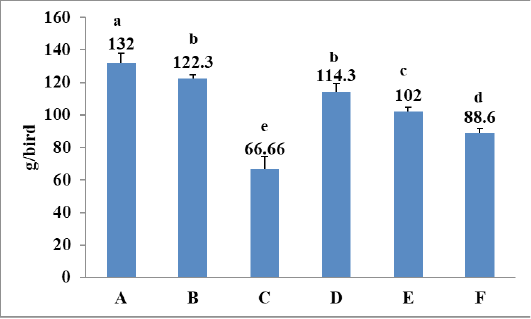
Effect of selenium on weight gain in lead induced Japanese quails.
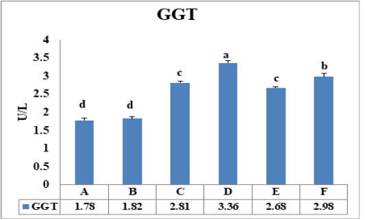
Effect of selenium on Gamma-glutamyltransferase (GGT) in lead induced Japanese quails.
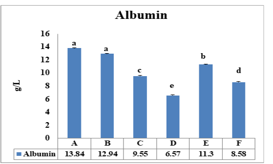
Effect of selenium on albumin level in lead induced Japanese quails
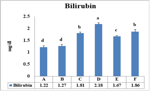
Effect of selenium on bilirubin level in lead induced Japanese quails.
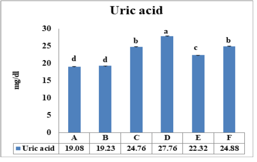
Effect of selenium on uric acid level in lead induced quails.
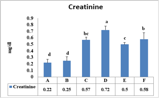
Effect of selenium on creatinine level in lead induced quails.
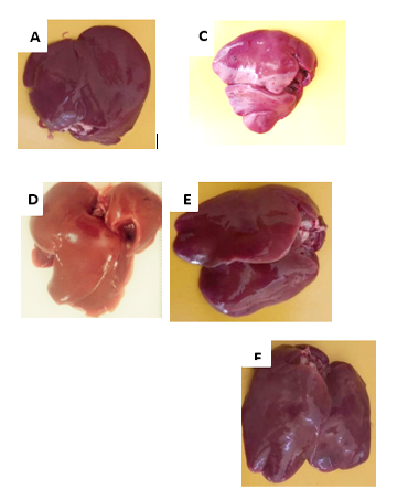
Gross pathology of liver of quail treated with lead acetate and selenium. A: Normal liver. C &D: Livers of lead-treated groups showing high intensity of discoloration and inflammation. E &F: Selenium-treated groups showing recovery towards normal color and texture.
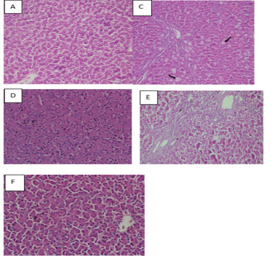
Histological observation of liver tissue. A: Liver tissue in the control group had normal histological appearance. C & D: Liver tissues in lead acetate groups showed that hepatocytes were severely swollen, and have granule denaturation and steatosis. E & F: Cell degeneration of liver tissue becomes mild and improvement was observed in the liver tissues.
References
- Abadin H, Taylor J, Buser MC, Scinicariello F, Przybyla J, Klotzbach JM., andMcIlroy LA (2019). Toxicological profile for lead: draft for public comment.
- Abdel-Hamid S.E., Shahin S.E,. Rehan I.F. (2020). Organic selenium supplementation: A convenient approach to improve behaviour, performance, and economics and to suppress stress in home-cage reared ducks. J. Anim. Health Prod, 9(s1): 9-16. https://doi.org/10.17582/journal.jahp/2020/9.s1.9.16[CrossRef]
- Abdel-Moneim WM, and Ghafeer HH (2007). The Potential Protective Effect of Natural honey against cadmium-induced hepatotoxicity and nephrotoxicity. Mansoura J. Foren. Med. Clin. Toxicol., 15(2): 75-98. https://doi.org/10.21608/mjfmct.2007.48851[CrossRef]
- Ahn J.H., Han, G.W., Nam I.S. (2021). Effects of selenium injection on the reproductive performance of holstein dairy cows. J. Anim. Health Prod., 9(3): 335-341. https://doi.org/10.17582/journal.jahp/2021/9.3.335.341[CrossRef]
- Ali S, Minhas M A, Awan UA, Mumtaz S, Shakir, HA, Tahir HM, Andleeb S (2021). Large scale toxicological evaluation of lead acetate in broiler chicken.
- Al-Wabel N.A., Mousa H.M., Omer O.H., Abdel-Salam A.M. (2007). Biological evaluation of synbiotic fermented milk against lead acetate contamination in rats. J. Food Agricult. Environ., 5(3/4): 169.
- Apaydin F.G., Kalender S., Bas H., Demir F, Kalender Y. (2014). Lead nitrate induced testicular toxicity in diabetic and non-diabetic rats: protective role of sodium selenite. Brazilian Archiv. Biol. Technol., 58: 68-74. https://doi.org/10.1590/S1516-8913201400025[CrossRef]
- Ashour A.E.R.A., Yassin M.M., AASI N.M., Ali R.M., (2007). Blood, serum glucose and renal parameters in lead-loaded albino rats and treatment with some chelating agents and natural oils. Turkish J. Biol., 31(1): 25-34.
- Ashrafizadeh M, Rafiei H, Ahmadi Z (2018). Histological changes in the liver and Biochemical Parameters of Chickens treated with lead acetate II. Iranian J. Toxicol., 12(6): 1-5. https://doi.org/10.32598/IJT.12.6.540.2[CrossRef]
- Bakker A B, Demerouti E (2017). Job demands–resources theory: taking stock and looking forward. J. Occupat. Health Psychol., 22(3): 273. https://doi.org/10.1037/ocp0000056[CrossRef]
- Binkowski ŁJ, Sawicka-Kapusta K, Szarek J, Strzyżewska E, Felsmann M (2013). Histopathology of liver and kidneys of wild living Mallards Anas platyrhynchos and Coots Fulica atra with considerable concentrations of lead and cadmium. Sci. Total Environ., 450: 326-333. https://doi.org/10.1016/j.scitotenv.2013.02.002[CrossRef]
- Boey A, Ho HK (2020). All roads lead to the liver: metal nanoparticles and their implications for liver health. Small., 16(21): 2000153. https://doi.org/10.1002/smll.202000153[CrossRef]
- Canaud B., Ye X., Usvyat L., Kooman J., van Der Sande F., Raimann J., Wang Y., Kotanko P. (2020). Clinical and predictive value of simplified creatinine index used as muscle mass surrogate in end-stage kidney disease haemodialysis patients—results from the international MONitoring Dialysis Outcome initiative. Nephrology Dialys. Transplant., 35(12): 2161-2171. https://doi.org/10.1093/ndt/gfaa098[CrossRef]
- Dioka CE, Orisakwe OE, Adeniyi FAA, Meludu SC (2004). Liver and renal function tests in artisans occupationally exposed to lead in mechanic village in Nnewi, Nigeria. Int. J. Environ. Res. Pub. Health., 1(1): 21-25. https://doi.org/10.3390/ijerph2004010021[CrossRef]
- Erdoğan Z, Erdoğan, S, Aksu T, Baytok E (2005). The effects of dietary lead exposure and ascorbic acid on Performance, lipid Peroxidation status and Biochemical parameters of broilers. Turkish J. Vet. Anim. Sci., 29(4): 1053-1059.
- Flora G, Gupta D, Tiwari A. (2012). Toxicity of lead: a review with recent updates. Interdiscipl Toxicol., 5(2): 47. https://doi.org/10.2478/v10102-012-0009-2[CrossRef]
- Francisco N De, Ruiz Troya J D, Estrella I Agüera. (2003). Lead and lead toxicity in domestic and free living birds. Avian Pathol. 32(1):3-13 https://doi.org/10.1080/0307945021000070660[CrossRef]
- Gani MU, Siddiqui MSI, Rashid MHO, Islam K, Moonmoon S, Ahmed S, Mostofa M (2016). Effect of lead acetate alone and in combination with whole milk (Star ship®) on body growth and liver functions in an experimentally induced lead toxicity in rat. Asian J. Med. Biolog. Res., 2(2): 183-189. https://doi.org/10.3329/ajmbr.v2i2.29009[CrossRef]
- Guignard JP, Drukker A (1999). Why do newborn infants have a high plasma creatinine? Pediatrics. Apr;103(4):e49 https://doi.org/10.1542/peds.103.4.e49[CrossRef]
- Haarhaus M, Brandenburg V, Kalantar-Zadeh K, Stenvinkel P, Magnusson P (2017). Alkaline phosphatase: a novel treatment target for cardiovascular disease in CKD. Nat. Rev. Nephrol., 13(7): 429-442. https://doi.org/10.1038/nrneph.2017.60[CrossRef]
- Jahromi MF, Liang JB, Ebrahimi R., Soleimani AF, Rezaeizadeh A, Abdullah N, Shokryazdan, P (2017). Protective potential of Lactobacillus species in lead toxicity model in broiler chickens. Animal., 11(5): 755-761. https://doi.org/10.1017/S175173111600224X[CrossRef]
- Jiayong Z., Shengchen W., Xiaofang H., Gang S, Shiwen, X. (2020). The antagonistic effect of selenium on lead-induced necroptosis via MAPK/NF-κB pathway and HSPs activation in the chicken spleen. Ecotoxicol. Environ. Safety., 204: 111049. https://doi.org/10.1016/j.ecoenv.2020.111049[CrossRef]
- Kalantar‐Zadeh K, Mehrotra R, Fouque D, Kopple JD (2004). POOR Nutritional Status and Inflammation: Metabolic Acidosis and Malnutrition ‐Inflammation Complex Syndrome in Chronic Renal Failure. In Semin. Dialysis. 17 (6): 455-465). Oxford, UK: Blackwell Science Inc. https://doi.org/10.1111/j.0894-0959.2004.17606.x[CrossRef]
- Khan S., Tanweer A.J., Rafiullah I., Abbas G., Khan J., Imran M.S., Kamboh A.A. (2022). Effect of Supplementation of Mealworm Scales (Tenebrio molitor) on Growth Performance, Carcass Traits and Histomorphology of Japanese Quails. J. Anim. Health Prod., 10(3): 273-411. https://doi.org/10.17582/journal.jahp/2022/10.3.381.389[Full text][CrossRef]
- Kou H., Ya J., Gao X., Zhao H. (2020). The effects of chronic lead exposure on the liver of female Japanese quail (Coturnix japonica): Histopathological damages, oxidative stress and AMP-activated protein kinase based lipid metabolism disorder. Ecotoxicol. Environ. Safety., 190: 110055. https://doi.org/10.1016/j.ecoenv.2019.110055[CrossRef]
- NRC (1994). Nutritional requirement of poultry. 8th ed. Washington (DC): National Research Council, National Academy of Science.
- Nissl J, Terra RP (2004). Creatinine and Creatinine Clearance. Health Wise (Medical Review) http:// www.bchealthguid.org/kbase/.
- Offor SJ, Mbagwu HO, Orisakwe, OE (2017). Lead induced hepato-renal damage in male albino rats and effects of activated charcoal. Front. Pharmacol., 8: 107. https://doi.org/10.3389/fphar.2017.00107[CrossRef]
- Olsvik P. A., Soft-land L., Hevrøy E. M., Rasinger J. D., Waagbø R. (2016). Fish pre-acclimation temperature only modestly affects cadmium toxicity in Atlantic salmon hepatocytes. J. Thermal Biol., 57: 21-34. https://doi.org/10.1016/j.jtherbio.2016.02.003[CrossRef]
- Qiao L., Dou X., Song X., Chang J., Pi S., Zhang X., Zhu L., Zeng X., Xu C. (2022). Protective effect of biogenic selenium nanoparticles against diquat-induced acute toxicity via regulation of gut microbiota and its metabolites. Food Chem. Toxicol., 170: p.113480. https://doi.org/10.1016/j.fct.2022.113480[CrossRef]
- Rice D., Silbergeld E. (2023). Lead neurotoxicity: Concordance of human and animal research. In Toxicol. Metals. I: 659-675. CRC Press.
- Saeeda H, Ali S, Shumaila M, Abdullah SH, Farooq A, Muhammad TH, Tariq ZM (2020). Dose and duration-dependent toxicological evaluation of lead acetate in chicks. Environ. Sci. Pollut. Res. Int., 27(13): 15149-15164. https://doi.org/10.1007/s11356-020-08016-8[CrossRef]
- Sant’Ana MG, Moraes R., Bernardi MM (2005). Toxicity of cadmium in Japanese quail: Evaluation of body weight, hepatic and renal function, and cellular immune response. Environ. Res., 99(2): 273-277. https://doi.org/10.1016/j.envres.2005.06.003[CrossRef]
- Savitha R, Suresh S, Kim HJ (2014). A Meta-Cognitive Learning Algorithm for an extreme learning machine classifier. Cognit. Computat., 6(2): 253-263. https://doi.org/10.1007/s12559-013-9223-2[CrossRef]
- Sharma S., Raghuvanshi B.P.S., Shukla, S. (2014). Toxic effects of lead exposure in rats: involvement of oxidative stress, genotoxic effect, and the beneficial role of N-acetylcysteine supplemented with selenium. J. Environ. Pathol. Toxicol. Oncol., 33(1). https://doi.org/10.1615/JEnvironPatholToxicolOncol.2014009712[CrossRef]
- Soliman MM, Baiomy AA, Yassin MH (2015).Molecular and Histopathological study on the ameliorative effects of curcumin against lead acetate-induced Hepatotoxicity and Nephrotoxicity in Wistar rats. Biolog. Trace Element Res., 167(1): 91-102. https://doi.org/10.1007/s12011-015-0280-0[CrossRef]
- Sonone SS, Jadhav S, Sankhla M S, Kumar R (2020). Water contamination by heavy metals and their toxic effect on aquaculture and human health through food Chain. Lett. Appl. Nano BioSci., 10(2): 2148-2166. https://doi.org/10.33263/LIANBS102.21482166[CrossRef]
- Suleman M, Khan A A, Hussain Z, Zia MA, Roomi, S, Rashid F, Ishaq R (2011). Effect of lead acetate administered orally at different dosage levels in broiler chicks. African J. Environ. Sci. Technol., 5(12): 1017-1026. https://doi.org/10.5897/AJEST10.278[CrossRef]
- Tahir M.A., Abbas A., Muneeb M., Bilal R.M., Hussain K., Abdel-Moneim A.M.E., Farag M.R., Dhama K., Elnesr S.S, Alagawany M. (2022). Ochratoxicosis in poultry: Occurrence, environmental factors, pathological alterations and amelioration strategies. World’s Poult. Sci. J., 78(3): 727-749. https://doi.org/10.1080/00439339.2022.2090887[CrossRef]
- Taha NM, Korshom MS, Mandoura EM, Lebdah MA, Aladham ES (2013). Effect of lead toxicity on mineral metabolism and immunological factors in rats. Alexandria J. Vet. Sci., 39(1): 64-73.
- Talpur BR, Nizamani ZA, Leghari IH, Tariq M, Rehman A, Kumbhar S (2022). Hepato-nephrotoxic effects of induced fluorosis in rabbits and broilers. J. Anim. Health Prod. 10(2): 214-220. https://doi.org/10.17582/journal.jahp/2022/10.2.214.220[CrossRef]
- Uyanik Fatma (2001). “The effects of dietary chromium supplementation on some blood parameters in sheep.” Biolog. Trace Element Res. 84.1 (2001): 93-101. https://doi.org/10.1385/BTER:84:1-3:093[CrossRef]
- Valko M, Rhodes CJB, Moncol J, Izakovic MM, Mazur M (2006). Free radicals, metals and antioxidants in oxidative stress-induced cancer. Chemico-biolog. Interact., 160(1): 1-40. https://doi.org/10.1016/j.cbi.2005.12.009[CrossRef]
- Vallverdú‐Coll N, Mougeot F, Ortiz‐Santaliestra ME, Rodriguez‐Estival J, López‐Antia A, Mateo R (2016). Lead exposure reduces carotenoid‐based coloration and constitutive immunity in wild mallards. Environ. Toxicol. Chem., 35(6): 1516-1525. https://doi.org/10.1002/etc.3301[CrossRef]
- Wang L., Jing L., Zhang Q., Li S., Wang Y, Zhao H. (2022). Lead induced thymic immunosuppression in Japanese quail (Coturnix japonica) via oxidative stress-based T cell receptor pathway signaling inhibition. J. Inorg. Biochem., 235: 111950. https://doi.org/10.1016/j.jinorgbio.2022.111950[CrossRef]
- Yazdanparast T., Strezov V., Wieland P., Lai,Y.J., Jacob D.E., Taylor M.P. (2022). Lead poisoning of backyard chickens: implications for urban gardening and food production. Environ. Pollut., 310: 119798.
- Yu A., Ji Y., Ma G., Xu J., Hu Q. (2023). Identification and preparation of selenium‐containing peptides from selenium‐enriched Pleurotus eryngii and their protective effect on lead‐induced oxidative damage in NCTC1469 hepatocytes. J. Sci. Food Agricult. https://doi.org/10.1002/jsfa.12529[CrossRef]
Services
Article usage statistics
Loading…