Research Article · Volume 10, Issue 5, 30 April 2022 · Open Access
Haemolysin, Catalase and Hydrogen Sulphide Production as Unique Phenotypic Virulence Determinants, Biofilm Formation and Defense against Antibiotics among Mycoplasma arginini and Mycoplasma ovipneumoniae Isolated from Pneumonic Lungs of Domestic Sheep (Ovis aries)
· · · · · · ·
Affiliations & Notes ⌄
1Mycoplasma Department, Animal Health Research Institute, Giza, ARC
2Microbiology Department, Faculty of Veterinary Medicine, Cairo University, Giza, Egypt
3Graduate Program in Health and Development in Midwest Region, Faculty of Medicine, Federal University of Mato Grosso do Sul, Brazil
4Food Hygiene Department, Animal Health Research Institute, Giza, Egypt
5Centro de Análises Proteômicas e Bioquímicas, Programa de Pós-Graduação em Ciências Genômicas e Biotecnologia, Universidade Católica de Brasília, Brasília-DF, Brazil
6S-inova Biotech, Programa de Pós-Graduação em Biotecnologia, Universidade Católica Dom Bosco, Campo Grande-MS, Brazil. *Correspondence | Heba N. Deif, Microbiology Department, Faculty of Veterinary Medicine, Cairo University, Giza, Egypt
Detection of mycoplasma’s DNA originated from recovered microbes of the nasal cavities of sheep marks carriage and possibly shedding, but may be without an active infection.
Antibiotic treatment may help tentatively, but response likely varies depending on the strain or/and species of mycoplasma involved.
More in vivo studies should be done for determination of the role of H2S production in the pathogenesis of M. arginini and M. ovipneumoniae.
These potent virulence factors could not be used in drug therapy and vaccine design because H2S produced by M. arginini was host associative.
Selected from the article's own text.
Abstract
This research is an endeavor to extend the understanding of factors implicated in the pathogenicity of Mycoplasma arginini (M. arginini) and Mycoplasma ovipneumoniae (M. ovipneumoniae). It shed light on the current knowledge gap in the role of two potential virulence determinants viz H2S production and catalase activity, for their possible roles in biofilm formation, antibiotic resistance and hemolytic activity of M. ovipneumoniae. The recovered sheep isolates of M. arginini and M. ovipneumoniae were examined bacteriologically and by PCR using Mycoplasma species-specific primers. MIC testing of the axenic isolates showed that 19 of them were 100% susceptible to tulathromycin, streptomycin, oxytetracycline and tylosin and highly resistant to danofloxacin, lincomycin and florfenicol. Some interactions demonstrated significance (p <0.05), such as hemolysis/ tulathromycin (-0.607), tylosin/streptomycin (0.519), lincomycin/ florfenicol (0.808), lincomycin/ oxytetracycline (0.47) and oxytetracycline/H2S (0.76). Our study distinctly elucidates that although H2S production and hemolysis are not relevant (0.26) yet, H2S and antibiotic resistance for oxytetracycline was relevant (0.76). On the other hand, the irrelevance between catalase and biofilm formation (-0,27) as summarized in our study brings to light quite clearly that the presence of catalase has a beneficial impact on biofilm formation. The xer virulence gene, the quinolone resistance-determining region (QRDR) genes parC, parE and gyrA and the macrolide and lincomycin resistance genes, rpID and rplV genes were undetected. The role of H2S production in the pathogenesis of M. arginini and M. ovipneumoniae needs further in vivo studies.
Mycoplasma is one of the most pathogenic bacterial species recorded in different animal species such as sheep , camels , chickens . Sheep can be infected with a variety of infectious microbes. Respiratory diseases are of the most important affections of sheep ; due to its impact on economic losses . This has been reported in many investigations in different geographic regions such as Jordan , Brazil , Argentina , Ethiopia , India , and in Egypt
M. ovipneumoniae was isolated from both sheep and goats andit has been recovered from the respiratory passages of both healthy and diseased sheep worldwide. Lamb pneumonia was also reported in Universidad Federal do Mato Grosso do Sul and in 1974 in New Zealand . Recently, McRae et al. (2016) reported pneumonic lesions in lambs at slaughter in New Zealand, and Swedish flocks .
M. ovipneumoniae is not normally highly pathogenic, but it makes sheep highly susceptible to other respiratory infections by lymphocyte quelling or interference with normal ciliary activity in the respiratory tract . The control of these microbes is encountering some difficulties of drug resistance. There is a great need for potent mycoplasma vaccines both in the veterinary and medical fields ; .
Pathogenic bacteria have multiple mechanisms which guarantee their survival and spread to consequently cause diseases in their hosts. Since infectious diseases represent a major cause of death worldwide, identifying and understanding their pathogenicity mechanisms is a key step for successful combating. Despite the existence of plenty of many pathogenic microbes’ infection strategies have turned out to be remarkably similar. However, diseases caused by mycoplasmas continue to be a serious problem for human health as well as livestock production with significant socio-economic divergence worldwide .
Effective control measures of mycoplasmas are wanting, mainly due to shortage of knowledge about their pathogenicity mechanisms. They lack typical pathogenicity features found in other bacteria, and despite the genomes of many important mycoplasma pathogens having been sequenced, questions related to their virulence and survival remain .
Hemolysis is an important aspect of bacterial pathogenicity which can have severe effects in both the human and animal hosts . Since H2S has been proven to play a role in hemolytic activity of other pathogenic bacteria , M. arginini and M. ovipneumoniae were also to be tested for production of additional hemolytic compounds, like hydrogen sulfide. Therefore, previous findings and reports of several researchers concerning the incorporation of certain Mycoplasma species with pneumonia in sheep and their possible hazardous influence on public health are to be considered and wildlife conservation ; ; ; ; . Hence, this research is an endeavor to evaluate two potential virulence determinants viz H2S production and catalase activity, for their possible roles in virulence, biofilm formation, resistance to antibiotics and hemolytic activity of M. arginine and M. ovipneumoniae, an unexplored issue until now.
Materials and Methods
Ethical issue: The Department of Microbiology, Faculty of Science, Cairo University approved all animal experiments (CU-04-20), which adopted all national standards for the use of animals in scientific testing, and the basic procedure defined by the OIE (2020 Edition).
Samples collection and processing
The samples were collected from domestic sheep (Ovis aries) at Cairo slaughterhouse during 2018 and transported while cold to the Animal Health Research Institute (AHRI), Dokki, Egypt as quick as possible. Nasal swabs were collected from live, apparently healthy animals (n=50); in addition to samples from pneumonic (n=200). Mycoplasma isolation, detection, characterization, DNA extraction and molecular typing were carried out in AHRI, Department of Mycoplasmology, following the standard mycoplasma isolation and identification procedures including PCR assays and 16S rRNA gene sequencing. Culture-enriched nasal swab eluates for mycoplasma culturing were inoculated into Friis’ medium and incubated at 37oC with a daily follow up for color change. Several PCR techniques were used to define the conformity of the culture isolates. As a primary step, conventional PCR for Mycoplasma species isolates were processed , then followed by an Acholeplasma laidlawii conventional PCR technique (Supplementary Table S1). To confirm the M. ovipneumoniae and M. arginini culture purity, 16S ribosomal (rRNA) genes were amplified using specific primers, as per previous reports (Supplementary Table S1) using previously described PCR assays (Supplementary Table S1) applied to the extracted DNA from broth culture media. After assuring that the isolates were mycoplasmas, the isolates were identified to the species level. The presence of M. ovipneumoniae and M. arginini were detected by previously described PCR methods (Supplementary Table S1) applied to the DNA extracted from broth culture media. PCR-positive M. ovipneumoniae and M. arginini extracts were genotyped by targeting genetic loci of the partial DNA sequences from the small ribosomal subunit (16S). Protocols and primers for PCR amplification of these loci was described previously (Supplementary Table S1). DNA sequencing of amplified PCR products was conducted by bidirectional Sanger sequencing. After that, each aligned sequence of the independent locus in MUSCLE72 were done, following the default parameter settings of Geneious R10.1.3 (http://geneious.com, Biomatters, Ltd.).
Phenotypic virulence traits
After species identification, each isolate was subjected to phenotypic virulence traits assays and antimicrobial susceptibility testing.
Hydrogen sulfide assays with lead acetate test strips
M. ovipneumoniae and M. arginini isolates were incubated in 1 ml 1x PBS, pH 7.4 or in MP-medium supplemented with different cysteine concentrations in 2 ml Eppendorf reaction tubes. Lead acetate test strips (Aldrich) were fixed under the lid of the tube (without contact to the liquid) to catch the produced hydrogen sulfide. The black PbS formed by the reaction between the lead covering the strips and the sulfide, results in dark staining of the test paper. After an overnight incubation at 37°C, the coloration of the test strips was examined and photographed .
Catalase enzyme activity
The procedure of Pritchard et al. (2014) was implemented. Mid-log phase cultures of each M. ovipneumoniae and M. arginini isolate were collected, centrifuged at 20,000×g for 20 minutes then washed 3 times with cold phosphate-buffered saline (PBS). Cells were spread onto a clean microscope slide then one drop of 3% H2O2 was added. Catalase activity was indicated by the production of bubbles.
Assays for detection of hemolytic activity
To analyze hemolytic activity of M. ovipneumoniae and M. arginini, the colonies were overlayed onto 5% sheep blood agar, after that the plates were incubated for 1-2 days at 37°C .
For analyzing the hemoxidative or hemolytic activities of the isolates of M. arginini and M. ovipneumoniae, the cultures were incubated with 5% defibrinated sheep blood or 2% washed sheep RBCs in PBS (pH 7.4) and various supplements (1 mM glycerol, 1 mM glycerol-3-phosphate and 1 or 10 mM L-cysteine) in a total volume of 1 ml followed by sample incubation at 37°C for several hours (100-110 rpm). After incubation, the tubes were centrifuged (1,400 x g at 4°C), the supernatant was transferred carefully into a new tube. The pellets were re-suspended and lysed in 1 ml dH2O. The spectra of both supernatant and pellets were recorded photometrically from 370 to 700 nm .
Analysis of biofilm production by crystal violet staining
Biofilm production was quantified by scoring the absorbance (560 nm) of 100 ml of the solubilized crystal violet in a microtiter plate .
Antimicrobial susceptibility testing
The broth microdilution method was used for determination of MIC of M. ovipneumoniae and M. arginini isolates recovered from the nasal and lung samples. The Broth microdilution method following the procedure of Hannan (2000) was implemented taking into consideration that the suggested breakpoint interpretations provided herein were derived from three sources: Clinical and Laboratory Standards Institute (CLSI) criteria for veterinary pathogenic bacteria in cattle , the MIC breakpoints of aminoglycosides according to , preceding publications and the MIC data generated in the current study. The M. ovipneumoniae (n=5) and M. arginini (n=14) isolates were examined for their sensitivity to seven antimicrobial agents by the disc diffusion method. The antimicrobial agents utilized in this study included: fluoroquinolone: danofloxacin (5 ug); macrolides: tulathromycin (30 µg) and tylosin (30 ug); phenicol: florfenicol (30 ug); aminoglycoside: streptomycin (10 ug); lincosamide: lincomycin (2 ug); tetracycline: oxytetracycline (30 ug). The type strain M. bovis ATCC 25523/ PG45 was used as quality control.
Detection of the virulence, QRDR, Macrolide and Lincomycin resistance genes
DNA was extracted by using the simple boiling procedure (100ºC for 5 min) to liberate the DNA. The supernatant was used as PCR templates to detect the presence of gyrB, ParC, ParE, rpID, rpIV and xer genes. Primers and PCR conditions for these genes were previously declared and summarized in Supplementary Table S1.
Nucleotide sequencing and sequence analysis
PCR products were purified using the Gene Jet PCR purification kit; Fermentas (Thermo Fisher Scientific). The purified amplicon of each isolate was sequenced in both forward and reverse directions using the amplification primers (Supplementary Table S1). Amplicons were sequenced in an automated sequencer (Macrogen Company 24, Gasan-dong, Geumchun-gu, Seoul 153-781, Korea). Sequence data similarity searches were analyzed by using NCBI-BLAST program (http://www.ncbi.nlm.nih.gov/BLAST). The comparisons of the obtained nucleotide sequences and associated multiple alignments were performed using the BioEdit sequence alignment editor (CLUSTALX software version 7.0.9.0) (6/27/07) for multiple sequence alignment.
Molecular phylogenetic analysis by Maximum Likelihood method
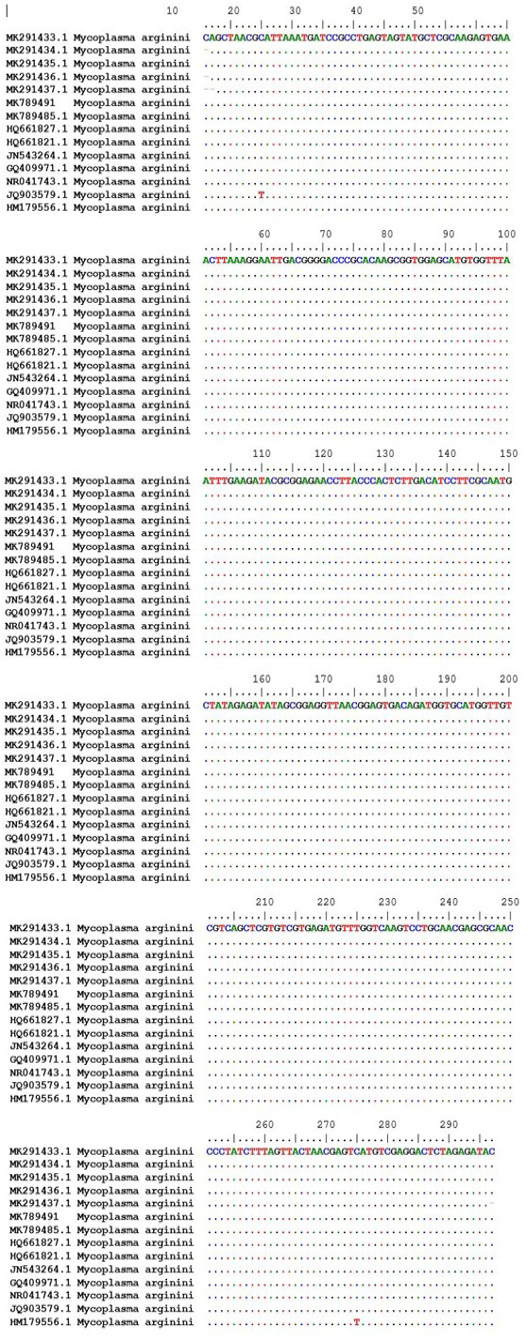
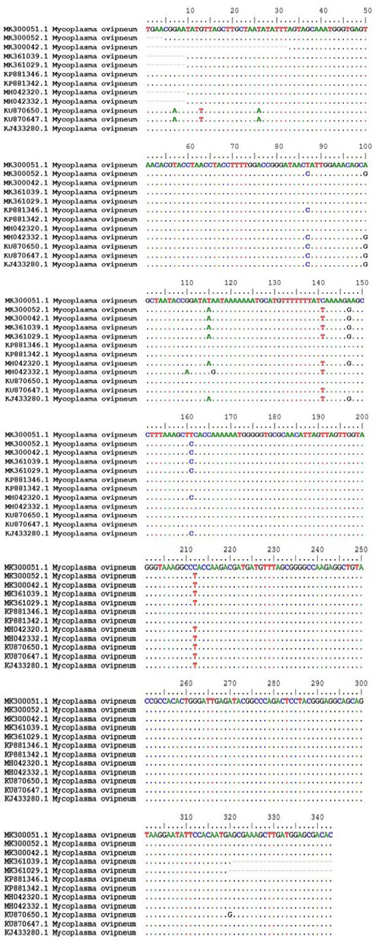
Based on Hasegawa et al. (1985), the evolutionary history was deduced by using the Maximum Likelihood method for revealing the tree. Next to the branches, the trees’ percentage with clustered associated taxa was shown. The Neighbour-Joining method was used to obtain the guiding Initial trees. The tree was drawn and the analysis involved 22 nucleotide sequences. Codon positions included were 1st+2nd+3rd+Noncoding. In the final dataset, there were a total of 2765 positions. The analyses of evolution was done using MEGA6 . Sequences (Supplementary Figure S1) were then submitted to NCBI GenBank using BankIt (http://www.ncbi.nlm.nih.gov/WebSub/?tool=genbank) under the accession numbers of Mycoplasma arginini: (MK640677.1*, MK640679.1*, MK643127.1*, MK774823*).
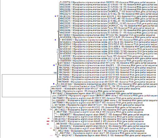
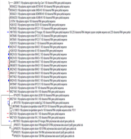
The online pipeline NGpyologeny.fr was used for constructing the 16S phylogenetic tree (Figures 3 and 4) . Within this pipeline, sequences were aligned using MUSCLE, ambiguous aligned regions were removed with GBLOCK, and a phylogenetic tree was constructed with PhyML+SMS using GTR model and bootstrap 100 times ; ; . The heatmap Phylogenetic tree was visualized and annotated with iTOL .
Statistical analysis
As a first step, the data were treated using descriptive statistics. After that, the heatmap graphs confirm the analyses performed by principal component analysis (PCA) as well as by the analysis of variance (ANOVA), using the outcome variables as factors (e.g., Virulence traits and antibiotics) for each tissue of interest (e.g., lung and nasal swab). In addition, Pearson’s correlation was verified for all variables. The results were analyzed using Microsoft Excel and GraphPad Prism 8.
Results and Discussion
Prevalence of Mycoplasma arginini and Mycoplasma ovipneumoniae in samples recovered from nasal swabs and lungs of sheep
Out of 50 nasal swabs of apparently healthy sheep and 200 samples of pneumonic lungs, 19 isolates were confirmed to be positive for putative mycoplasmas identified to the species level as five Mycoplasma ovipneumoniae (5/19) isolated from the pneumonic lungs and 14 M. arginini isolated from the nasal swabs (5/14) and the pneumonic lungs of the sheep (9/14) (Supplementary Table S2).
Phenotypic virulence traits of Mycoplasma arginini and Mycoplasma ovipneumoniae
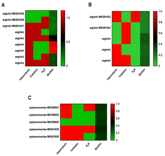
The four phenotypic virulence traits are recorded in Supplementary Table S2. Regarding the virulence traits, Figure 1 demonstrates the effect of M. arginini (A) and M. ovipneumoniae (C) on the lungs and M. arginini for the nasal swab (B). Be that as it may, no significant differences were recognized (p> 0.05).
Hydrogen sulphide production
The recorded hemolytic activities of the M. arginini and M. ovipneumoniae isolates in the presence of cysteine suggested that the bacteria produce H2S from cysteine which in turn causes hemolysis. Yet, 8/14 of the M. arginini isolates (4/5, from the nasal swabs and 4/9, from the pneumonic lungs) were unable to produce H2S. In addition, 3/5 Mycoplasma ovipneumoniae isolated from pneumonic sheep lungs were also unable to produce H2S.
Detection of hemolytic activities
After two days of incubation on blood agar it was revealed that 14 isolates (10 Mycoplasma arginini and 4 Mycoplasma ovipneumoniae) exhibited clear zones of α-hemolysis around their colonies. The effect of M. arginini and M. ovipneumoniae on RBCs was also examined in a liquid environment.
Catalase activity
Five out of nine M. arginini isolated from pneumonic sheep lungs were impuissant to indicate any catalase activity. Two out of the five M. arginini isolated from nasal swabs of apparently healthy sheep were unable to indicate any catalase activity. The catalase activity was not obvious in 3/5 of the M. ovipneumoniae that were isolated from the pneumonic lungs of sheep.
Biofilm formation capability
The ability of M. arginini strains adherence to polystyrene multiwall plates were found to be weak. A very low spectrophotometric values (A560 (0.187-0.292)) were recorded for the isolates from the nasal swabs of apparently healthy sheep indicating poor adhesion and biofilm formation abilities. On the other hand, two isolates had a relatively moderate ability for biofilm formation ((A560 (1.099-1.153)) while the remaining seven isolates isolated from pneumonic lungs, were in their normal range of weak biofilm formation (A560, 0.216-0.564). The five M. ovipneumoniae isolated from the pneumonic lungs also exhibited a weak tendency for biofilm formation (A560, (0.347-0.400)).
Antimicrobial resistance among the Mycoplasma arginini and Mycoplasma ovipneumoniae isolates
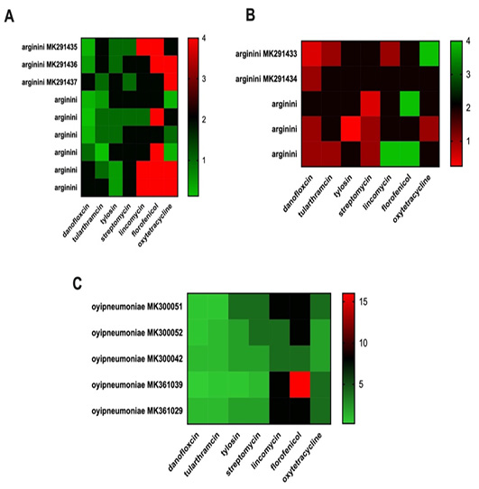
The susceptibility of 19 field isolates of M. ovipneumoniae and M. arginini isolated from sheep, to seven different antimicrobial agents representing six antimicrobial groups is displayed in Supplementary Table S2. MIC testing of the seven antimicrobials showed that the 19 mycoplasmas were 100% susceptible to tulathromycin, streptomycin, oxytetracycline and tylosin and highly resistant to danofloxacin, lincomycin and florfenicol. Moreover, the antimicrobial resistance combinations of M. arginini and M. ovipneumoniae to various antibiotics displayed six different combinations of antibiotics representing three classes, with the lincosamide (lincomycin) being the most obviously seen in four out of the six combinations (Table 1). Danofloxacin, lincomycin, florfenicol combination was the most recorded in ten out of fourteen isolates; furthermore, the multidrug-resistant (MDR), and extensively drug-resistant (XDR) isolates were apparent in three (21.4%) and five (35.7%) isolates, respectively.
In this sense, Figure 2A demonstrates the role of different antibiotics on M. arginini in the lungs, which showed other actions among the tested antibiotics [F (6, 48) = 8,409, p <0.0001]. About the effects on the nasal swabs, no significant differences were identified (Figure 2B, p = 0.06). When M. ovipneumoniae was verified in the lungs (Figure 2C), differences between the studied antibiotics were demonstrated [F (6, 48) = 11.48, p <0.0001].
Table 1: The antimicrobial resistance combinations of Mycoplasma arginini and Mycoplasma ovipneumoniae to various antibiotics.
Antibiotics
n= resistant antibiotics
n= antibiotic classes
n= of isolates
Extensive drug resistance (XDR)
Multiple drug resistance (MDR)
Pan drug resistance (PDR)
Danofloxacin
1
1
1
0
0
0
Lincomycin
1
1
2
0
0
0
Florfenicol
1
1
1
0
0
0
Danofloxacin, lincomycin
2
2
3
3
0
0
Lincomycin, florfenicol
2
3
2
2
0
0
Danofloxacin, lincomycin, florfenicol
3
3
10
0
3
0
Table 2: Pearson’s correlations matrix from PCA analysis from nasal swabs and lung isolates.
Tulathr
omycin
Tylosin
Streptomycin
Lincomycin
Florfenicol
Oxytetr
acycline
Hemolysis
Catalase
H2S
Biofilm
Dano
floxacin
0.288
-0.077
-0.223
-0.069
-0.243
0.281
-0.045
0.248
0.110
-0,204
Tulathr
omycin
-0.398
-0.323
-0.292
-0.352
0.092
-0.607*
0.050
-0.094
-0.454
Tylosin
0.519*
0.225
0.013
0.014
-0.024
-0.011
-0.126
-0.058
Streptomycin
0.320
0.179
0.127
0.208
-0.447
0.056
0.088
Lincomycin
0.808*
0.470*
0.182
0.083
0.107
-0.109
Florfenicol
0.389
0.186
0.137
0.224
-0.005
Oxytetr
acycline
0.068
0.100
0.760*
-0.455
Hemolysis
0.088
0.268
0.281
Catalase
0.045
-0.271
H2S
-0.203
Distribution of the virulence, QRDR and macrolide and lincomycin resistance genes
Again, to our surprise, we were unable to detect any of the investigated genes, the xer virulence gene, the QRDR genes parC, parE and gyrA and the Macrolide and Lincomycin resistance genes, rpID and rplV genes by PCR in any of the 19 Mycoplasma arginini and Mycoplasma ovipneumoniae isolates.
When performing the PCA analyses, Pearson’s correlation matrix was generated (Table 2). Thus, it is observed that some interactions demonstrated significance (p <0.05), such as Hemolysis/ Tulathromycin (-0.607), tylosin/ streptomycin (0.519), lincomycin/ florfenicol (0.808), lincomycin/ oxytetracycline (0.47) and oxytetracycline/ H2S (0.76).
Phylogenetic analysis of 16S rRNA of M. arginini isolates from sheep, goat, cattle, camel and human in Egypt with M. arginini isolates recovered from different countries
M. arginini cattle isolate (MF101758.1*): show no 100% identity to any other sequence but show high similarity (98%) to: MK789480.1 (sheep M. bovigenitalium from Turkey), LC158833.1 (Bovine M. bovigenitalium from Japan) and HQ661819.1 (sheep M. bovigenitalium from South Africa).
M. arginini sheep isolates (MK291433.1*, MK291434.1*, MK291435.1*, MK291436.1*, MK291437.1*) show 100% identity to each other’ and to goat M. arginini, Egypt sequences (MK774823.1*, MK643127.1*, MK640679.1*), sheep M. arginini from Turkey), (MK789487.1,) and HQ661822.1 (sheep M. arginini from South African) and show high similarity 99.66% to Camel M. arginini from Egypt (MK271638.1*, MK271640.1*, MK271641.1*), human M. arginini from Hungary (HM179556.1), and Feline, M. arginini, USA (U15794.1). All goat M. arginini isolates with accession numbers (MK640677.1*, MK640679.1*, MK643127.1*, MK774823*) show 100% identity to each other’s and to sheep M. arginini isolates from Turkey (MK789491.1 and MK789487.1) but show 99.66% similarity to: Camel M. arginini, Egypt (MK271638.1*, MK271640.1*, MK271641.1*), human M. arginini from Hungary (HM179556.1), and Feline, M. arginini, USA (U15794.1).
The human M. arginini from Hungary (HM179556.1) show 100% identity to M. arginini isolated from tick infesting Camelus dromedarius, India (MG564230.1, MG564231.1, MG564232.1, MG564233.1) but show similarity with sheep M. arginini from Turkey (MK789485.1); 99.71%, (MK789486.1); 99.70%, and 99.66% to HQ661819.1 (sheep M. arginini, South Africa), 99.58% to U15794.1 (Feline, M. arginini, USA), 99.52% to HQ661828.1 (sheep M. arginini, South Africa), 99.33% to MK271641.1 (camel M. arginini, Egypt), 99.30% to (KP742976.1) M. arginini of Dromedary Camel from Sudan and 98.31% to camel, M. arginini, Egypt (JQ859817.1 1). The human M. arginini from Hungary (HM179555.1) show no 100% identity to any other published sequence but show similarity to goat M. arginini sequences: 97.85% (KP685373.1), and 97.71% to (KP685376.1, KP685375.1 and KP685374.1). Camel M. arginini isolates (MK271638.1*, MK271640.1*, MK271641.1*) show 100% identity to each other and show high similarity 99.66% to sheep M. arginini, turkey (MK789491.1, MK789487.1, sheep, M. arginini, Egypt (MK291434.1*, MK291435.1*, MK291436.1*, MK271639.1*), camel M. arginini, Egypt (MK640677.1*), goat M. arginini, Egypt, and sheep M. arginini, South Africa (HQ661825.1 and HQ661826.1). Camel M. arginini (M0K271639.1*) show no 100% identity with any other published sequences but show 99.66% similarity to: MK271640.1*, MK271641.1*, 99.32% to: sheep M. arginini, turkey (MK789491.1, MK789487.1), sheep M. arginini, Egypt (MK291433.1*, MK291434.1*, MK291435.1*, MK291436.1*), and goat M. arginini, Egypt (MK640677.1*, MK640679.1*) (Figures 3 and 4).
N.B. *(these sequences are from our preceding studies).
This research aimed to look into and evaluate three potential virulence determinants: viz hemolysis, H2S production and catalase activity for their possible roles in virulence, biofilm formation, antibiotic resistance, and hemolytic activity of M. arginini and M. ovipneumoniae, an issue unexplored until now. M. ovipneumoniae is the most commonly isolated mycoplasma from the sheep respiratory tract and is associated with atypical pneumonia affecting sheep in their first year of life. Its first recorded isolation was in New Zealand in 1974, and from this time, it has been recovered from the respiratory tracts of healthy and symptomatic sheep worldwide . Its association with M. arginini has been often reported in sheep ; ; ; ; leading to paroxysmal coughing, rectal prolapse and reduced weight gain in lambs. The temperature, velocity of wind, clouds, precipitation, and explosion of volcanoes usually incorporate in the aerographic conditions. The prevalent climatic conditions have a considerable influence on the pathogens survival . An alteration in weather conditions of a geographical area has always witnessed an outburst of infectious diseases and has been labeled as predisposed to disease epidemics. Small ruminants are well adapted to extreme temperatures, with their body hair coats providing insulation against cold and heat . Any modulation in the environmental temperature influence the incubation period, the life cycle (the time between infection and sporulation), and the contagious period (the time during which the pathogen continues to propagate the disease). At higher temperatures, the pathogen’s life cycle usually gets hastened consequently, epidemics develop at a faster rate. In colder conditions, the pathogens produce dormancy, and the progress of the pandemic is slower, leading to a decline in the disease incidence and seriousness. During times of stress or hot weather, the subclinical infection may predispose sheep to acute fibrinous pneumonia, pulmonary abscessation, or pleurisy leading some investigators to suggest that M. ovipneumoniae is actually a commensal of sheep .
Transmission occurs by means of aerosol spread and close contact with infected animals. The nasal cavity of adult animal is considered the predilection seat of M. ovipneumoniae, from where it spreads to lambs, causing transitory, mild respiratory affections . Stressors such as shipping, weather, unbalanced rations, sub-clinically infected sheep may suffer from acute pneumonia . Solo, it isn’t usually highly pathogenic, but make sheep highly susceptible to other respiratory microbes by interfering with regular respiratory tract ciliary activity and by quelling lymphocytes ; ; . It is documented that M. arginini isn’t the main causative agent of pneumonia but the condition can be worsened in the presence of other microorganisms . Mycoplasma isolation rate in our study (37 %) is close to the 36 % reported by Güler (1993). In the present study, most of the isolates were identified as M. ovipneumoniae, and that agrees with Sheehan et al. (2007). These results elucidate that the former is more persistent than the latter in our geographic area. H2S increases the bacterial resistance to oxidative stress and antibiotics by a dual mechanism of suppressing the DNA-damaging Fenton reaction via Fe2+ sequestration and animating the major antioxidant enzymes catalase and SOD. This cytoprotective mechanism of H2S parallels that of NO , suggesting that bacteria capable to produce both gases may benefit from their synergistic action.
In agreement with Gusarov et al. (2009), these results demonstrate the dependent, and specific antagonistic effects of NO and H2S against antibiotics. Impressively, in contrary to bNOS, which is present in only a few Gram-positive species, H2S enzymes are essentially universal. Because endogenous H2S diminishes the effectiveness of many clinically used antibiotics, the inhibition of this gas keeper should be considered as an increment therapy against many pathogens.
The same as nitric oxide, H2S, formerly known to be a toxic gas, is now considered as a signaling molecule related to constructive functions in mammals from vaso-relaxation, cardio-protection, and neurotransmission to anti-inflammatory action in the gastrointestinal tract ; ; . Three H2S generating enzymes have been recognized in mammals: Cystathionine b-synthase (CBS), cystathionine g-lyase (CSE) and 3-mercaptopyruvate sulfur-transferase (3MST). CBS and CSE produce H2S predominantly from L-cysteine (Cys). 3MST does so via the intermediate synthesis of 3-mercaptopyruvate produced by cysteine aminotransferase (CAT), which is inhibited by aspartate (Asp) competition for Cys on CAT. The bacterial combating for iron is often accompanied by hemolysis. This is a convenient effect since the bacteria gain access to many nutrients that are released from the lysed RBCs.
Most importantly, they gain access to iron-bound inside the RBCs hemoglobin molecules. In vivo, oxidized hemoglobin has a lowered binding affinity towards oxygen and also the release of oxygen is hindered. However, this process is reversible. Like oxidation, hemoglobin can also be sulfonated, resulting in so-called sulfhemoglobin (sulfHb), which appears as a greenish-brownish discoloration of blood . As a result, hemoglobin loses its ability to bind oxygen in a non-reversible manner. Both, the formation of sulfHb and metHb are forms of alpha hemolysis. Secretion of H2O2 or H2S is the prime cause for bacterial oxidation or sulfenylation of hemoglobin. Alpha-hemolysis following hydrogen peroxide production is used for typing of bacterial species and is typically seen in S. pneumoniae and S. mutans . Hemoglobin alteration and hemolysis as result of hydrogen sulfide production has been studied in several oral pathogens. Among them, the cystalysin of Treponema denticola, E. coli and its hemoxidative and hemolytic activity have been elaborately studied , 1997). The production of hydrogen sulfide is a prevalent feature of oral pathogenic bacteria and responsible for periodontal diseases and oral malodor . The genera Fusobacterium, Prevotella and Porphyromonas are amongst the predominant H2S producers . Hemolytic activity associated with the production of H2S has not only been reported in T. denticola, but also for Fusobacterium nucleatum, Streptococcus anginosus, Streptococcus intermedius, Prevotella intermedia, M. pneumonia, Mycoplasma arginini and M. bovis ; . Shatalin et al. (2011) announced that putative cystathionine b-synthase, cystathionine g-lyase, or 3-mercaptopyruvate sulfur transferase suppression in B. anthracis, P. aeruginosa, S. aureus and E. coli repress H2S production, requisition these pathogens highly sensitive to a multitude of antibiotics. Exogenous H2S restrains this effect. The resistance mechanism of gas-mediated antibiotic count on alleviation of oxidative stress enjoined by antibiotics. Our study provides prudence into the mechanism responsible for M. arginini and M. ovipneumoniae host cell cytotoxicity, distinctly elucidating that although H2S production and hemolysis are not relevant (0.26) yet, H2S and antibiotic resistance for oxytetracycline was relevant (0.76). On the other hand, the irrelevance between catalase and biofilm formation (-0,27) as summarized in our study bring to light quite clearly that the presence of catalase has a beneficial impact on biofilm formation .
For centuries and in contrast to mammal-derived H2S, bacteria derived H2S has been considered to be only a byproduct of sulfur metabolism, without specific physiological function in non-sulfur microorganisms. It has been concerned with the role played by these enzymes . Considering some functional correspondence between mammalian gasotransmitters, ; ; ; supposed that bacterial H2S may, be cytoprotective. Furthermore, not too much is known about the metabolic pathways involving H2S in mesophilic bacteria . The virulence role played by hydrogen peroxide production in pathogenic Mycoplasma species is still debatable ; , M. arginini and M. ovipneumoniae could also be under the same conclusion and the role of H2S production in their pathogenesis needs more in vivo studies.
Some Mycoplasma species such as M. genitalium and M. pneumoniae lack xer-like site-specific recombinase genes, supposedly due to reductive evolution of their minimum genomes and thus has no need to Xer-like recombinase for resolving their dimer chromosomes during cell division. Recchia and Sherratt (1999); Glew et al. (2002); Chopra-Dewasthaly et al. (2017) supposed that Vpma antigenic divergence is fundamental for survival and continuation inside the immunocompetent host. Despite that Xer1 is substantial for causing Vpma variation in vitro, it is not considered as a virulence factor as alternative Xer1-independent mechanisms operate in vivo, probably under the selection pressure of the immune response induced by the host. This study highlights new aspects of mycoplasma antigenic variation systems, including the expression mechanisms regulated by host factors . The data introduced by Chopra-Dewasthaly et al. (2017) summarized that in spite of not being a virulence factor of M. agalactiae, Vpma phase variation of Xer1 recombinase might critically have impact on its surveillance during natural infections, a situation that could explain the absence of the xer gene in our M. arginini and M. ovipneumoniae isolates.
Conclusions and Recommendations
The scenario of globalization and regulations concerned with international trades, continuous monitoring of enlisted diseases is obligatory. Consequently, a standard protocol for sampling, isolation, and identification processes is a must. Effective control of the respiratory diseases of sheep, through accurate diagnosis, and detailed genetic analysis of the causative agents is needed. Detection of mycoplasma’s DNA originated from recovered microbes of the nasal cavities of sheep marks carriage and possibly shedding, but may be without an active infection. Antibiotic treatment may help tentatively, but response likely varies depending on the strain or/and species of mycoplasma involved. More in vivo studies should be done for determination of the role of H2S production in the pathogenesis of M. arginini and M. ovipneumoniae. These potent virulence factors could not be used in drug therapy and vaccine design because H2S produced by M. arginini was host associative.
Novelty Statement
Our study distinctly elucidates that; although H2S production and hemolysis are not relevant yet, H2S and antibiotic resistance for oxytetracycline was relevant. On the other hand, the irrelevance between catalase and biofilm formation clears that the presence of catalase has a beneficial impact on biofilm formation. The interactions between haemolysis/tulathromycin demonstrated significance: between oxytetracycline/H2S; tylosin/streptomycin; lincomycin/florfenicol; and between lincomycin/oxytetracycline. The xer virulence gene, the QRDR genes parC, parE and gyrA and the Macrolide and Lincomycin resistance genes; rpID and rplV genes; were undetected. As a result, Although Haemolysin, catalase and hydrogen sulphide production represent a unique potential phenotypic virulence determinants, yet, they couldn’t be used as candidates for drug therapy and vaccine design, as H2S production by M. arginini is host dependent.
Author’s Contribution
MO data curation, Investigation. KMO supervision, data curation, writing original draft, review and editing. MH supervision, data curation, writing original draft, review and editing. MAEM Supervision. JAA data analysis, data interpretation. AS data curation. OLF data analysis, data interpretation. HND data curation, investigation and review final version.
Conflict of interest
The authors have declared no conflict of interest.
Figures
Figure 1. Figure 2. Figure 1.
Clustering heatmap of virulence traits phenotypes by lungs and nasal swab.
Supplementary Figure S1.
Sequence results submitted to the Genbank.
Figure 2.
Clustering heatmap of antimicrobial resistance by lungs and nasal swab.
Figure 3.
Maximum Likelihood phylogenetic tree of 16S ribosomal RNA gene, partial sequence of sheep Mycoplasma arginini (five isolates) and M. ovipneumoniae (four isolates) in comparison with other representative strains of M. arginini and M. ovipneumoniae available on NCBI.
Figure 4.
Maximum Likelihood phylogenetic tree of 16S ribosomal RNA gene, partial sequence of goat M. arginini (four strains) and sheep M. arginini (five strains), Cattle M. arginini (one strain), Camel M. arginini (four strains) in comparison with human M. arginini (two strains) on Genbank, and other representative strains of M. arginini available on NCBI. Camel â”, Sheep â—, Goat â–¡, Human â” and cattle â–¡ isolates.
References
Abdelazeem WM, Zolnikov TR, Mohammed ZR, Saad A, Osman KM (2020). Virulence, antimicrobial resistance and phylogenetic analysis of zoonotic walking pneumonia Mycoplasma arginini in the one-humped camel (Camelus dromedarius). Acta Trop., 207: 105500. https://doi.org/10.1016/j.actatropica.2020.105500[CrossRef]
Alley MR, Ionas G, Clarke JK (1999). Chronic non-progressive pneumonia of sheep in New Zealand. A review of the role of Mycoplasma ovipneumoniae. N. Z. Vet. J., 47(5): 155- 215. https://doi.org/10.1080/00480169.1999.36135[CrossRef]
Almeida TL, Brum KB, Lemos RAA, Leal CRB, Borges FA (2013). Doenças de ovinos diagnosticadas no laboratório de anatomia patológica animal da Universidade Federal de Mato Grosso do Sul, Brasil. (1996-2010). Pesq. Vet. Bras., 33: 21-29. https://doi.org/10.1590/S0100-736X2013000100005[CrossRef]
Al-Momani W, Halablab MA, Abo-Shehada N, Miles K, Mcauliffe L, Nicholas RAJ (2006). Isolation and molecular identification os small ruminant mycoplasmas in Jordan. Small Rumin. Res., 65: 106-112. https://doi.org/10.1016/j.smallrumres.2005.05.022[CrossRef]
APHIS (2015). Animal and plant health inspection service, veterinary services, center for epidemiology and animal health, United States Department of Agriculture, USDA–APHIS–VS–CEAH–NAHMS NRRC Building B, M.S. 2E7 2150 Centre Avenue Fort Collins, CO 80526-8117 970.494.7000. http://www.aphis.usda.gov/nahms#708.0615
Ayling RD, Nicholas RAJ (2007). Mycoplasma respiratory infections. In: Aitken, I. D. (Ed.), Diseases of Sheep, Fourth Edition. Blackwell Publishing, Moredun., 33: 231-235. https://doi.org/10.1002/9780470753316.ch33[CrossRef]
Bashiruddin JB, Taylor TK, Gould AR (1994). A PCR-based test for the specific identification of Mycoplasma Mycoides Subspecies mycoides SC. J. Vet. Diag. Invest., 6(4): 428-434. https://doi.org/10.1177/104063879400600405[CrossRef]
Basic A, Blomqvist M, Dahlén G, Svensäter G (2017). The proteins of Fusobacterium spp. involved in hydrogen sulfide production from L-cysteine. BMC Microbiol., 17(1): 61. https://doi.org/10.1186/s12866-017-0967-9[CrossRef]
Besser TE, Cassirer EF, Yamada C, Potter KA, Herndon C, Foreyt WJ, Knowles DP; Srikumaran S (2012). Survival of bighorn sheep (Ovis canadensis) commingled with domestic sheep (Ovis aries) in the absence of Mycoplasma ovipneumoniae. J. Wildl. Dis., 48(1): 168-172. https://doi.org/10.7589/0090-3558-48.1.168[CrossRef]
Chatfield MJ, GN La Mar H (1992). Nuclear magnetic resonance study of the prosthetic group in sulfhemoglobin. Arch. Biochem. Biophys., 295: 289-296. https://doi.org/10.1016/0003-9861(92)90520-7[CrossRef]
Chopra-Dewasthaly R, Christine C, Michelle DG, Zimmermann M, Rosengarten R, Jechlinger W (2008). Phase-locked mutants of Mycoplasma agalactiae: defining the molecular switch of high-frequency. VPMA Antigen. Variat., 67(6): 1196-1210. https://doi.org/10.1111/j.1365-2958.2007.06103.x[CrossRef]
Chopra-Dewasthaly R, Spergser J, Zimmermann M, Citti C, Jechlinger W, Rosengarten R (2017). Vpma phase variation is important for survival and persistence of Mycoplasma agalactiae in the immunocompetent host. PLoS Pathog., 13: e1006656. https://doi.org/10.1371/journal.ppat.1006656[CrossRef]
Chu L, Burgum A, Kolodrubetz D, Holt SC (1995). The 46-kilodalton-hemolysin gene from Treponema denticola encodes a novel hemolysin homologous to aminotransferases. Infect. Immun., 63: 4448-4455. https://doi.org/10.1128/iai.63.11.4448-4455.1995[CrossRef]
Chu L, Ebersole J, Kurzban GP, Holt SC (1997). Cystalysin, a 46-kilodalton cysteine desulfhydrase from Treponema denticola, with hemolytic and hemoxidative activities. Infect. Immun., 65: 3231-3238. https://doi.org/10.1128/iai.65.8.3231-3238.1997[CrossRef]
Citti C, Blanchard A (2013). Mycoplasmas and their host: emerging and re-emerging minimal pathogens. Trends Microbiol., 21: 196-203. https://doi.org/10.1016/j.tim.2013.01.003[CrossRef]
CLSI (2002). Development of in vitro susceptibility testing criteria and quality control parameters for veterinary antimicrobial agents, approved guideline. pp. 22.
Deeney AS, Collins R, Ridley AM (2021). Identification of Mycoplasma species and related organisms from ruminants in England and Wales during 2005–2019. BMC Vet. Res., 17: 325. https://doi.org/10.1186/s12917-021-03037-y[CrossRef]
Duane PG, Rubins JB, Weisel HR, Janoff EN (1993). Identification of hydrogen peroxide as a Streptococcus pneumoniae toxin for rat alveolar epithelial cells. Infect. Immun., 61: 4392-4397. https://doi.org/10.1128/iai.61.10.4392-4397.1993[CrossRef]
Edgar RC (2004). MUSCLE: Multiple sequence alignment with high accuracy and high throughput. Nucl. Acids Res., 32: 1792-1797. https://doi.org/10.1093/nar/gkh340[CrossRef]
El-Ashram S, Hashad ME, Abdel-Alim GA, Abdelhamid T, Deif HN (2021). Seroprevalence of mycoplasmosis in broiler, layer, and native chickens in Giza, Egypt. PLoS One, 16(7): e0254220. https://doi.org/10.1371/journal.pone.0254220[CrossRef]
Gadalla MM, Snyder SH (2010). Hydrogen sulfide as a gasotransmitter. J. Neurochem., 13: 14-26. https://doi.org/10.1111/j.1471-4159.2010.06580.x[CrossRef]
Garedew L, Gelagay A, Yilma R, Zelek A, Gelaye E (2010). Isolation of diverse bacterial species associated with Maedi-Visna infection of sheep in Ethiopia. Afr. J. Microbiol. Res., 4: 14-21. https://doi.org/10.1128/microbiolspec.ARBA-0030-2018[CrossRef]
Gautier-Bouchardon AV (2018). Antimicrobial resistance in Mycoplasma spp. Microbiol. Spect., 6: ARBA-0030.
Glew MD, Marenda M, Rosengarten R and Citti C (2002). Surface diversity in Mycoplasma agalactiae is driven by site-specific DNA inversions within the vpma multigene locus. J. Bacteriol., 184: 5987-5998. https://doi.org/10.1128/JB.184.21.5987-5998.2002[CrossRef]
Goodwin-Ray KA, Stevenson MA, Heuer C and Cogger N (2008). Economic effect of pneumonia and pleurisy in lambs in New Zealand. N. Z. Vet. J., 56: 107-114. https://doi.org/10.1080/00480169.2008.36818[CrossRef]
Großhennig S, Ischebeck T, Gibhardt J, Busse J, Feussner I, Stulke J (2016). Hydrogen sulfide is a novel potential virulence factor of Mycoplasma pneumoniae: characterization of the unusual cysteine desulfurase/ desulfhydrase HapE. Mol. Microbiol., 100(1): 42-54. https://doi.org/10.1111/mmi.13300[CrossRef]
Güler L (1993). The isolation and identification of Mycoplasmas in sheep and goats with pneumonia and determination of antibiotic susceptibility (unpublished PhD thesis, University of Selcuk, Faculty of Veterinary Medicine).
Gusarov I, Shatalin K, Starodubtseva M, Nudler E (2009). Endogenous nitric oxide protects bacteria against a wide spectrum of antibiotics. Science, 325: 1380-1384. https://doi.org/10.1126/science.1175439[CrossRef]
Halium M, Salib FA, Marouf SA, Massieh E (2019). Isolation and molecular characterization of Mycoplasma spp. in sheep and goats in Egypt. Vet. World, 12(5): 664-670. https://doi.org/10.14202/vetworld.2019.664-670[CrossRef]
Hannan PCT (2000). Guidelines and recommendations for antimicrobial minimum inhibitory concentration (MIC) testing against veterinary Mycoplasma species. Vet. Res., 31: 373-395. https://doi.org/10.1051/vetres:2000100[CrossRef]
Hasegawa M, Kishino H, Yano T (1985). Dating the human-ape split by a molecular clock of mitochondrial DNA. J. Mol. Evol., 22: 160-174. https://doi.org/10.1007/BF02101694[CrossRef]
Himmelreich R, Plagens H, Hilbert H, Reiner B, Herrmann R (1997). Comparative analysis of the genomes of the bacteria Mycoplasma pneumoniae and Mycoplasma genitalium. Nucl. Acids Res., 25: 701-712. https://doi.org/10.1093/nar/25.4.701[CrossRef]
Jelinski M, Kinnear A, Gesy K, Andrés-Lasheras S, Zaheer R, Weese S, McAllister TA (2020). Antimicrobial sensitivity testing of Mycoplasma bovis isolates derived from Western Canadian Feedlot cattle. Microorganisms, 16, 8(1): 124. https://doi.org/10.3390/microorganisms8010124[CrossRef]
Kamath PL, Manlove K, Cassirer EF, Cross PC, Besser TE (2019). Genetic structure of Mycoplasma ovipneumoniae informs pathogen spillover dynamics between domestic and wild Caprinae in the western United States. Sci. Rep., 9: 15318. https://doi.org/10.1038/s41598-019-51444-x[CrossRef]
Kimura H (2010). Hydrogen sulfide from brain to gut. Antioxid. Redox. Signal., 12: 1111-1123. https://doi.org/10.1089/ars.2009.2919[CrossRef]
Kirby T (2018). Mycoplasma genitalium: A potential new superbug. Lancet Infect. Dis., 18: 951-952. https://doi.org/10.1016/S1473-3099(18)30506-1[CrossRef]
Lefort V, Longueville JE, Gascuel O (2017). SMS: Smart model selection in PhyML. Mol. Biol. Evol., 34: 24222424. https://doi.org/10.1093/molbev/msx149[CrossRef]
Lemoine M, Moens T, Vafeiadou AM, Bezerra LAV, Lana P (2019). Resource utilization of puffer fish in a subtropical bay as revealed by stable isotope analysis and food web modeling. Mar. Ecol. Prog. Ser., 626: 161-175. https://doi.org/10.3354/meps13045[CrossRef]
Letunic I, Bork P (2016). Interactive tree of life (iTOL) v3: An online tool for the display and annotation of phylogenetic and other trees. Nucl. Acids Res., 44(W1): W242-W245. https://doi.org/10.1093/nar/gkw290[CrossRef]
Manlove K, Almberg ES, Kamath PL, Plowright RK, Besser TE, Hudson PJ (2019). Wildlife disease ecology, linking theory to data and application edited by Wilson, K., Lancaster University, Andy Fenton, University of Liverpool, Dan TompkinsPublisher: Cambridge University Press, pp. 368-396. https://doi.org/10.1017/9781316479964.013[CrossRef]
Marcondes JS, Martins MTA, Silva AA, Rodrigues MMP, Ferreira DOL, Amorim RL, Dias A and Gonçalves RC (2011). Lavado traqueobrônquico por via nasotraqueal como metodologia de colheita de células do trato respiratório de ovinos sadios e portadores de afecções pulmonares. Pesq. Vet. Bras., 31: 281-286. https://doi.org/10.1590/S0100-736X2011000400002[CrossRef]
McAuliffe L, Ellis RJ, Miles K, Ayling RD, Nicholas RAJ (2006). Biofilm formation by Mycoplasma species and its role in environment persistence and survival. Microbiology, 152: 913-922. https://doi.org/10.1099/mic.0.28604-0[CrossRef]
McAuliffe L, Hatchell FM, Ayling RD, King AIM (2003). Nicholas RAJ. Detection of Mycoplasma ovipneumoniae in Pasteurella-Vaccinated sheep flocks with respiratory disease in England. Vet. Rec., 153: 687-688. https://doi.org/10.1136/vr.153.22.687[CrossRef]
McRae KM, Baird HJ, Dodds KG, Bixley MJ, Clarke SM (2016). Incidence and heritability of ovine pneumonia, and the relationship with production traits in New Zealand sheep. Small Rumin. Res., 145: 136-141. https://doi.org/10.1016/j.smallrumres.2016.11.003[CrossRef]
Mellau LS, Nonga HE, Karimuribo ED (2010). A slaughterhouse survey of lung lesions in slaughtered stocks Arusha, Tanzania. Prev. Vet. Med., 97: 77-82. https://doi.org/10.1016/j.prevetmed.2010.08.008[CrossRef]
Niang M, Rosenbusch RF, Andrews JJ, Lopez-Virella J, Kaeberle ML (1998a). Occurrence of autoantibodies to cilia in lambs with a coughing syndrome. Vet. Immunol. Immunopathol., 64: 191-205. https://doi.org/10.1016/S0165-2427(98)00133-0[CrossRef]
Niang M, Rosenbusch RF, DeBey MC, Niyo Y, Andrews JJ, Kaeberle ML (1998b). Field isolates of Mycoplasma ovipneuimoniae exhibit distinct cytopathic effects in ovine tracheal organ cultures. Zentralbl. Veterinarmed. A., 45: 29-40. https://doi.org/10.1111/j.1439-0442.1998.tb00798.x[CrossRef]
Nicholas R, Aylin R, Loria G (2008). Ovine mycoplasmal infections. Small Rumin. Res., 76: 92-98. https://doi.org/10.1016/j.smallrumres.2007.12.014[CrossRef]
Nicholas RAJ, Baker SE (1998). Recovery of mycoplasmas from animals. In: Miles RJ, Nicholas RAJ eds. Mycoplasma Protocols. Totowa, USA: Humana Press. pp. 37-44. https://doi.org/10.1385/0-89603-525-5:37[CrossRef]
Oliveira RC (2008). Isolamento de ureaplasma e micoplasma dotrato reprodutivo de ovinos e caprinos e tipificação geno-típica por meio da PFGE e seqüenciamento do gene 16SrRNA. São Paulo, Brasil., pp. 138. (Tese de Doutorado, Uni-versidade de São Paulo, FMVZ).
Orf K, Cunnington AJ (2015). Infection-related hemolysis and susceptibility to Gram-negative bacterial co-infection. Front. Microbiol., 6: 666. https://doi.org/10.3389/fmicb.2015.00666[CrossRef]
Osman M, Abu Elmakarem Mohamed, M, Heba ND, Osman KM (2021). Virulence traits and antimicrobial sensitivity testing of untyped Mycoplasma species recovered from sheep and goat in Egypt. J. Appl. Vet. Sci., 6(4): 39-45.
Parker AM, House JK, Hazelton MS, Bosward KL, Sheehy PA (2017). Comparison of culture and a multiplex probe PCR for identifying Mycoplasma species in bovine milk, semen and swab samples. PLoS One, 12(b, 28264012): e0173422. https://doi.org/10.1371/journal.pone.0173422[CrossRef]
Prats-van der Ham M, Tatay-Dualde J, de la Fe Ch, Paterna A, Sánchez A, Corrales JC, Contreras A, Gómez-Martín A (2017). Molecular resistance mechanisms of Mycoplasma agalactiae to macrolides and lincomycin, Vet. Microbiol., 211: 135-140. https://doi.org/10.1016/j.vetmic.2017.10.012[CrossRef]
Pritchard RE, Prassinos AJ, Osborne JD, Raviv Z, Balish MF (2014). Reduction of hydrogen peroxide accumulation and toxicity by a Catalase from Mycoplasma iowae. PLoS One, 9: e105188. https://doi.org/10.1371/journal.pone.0105188[CrossRef]
Radostits OM, Gay C, Blood DC, Hinchcliff KW (2017). Clinica Veterinária: um tratado de doenças dos bovinos, ovinos, suínos, caprinos e equinos. 10ª ed. Guanabara Koogan, Rio de Janeiro. pp. 1737.
Rahal A, Ahmad AH, Prakash A, Mandil R, Kumar AT (2014). Environmental attributes to respiratory diseases of small ruminants. Vet. Med. Intvol., Article ID 853627, 10 pages. https://doi.org/10.1155/2014/853627[CrossRef]
Recchia GD, Sherratt DJ (1999). Conservation of xer site specific recombination genes in bacteria. Mol. Microbiol., 34: 1146-1148. https://doi.org/10.1046/j.1365-2958.1999.01668.x[CrossRef]
Schumacher M, Nicholson P, Stoffel MH, Chandran S, D’Mello A, Ma L, Vashee S, Jores J, Labroussaa F (2019). Evidence for the cytoplasmic localization of the L-α-glycerophosphate oxidase in members of the Mycoplasma mycoides Cluster. Front. Microbiol., 10: 1344. https://doi.org/10.3389/fmicb.2019.01344[CrossRef]
Shahzad W, Ajuwape AT, Rosenbusch RE (2010). Global suppression of mitogen-activated ovine peripheral blood mononuclear cells by surface protein activity from Mycoplasma ovipneumoniae. Vet. Immunol. Immunopathol., 136: 116-121. https://doi.org/10.1016/j.vetimm.2010.02.001[CrossRef]
Shatalin K, Shatalina E, Mironov A, Nudler E (2011). H2S: A Universal Defense against Antibiotics in Bacteria. Science, 334: 986. https://doi.org/10.1126/science.1209855[CrossRef]
Sheehan M, Casidy JP, Brady J, Ball H, Doherty ML, Quinn PJ, Nicholas RAJ, Markeya BK (2007). An aetiopathological study of chronic bronchopneumonia in lambs in Ireland. Vet. J., 173: 630-637. https://doi.org/10.1016/j.tvjl.2006.01.013[CrossRef]
Simmons WL, Dybvig K (2015). Catalase enhances growth and biofilm production of Mycoplasma pneumoniae. Curr. Microbiol., 71: 190-194. https://doi.org/10.1007/s00284-015-0822-x[CrossRef]
Song Y, Ahn Y-B, Shin M-S, Brennan D, Kim H-D (2021). Association of periodontitis with oral malodor in Korean adults. PLoS One, 16(3): e0247947. https://doi.org/10.1371/journal.pone.0247947[CrossRef]
Suárez VH, Busetti MR (2009). Práticas de manejo sanitário e frequência de doenças em ovinos leiteiros na Argentina. Pesq. Vet. Bras., 29: 931-937. https://doi.org/10.1590/S0100-736X2009001100012[CrossRef]
Sumithra TG, Chaturvedi VK, Susan C, Siju SJ, Rai AK, Harish C, Sunita SC (2013). Mycoplasmosis in wildlife: A review. Eur. J. Wildl. Res., 59: 769-781. https://doi.org/10.1007/s10344-013-0769-9[CrossRef]
Szczepanek SM, Boccaccio M, Pflaum K, Liao X, Geary SJ (2014). Hydrogen peroxide production from glycerol metabolism is dispensable for virulence of Mycoplasma gallisepticum in the tracheas of chickens. Infect. Immun., 82: 4915-4920. https://doi.org/10.1128/IAI.02208-14[CrossRef]
Talavera G, Castresana J (2007). Improvement of phylogenies after removing divergent and ambiguously aligned blocks from protein sequence alignments. Syst. Biol., 56: 564-577. https://doi.org/10.1080/10635150701472164[CrossRef]
Tamura K, Stecher G, Peterson D, Filipski A, Kumar S (2013). MEGA6: Molecular evolutionary genetics analysis version 6.0. Mol. Biol. Evol., 30: 2725-2729. https://doi.org/10.1093/molbev/mst197[CrossRef]
Tatay-Dualde J, Prats-van der Ham M, de la Fe Ch, Paterna A, Sánchez A, Corrales JC, Contreras A, Gómez-Martín A (2017). Mutations in the quinolone resistance determining region conferring resistance to fluoroquinolones in Mycoplasma agalactiae. Vet. Microbiol., 207: 63-68. https://doi.org/10.1016/j.vetmic.2017.06.003[CrossRef]
Tauni FA (2017). Association of Mycoplasma ovipneumoniae infection with respiratory disease in Swedish sheep. Degree Project in Veterinary Medicine, Swedish University of Agricultural Sciences, Uppsala, Sweden. pp. 33.
Vahid MK, Shokrgozar MA, Arabestani MR, Moghadam MS, Azari S, Maleki S, Amanzadeh A, Tehrani MJ, Shokri F (2009). PCR-based detection and eradication of mycoplasmal infections from various mammalian cell lines. Local Exp., 61(3): 117-124. https://doi.org/10.1007/s10616-010-9252-6[CrossRef]
van Kuppeveld FJ, van der Logt JT, Angulo AF, van Zoest MJ, Quint WG, Niesters HG, Galama JM, Melchers WJ (1992). Genus- and species-specific identification of mycoplasmas by 16S rRNA amplification. Erratum Appl. Environ. Microbiol., 58(8): 2606-2615. https://doi.org/10.1128/aem.58.8.2606-2615.1992[CrossRef]
Viana L, Gonçalves RC, Oliveira Filho JP, Paes AC, Amorim RM (2007). Ocorrência de Mannheimia haemolytica e de Pasteurella multocida em ovinos sadios e com enfermidade respiratória. Arq. Bras. Med. Vet. Zootec., 59: 1579-1582. https://doi.org/10.1590/S0102-09352007000600035[CrossRef]
Wang R (2010). Hydrogen sulfide: the third gasotransmitter in biology and medicine. Antioxid. Redox. Signal., 12: 1061-1064. https://doi.org/10.1016/j.ijhydene.2010.07.008[CrossRef]
Watanabe M, Hitomi S, Goto M, Hasegawa Y (2012). Blood stream infection due to Mycoplasma arginini in an Immunocompromised Patient. J. Clin. Microbiol., 50: 3133-3135. https://doi.org/10.1128/JCM.00736-12[CrossRef]
Wolff PL, Blanchong JA, Nelson DD, Plummer PJ, McAdoo C, Cox M, Besser TE, Muñoz-Gutiérrez J, Anderson CA (2019). Detection of Mycoplasma ovipneumoniae in pneumonic mountain goat (Oreamnos americanus) kids. J. Wildl. Dis., 5: 206-212. https://doi.org/10.7589/2018-02-052[CrossRef]
Woubit S, Lorenzon S, Peyraud A, Manso-Silván L, Thiaucourt F (2004). A specific PCR for the identification of Mycoplasma capricolum subsp. capripneumoniae, the causative agent of contagious caprine pleuropneumonia (CCPP), Vet. Microbiol., 104(1–2): 125-132. https://doi.org/10.1016/j.vetmic.2004.08.006[CrossRef]
Supplementary Table S1: Oligonucleotide primers used for detection of Mycoplasma species, virulence genes, and Quinolones, Macrolide and Lincosamide resistance (QRDR) genes.
van Kuppeveld et al. (1992)
GPO3F (5’-TGGGGAGCAAACAGGATTAGATACC-3’)
MGSO 5’-TGCACCATCTGTCACTCTGTTAACCTC-3’)
Mycoplasma group specific
(5’-TGATCATTAGTCGGTGGAGAGTTC-3’)
(5’-TATCTCTAGAGTCCTCGACATGACTC-3’)
94ºC 5min (94oC for 30 sec, 55oC for 30 sec., and 72oC for 30 sec.) x 30, 72oC for 3 min.
LMFI (5’-TGAACGGAATATGTTAGCTT-3’)
LMRI (5’-GACTTCATCCTGCACTCTGT-3’)
Bashiruddin et al. (1994)
(5’-TAG AGG TAC TTT AGA TAC TCA AGG-3’)
(5’-GAT ATC TAA AGG TGA TGGT-3’)
(5’-ATCATTTTTAATCCCTTCAAG-3’)
(5’-TACTATGAGTAATTATAATATATGCAA-3’)
M. capricolum subspecies capripneumoniae (Mccp)
(5’-CCT TTT AGA TTG GGA TAG CGG ATG-3’)
(5’- CCG TCA AGG TAG CGT CAT TTC CTA C-3’)
Tatay-Dualde et al. (2017)
(5’-CACATCAACCTTCCAACCAA-3’)
(5’-GACGTCGGCATCAGTCATAA-3’)
(5’-TGATGGTCTTAAACCTGTGCAA-3’)
(5’-TGTTGGAAAATCTGGTCCTTG-3’)
(5’-GGCACACCTGAGGCTAAGAG-3’)
(5’-TATCGCCCATCAGTGTTGAA-3’)
Prats-van der Ham et al. (2017)
3 min at 94°C, 30 cycles of 94°C for 45 sec ,annealing at (57.5 oC) for 45 sec ,extension at 72°C for 1min , and a final cycle of 10 min at 72°C
(5’-CGTTTTTGAGCGCGTTATTA-3’)
(5’-GGCCATTCCATATTCAGTGC-3’)
3 min at 94°C, 30 cycles of 94°C for 45sec, annealing ( 58.5 oC) for 45 sec ,extension at 72°C for 1min , and a final cycle of 10 min at 72°C
(5’-TCTCTGCTAAACCGCAAGGT-3’)
(5’-CCATTCGCCGCTATGATATT-3’)
3 min at 94°C, 30 cycles of 94°C for 45 sec, annealing at (56.9oC) for 45 sec ,extension at 72°C for 1min , and a final cycle of 10 min at 72°C
(5’-AACCAAGAAATCTACTTCAACTGC-3’)
(5’-AACAGCTTTGCTTTGTGCT-3’)
3 min at 94°C, 30 cycles of 94°C for 45 sec ,annealing at (57.5oC) for 45 sec ,extension at 72°C for 1min , and a final cycle of 10 min at 72°C
(5’-CTCAGGTCATGGTGCTGAAA-3’)
(5’-CGGAAGCCATTTGGATTAAC-3’)
Chopra-Dewasthaly et al. (2008)
950C for 7 min, 30 cycles of 950C for 43 sec, annealing at 560C for 43 sec and extension at 720C for 43 sec with the final extension step at 720C for 10 min
(5’-GCTAGGTCTAGATAGAGTGATATACGACAC-3’)
(5’-TACTGTGGTACCTAGACTATTGATGCTTAC-3’)
Supplementary Table S2: Investigation of phenotypic antibacterial resistance, phenotypic virulence traits, virulence and Fluoroquinolone Resistance genes in M. arginini and ovipneumoniae species isolated from pneumonic sheep lungs.
Haemolysin, Catalase and Hydrogen Sulphide Production as Unique Phenotypic Virulence Determinants, Biofilm Formation and Defense against Antibiotics among Mycoplasma arginini and Mycoplasma ovipneumoniae Isolated from Pneumonic Lungs of Domestic Sheep (Ovis aries) · ResearchersLinks