A total of 16 species of fungi belonging to ten genera were isolated from the guano samples of R. hardwickii and R. microphyllum. Majority of them belonged to ascomycota (13 species), followed by zygomycota (2 species) and a yeast species (Table 1). Fungal species such as Aspergillus versicolar gr., A. flavus, A. niger, Absidia corymbifera, Paecilomyces varitii, Cladosporium cladosporiodes, C. resinae, Chrysosporium tropicum and Penicillium funiculosum were isolated from the guano samples of R. hardwickii. In addition, two species of genus Penicillium, and one species of each of the genera Aspergillus, Malbranchea, Trichoderma and yeast were also isolated from the guano samples of R. hardwickii. The guano samples of R. microphyllum which collected from Godavari cave temple at Chitrakoot had only Absidia corymbifera and sterile mycelia.
The colony of Aspergillus versicolar was antique bronze, rounded and velvety (Figure 1A). It attained a diameter of 14.4 mm on seventh day (Table 1). The hyphae boar chains of rough conidia on terminal ends (Figure 2A). The colony of A. flavus was pale green with white margin, circular and floccose (Figure 1B). The hyphae of A. flavus were septate and dichotomously branched. Conidial heads were radiated, uni-and biseriate. The conidium was pale green and
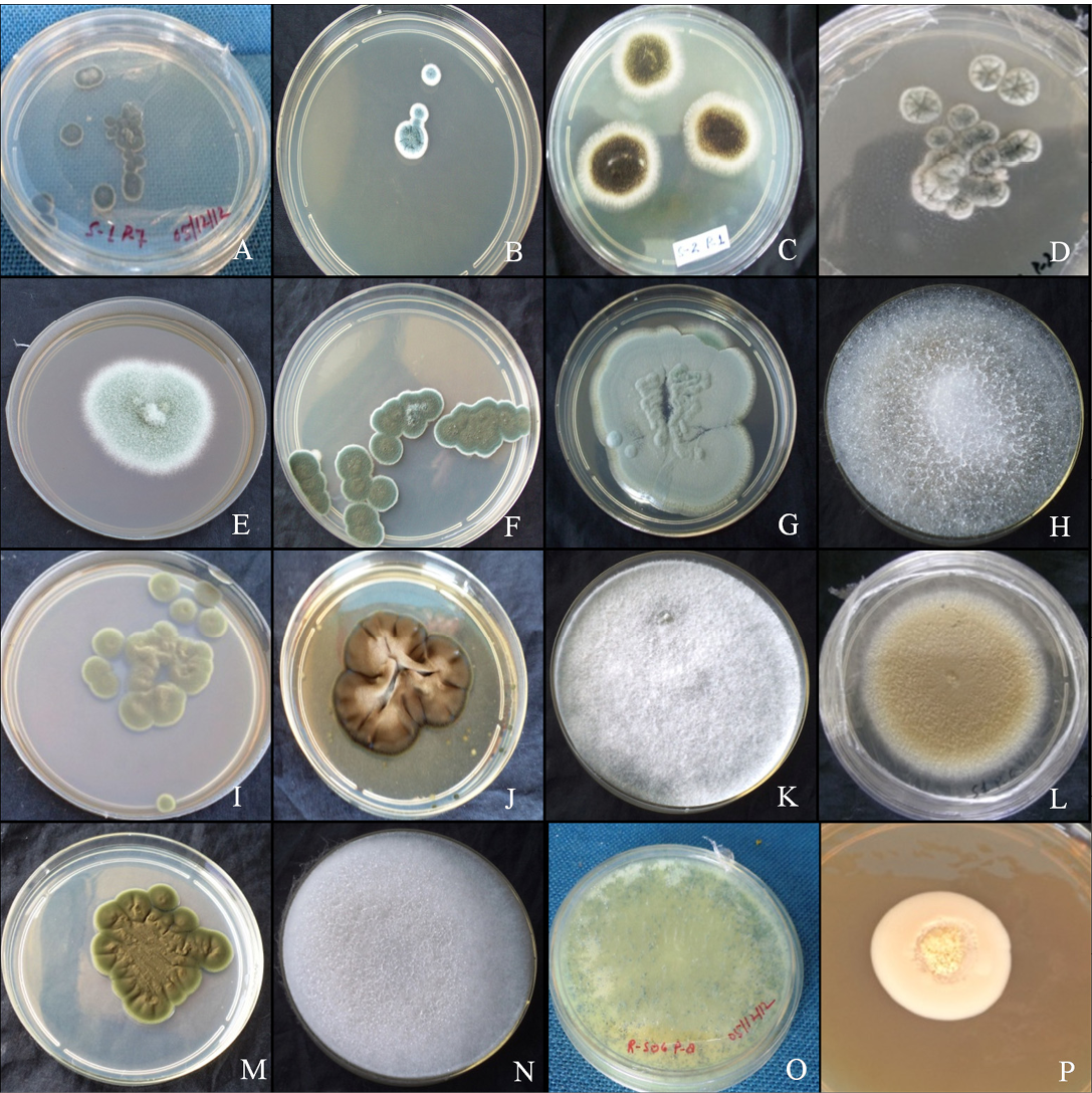
Figure 1: Colony morphology
A. versicolar gr (A), A. flavus (B) and A. niger (C). Aspergillus sp. (D), Penicillium funiculosum (E), Penicillium sp. (F), Penicillium sp. (G), Absidia corymbifera (H), Cladosporium cladosporiodes (I), Cladosporium resinae (J), Chrysosporium tropicum (K), Paecilomyces varitii (L), Malbranchea sp. (M), Mucor sp. (N), Trichoderma sp. (O) and yeast sp. (P)
conspicuously echinulate, smooth to very finely roughened and spherical (Figure 2B). Conidiophores were coarsely roughened and uncolored. Colonies of A. niger were isolated from the guano samples of R. hardwickii collected from a monument at Khusrubagh, Allahabad and at building roost in Sultanpur. The round and powdery colony with inner black and white margin (Figure 1C) attained 50.08 mm (Table 1). The conidium of A. niger was brown to black, very rough and globose (Figure 1C). The conidiophores bore numerous black dot-like spores at the terminal end (Figure 2C). The hyphae were translucent and septate. The conidial heads of A. niger were radiated initially and split into columns at maturity. A colony of unknown species belongs to the genus Aspergillus was isolated from the guano of R. hardwickii collected at Atala mosque, Jaunpur (Figure 2D). The rounded colony was downy; with inner army green and white margin attained 16.5 mm at maturity (Figure 1D).
A colony of Penicillium funiculosum was isolated from the guano sample collected from the Thar Ganga Ghat, Varanasi. The colony was cottony, white, rounded and attained
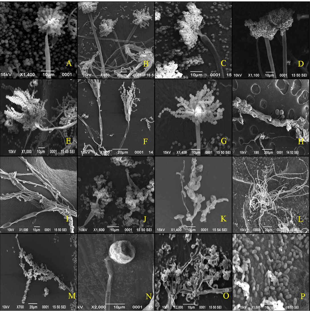
Figure 2: Scanning electron micrographs
A. versicolar gr (A), A. flavus (B) and A. niger (C). Aspergillus sp. (D), Penicillium funiculosum (E), Penicillium sp. (F), Penicillium sp. (G), Absidia corymbifera (H), Cladosporium cladosporiodes (I), Cladosporium resinae (J), Chrysosporium tropicum (K), Paecilomyces varitii (L), Malbranchea sp. (M), Mucor sp. (N), Trichoderma sp. (O) and yeast sp. (P)
32.35 mm (Figure 1E). A large number of whip-like conidial chains observed at the terminal end of conidiophore with spherical spores (Figure 2E). In addition, two isolates of Penicillium were collected from the guano of R. hardwickii. A dark moss green, velvety and round colony was isolated from the guano samples collected from Diyara, Sultanpur attained 28.42 mm (Figure 1F). The conidiophores consist long and oval spores at the terminal end (Figure 2F). Another rounded and velvety colony with Cambridge blue was isolated from the sample collected from Khusrubagh fort, Allahabad (Figure 1G). The matured colony attained 6.13 mm. The conidiophore was branched and bore chains of conidia at the terminal end (Figure 2G).
Absidia corymbifera was isolated from the guano samples of R. hardwickii collected at Khusrubagh fort, Allahabad and R. micorphyllum samples from Godavari cave temple, Chitrakoot. The colony of A. corymbifera was white, wooly, rounded and attained 37.93 mm at maturity (Figure 1H). The branched conidiophores bore few numbers of conidia (Figure 2H). Two species of Cladosporium were isolated from the guano samples of R. hardwickii (Table 1). Cladosporium cladosporoides was isolated from a roost at the riverbank of Ganga, Varanasi and C. resinae was isolated from a monument at Sultanpur. The colony of C. cladosporoides was bronze yellow, velvety, rounded and attained 9.39 mm (Figure 1I). The septate hyphae had spherical conidia. The conidia were sparsely attached on the hyphae (Figure 2I). The colony of C. resinae was moderately sporulated, dark brown, velvety and attained 16.43 mm (Figure 1J). The first conidium was developed in to ramoconidia with three protuberant scars and conidiophores arose laterally from vegetative hyphae (Figure 2J). Chrysosporium tropicum was isolated from the guano samples of R. hardwickii collected from Jhusi fort, Allahabad. The colony of C. tropicum was white, wooly, rounded and attained 7.58 mm at maturity (Figure 1K). The thin walled hyphae were hyaline, branched and divided by septa. The barrel-shaped conidium was solitary, terminal and stalked (Figure 2K). The colony of P. varitii was wooly, rounded, burly wood colour and attained a diameter 66.29 mm (Figure 1L). The hyphae were septate, branched and bore chains of spores (Figure 2L).
In addition, one species belongs to each of genera Malbranchea, Mucor, Trichoderma and yeast was isolated. The colonies of Malbranchea sp. and Mucor sp. were isolated from the guano samples of R. hardwickii collected from Khusrubagh and Allahabad. The colony of Malbranchea sp. was bronze colored, velvety textured and attained 14.62 mm (Figure 1M). The branched and segmented hyphae bore spores (Figure 2M). The colony of Mucor sp. was white, wooly, rounded and attained 58.01 mm (Figure 1N). The hyphae were hyaline with terminal conidia (Figure 2N). The colony of Trichoderma sp was acid green, wooly and attained 58.01 mm (Figure 1O). The hyphae were highly branched and bore spores (Figure 2O). The yeast colony was pink, wooly, rounded, non-sporulating and attained 7.72 mm with capsule-like individual yeast (Figure 1P, 2P). In addition, sterile mycelia were isolated from the guano samples of R. hardwickii collected from Atla mosque, Jaunpur, Jhunsi fort, Allahabad and Godavari cave, Chitrakoot. The colonies of sterile mycelia were white, cottony, rounded and non-sporulating.