Special Issue: Emerging and Re-Emerging Animal Health Challenges in Low and Middle-Income Countries · Volume 12, Issue 1, 18 December 2024 · Open Access
Genetic Analysis and Phylogenetic Study of IL-10-Like Protein and ORF103 Encoded Genes of Sheep Poxvirus Infecting Sheep from Iraq
· · · ·
Affiliations & Notes ⌄
1Department of Internal Medicine, College of Veterinary Medicine, University of Al-Qadisiyah, Al-Diwaniyah, Iraq
2Department of Veterinary Microbiology, College of Veterinary Medicine, University of Al-Qadisiyah, Al-Diwaniyah, Iraq
3Laboratory of Microbiology, National School of Veterinary Medicine of Sidi Thabet, University of Manouba, Manouba, Tunisia. *Correspondence | Mohammed A. Al-Saadi, Department of Internal Medicine, College of Veterinary Medicine, University of Al-Qadisiyah, Al-Diwaniyah, Iraq
The evolutionary analysis by maximum likelihood method revealed high sequence similarity in both IL-10 like protein and ORF103 genes with no significant genetic variation.
The genetic analysis of both IL-10 and ORF103 genes showed high sequence similarity with sequences from Turkey and Saudi Arabia (Figure 4).
This study provides information about the molecular characteristics of SPV after the genetic analysis of the important genes of this virus.
These findings continue to emphasize the importance of doing a continuous genetic monitoring of SPV on field, that will help us to keep the animal health status, in addition to controlling outbreaks.
Selected from the article's own text.
Abstract
This study aimed to investigate the genetic and phylogenetic characteristics of IL-10-like protein and ORF103 encoded genes of Sheep poxvirus (SPV) in sheep in Iraq. DNA samples of three sheep farms with clinical signs of pox infection were employed to collect the specimens from Al-Qadisiyah Province, Iraq, and the DNA was extracted and amplified by PCR targeting IL-10 and ORF103 genes. After purifying the PCR products, the amplicons were sequenced by Sanger method. Following the sequencing, the Data were studied using the MEGA11 software. The findings revealed that infected sheep showed typical pox lesions in various parts of the body. while lambs infected with SPV, showed more severe symptoms of this infection. The genetic analysis of both IL-10 and ORF103 genes showed high sequence similarity with sequences from Turkey and Saudi Arabia. This study provides information about the molecular characteristics of SPV after the genetic analysis of the important genes of this virus. This gives us ideas to develop better diagnostic tools and control measures and help to decrease the effect of this infection on sheep populations. These findings continue to emphasize the importance of doing a continuous genetic monitoring of SPV on field, that will help us to keep the animal health status, in addition to controlling outbreaks.
Keywords | Cytokines, Dermatitis, Itch
Introduction
Sheep poxvirus (SPPV) is a highly virulent virus that causes common sheep pox with significant exposure. The disease presents pox skin lesions on the dermal layer of the sheep. The virus is a member of the family Poxviridae that belongs to the Capripoxvirus, which also includes Goat pox virus (GTPV) and lumpy skin disease virus (LSDV). Together with swine pox virus (SWPV), these are mainly responsible for the infection and are the main agents of poxvirus diseases in animals ; ; .
The disease is often manifested in the dusky-hued areas of the world and is characterized by circular skin rash and nodal autolysis. Animals are often infectious via urine, stool, and secretions. The virus enters the body through the conjunctiva, or the mucous membranes of the nasal cavity, tongue, oral cavity, and infiltration of lung tissues ; ; .
Sheep poxvirus (SPPV) causes one of the most contagious diseases of sheep, goats, and various wild mouse species. Sheep pox tends to cause high morbidity and mortality epidemics in countries from the East, Middle East, Asia, and Africa (Tuppurainea et al., 2021; ; . The disease is transmitted through aerosols and direct or indirect contact with infected animals. Due to its highly contagious nature, the disease has to be reported to the World Organization for Animal Health (OIE). The main features of this disease are fever, generally occurring skin eruption, and enlarged lymph nodes. The economic losses caused by epidemics can be extensive; they are both direct due to death and carriage of sheep, and meat and wool production losses. Indirect losses favor the illicit transboundary movement of animals ; ; .
Some researchers suggested that sheep poxvirus should belong to the genus Suipoxvirus and rename it Ovipo virus, which was avoided to be widely accepted. A study focused on the whole-genome target enrichment hybridization indicated that the established phylogenetic three-species paradigm is not consistent with the evolutionary history of the viruses, proposing that all host-virus combinations may be able to occur in the wild ; ; .
The virus is categorized as an established member of the family Poxviridae and the subfamily Chordopoxvirinae. Sheeppox is an exceptionally high-profile consequence of a long-standing cross-species infection by an ancestral sheep poxvirus ; .
Sheep poxvirus exhibits minimal serologic cross-reactivity with closely related goat poxvirus. Both sheep and goat poxviruses occur in a wide variety of natural hosts in their homeland, and disease in a small subset of these animals can sometimes be very severe. Disease reports have involved wild and captive animals, including species from the Order Artiodactyla: Deer, oryx, and buffalo, and many Chiroptera species: Egyptian fruit bats, serotine bats, and mouse-eared bats (Berguuido et al., 2022).
SPV is transmitted mainly through the association of animals, but mechanical transmission by people, equipment, and vectors (in particular blood-feeding diptera like the genus Culicoides) are also relevant routes of transmission and can be important route amplifiers ; .
The primary organs involved are the lungs, oral cavity, esophagus, and digestive tract, especially the hard corneal nodules and trodden ulceration. Bacterial infections occur frequently and may exacerbate clinical disease. Other organs with initial disease participation include udders and teats, genital orifices, and liver, lung, and kidney in the internal organs. The disease is usually fatal, mainly due to severe enteric lymphadenopathy and pneumonia with secondary bacterial infections ; Baker et al., 2021).
This study aimed to investigate the genetic and phylogenetic characteristics of IL-10-like protein and ORF103 encoded genes of Sheep poxvirus (SPV) in sheep in Iraq.
Materials and Methods
All the details related to the materials and methods are illustrated in Table 1.
Results and Discussion
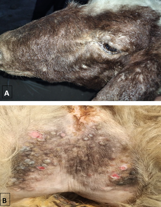
Clinical observations of affected sheep revealed benign pox lesions on the face, inner side of lips, inner thighs, and under tail. Symptoms included weakness and lack of appetite. Lambs were more severely affected than adults. Fever was only noted during the first 3 days with no other systemic involvement (Figure 1).
Table 1: Materials and Methods.
Section
Details
Animals and sample collection
- Locations: Three herds in Al-Qadisiyah province (2022) - Samples: 30 skin nodules/crusts (20 mg each) - Condition: Unvaccinated sheep showing pox symptoms - Storage: Collected aseptically, stored at -20 ºC
DNA extraction
- Procedure: Add Bio (South Korea) - Steps: - Tissue lysis: 200 µl lysis buffer, 20 µl - Measurement: Quantus fluorometer (Promega USA) - Storage: -20°C
PCR amplification
- Volume: 20 µl - Components: - Template DNA: 2 µl - Primers: 1 µl each - Master mix: 10 µl - PCR water: Remaining volume - Cycler: T100 thermal cycler (BioRad USA) - Conditions: - Initial denaturation: 95°C, 5 min - Denaturation: 95°C, 30 sec - Annealing: 55°C, 35 sec - Extension: 72°C, 40 sec - Final extension: 72°C, 5 min - Analysis: 1.5% agarose gel electrophoresis, ethidium bromide staining, gel documentation system
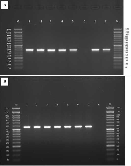
Genetic analysis using gel electrophoresis confirmed the presence of positive samples for IL-10 like protein gene (387 bp) and ORF103 gene (570 bp). Molecular markers (50-1500 bp) were also identified upon presence of positive bands/smear. Finally, controls were investigated by keeping template DNA and replacing it with H2O. This approach indicated the consistent presence of positive samples in all confirmatory tests (Figure 2).
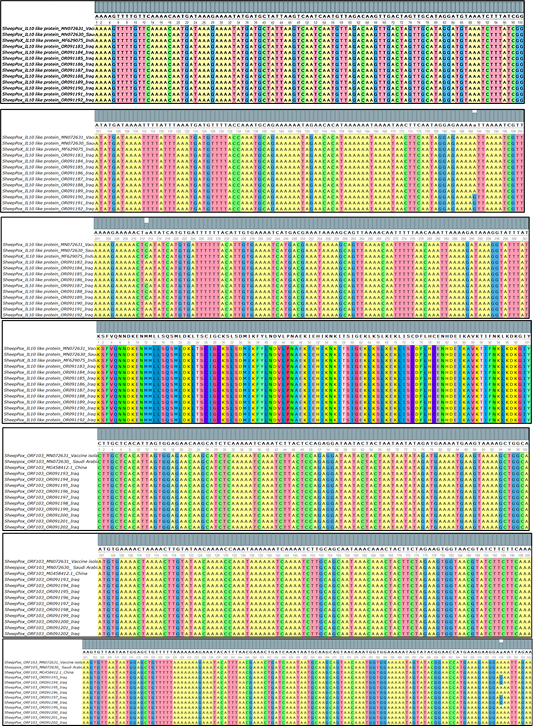
A mutation was induced by point mutations and detected in the probe through Sanger sequencing (Figure 3). Multiple sequence alignment analysis was then used to identify single nucleotide polymorphisms (SNP) in IL-10 gene. It is noteworthy to mention the high level of sequence similarity with some apparent change resulted from multiple mutations. More precisely, two (78%) SNPs were identified in ORF130 gene in the form of three (Figure 3).
The results of the present analysis identified a range of changes resulting from point mutations in the probe. Comparison analysis showed a percentage of minimum 39.3-86.8%, with no significant alteration in the encoded amino acids. Similarly, ORF103 gene revealed high level of sequence similarity with point mutation found in the form of five. Detailed analysis showed mutations that led to minor alteration in amino acid (lysine K is mutated to glutamic acid E).
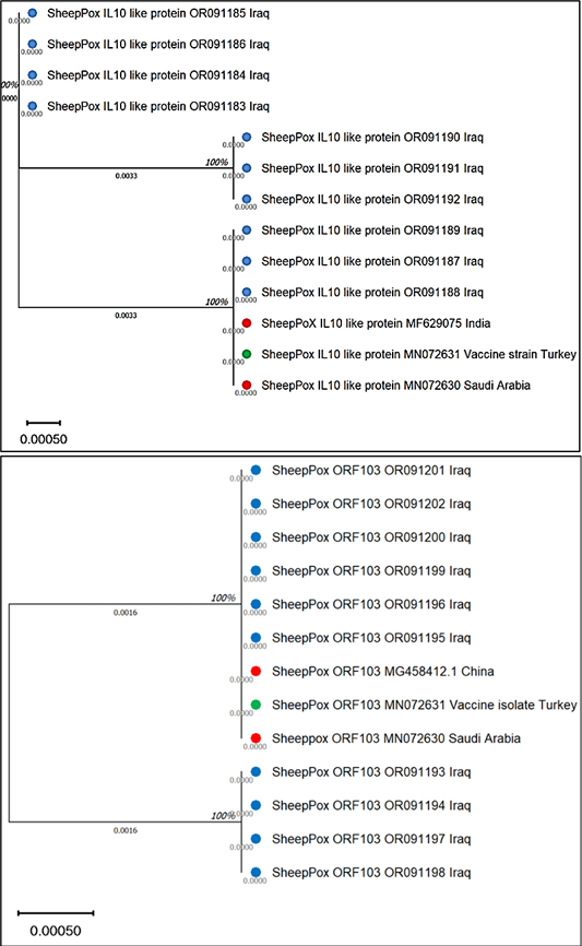
Understanding phylogenetic evolution is relevant to evaluate the variety of bacteria and spread of disease. The evolutionary analysis by maximum likelihood method revealed high sequence similarity in both IL-10 like protein and ORF103 genes with no significant genetic variation. This indicated that the Sheep poxvirus strains in the concerned region are highly conserved and less prone to changes. Consequently, such information will help to understand the genome and develop diagnosis and control methods of the disease. The genetic analysis of both IL-10 and ORF103 genes showed high sequence similarity with sequences from Turkey and Saudi Arabia (Figure 4).
SPV is a severe, highly infectious infection in sheep, goats, and other cloven-hooved animals, causing substantial damage to the livestock industry. Members of SPV belong to the genus Capripoxvirus (family Poxviridae), the same class of readily transmitted viruses that includes the infectious viruses behind smallpox and monkeypox. SPV itself encodes several immunomodulatory proteins that boost the viral immune evasion mechanisms, including a protein like interleukin-10 (IL-10) and the ORF103 protein. The role of these host-range proteins in viral pathogenicity, and particularly SPV immune evasion mechanisms, has been established ; .
The sequence data was then analyzed using MEGA11 software which showed lesions in the infected sheep and that a lamb infected with SPV showed severe symptoms of the infection. This finding was supported by previous reports that suggested young animals are more susceptible to SPV infection .
More genetic analysis of both the IL-10-like protein-homologous and ORF103 genes showed the highest similarity with sequences, respectively, from Turkey and Saudi Arabia . This is not unexpected because SPV strains exhibit a genetic variability when compared across different continents ; .
SPV encodes an IL-10-like protein because it is a viral homolog of the cellular and well-studied cytokine IL-10, an essential immune-modulator of the host response ; . The viral IL-10-like mimicry assists viral escape from the immune system by acting like cellular IL-10 to halt the production of numerous pro-inflammatory cytokines and prevent the activation of antigen-presenting cells.
In contrast, the ORF103 protein of SPV serves as a virulence factor associated with pathogenesis ; . The ORF103 protein is present in the surface of the viral envelope and is critical for viral morphogenesis and egress . The genetic diversity of these viral proteins forms the backdrop for elucidating evolutionary mechanisms leading to strains causing various SPV-related diseases and their pathogenic variation.
The results of the genetic analysis performed in this study agree with some of the previous studies done elsewhere. They show high level of similarity with other researchers’ results based on recognition of some similar findings, by which we have identified some unique observations. In recently published studies, the IL-10 like protein gene of SPV shows a high level of conservation in the sequence identity across different geographical regions. Our study also exhibited this feature by showing high similarity level within the Al-Qadisiyah province samples with some minor mutations that lead to few non-helped changes in the amino acid set. A previous study by Bowden et al. (2008) showed the IL-10 genes in SPV from Caracas, Turks and Caicos Islanders and England showed highly conservative in the genetic level where two samples from England were highly identical in their sequences.
Correspondingly, the ORF103 gene, having the potential to encode for the viral glycoprotein, also displayed the highest overall sequence similarity with minor amino acid variations. Initially, we found point mutations such as a lysine (K) to a glutamic acid (E) substitution. Instead of exhibiting much genetic instability as previously reported by Balinsyg et al. (2008), who enumerated typical point mutations in various SPV strains.
In addition to the virus virulence seen, we observed that evolutionary analyses previously performed by others such as by Tulman et al. (2002) have shown that SPV genes, including the IL-10 and ORF103 genes, show high degrees of conservation.
Therefore, our results on the variation of IL-10 and ORF103 genes in SPV are consistent with other researchers that IL-10 and ORF103 genes were under strong selection and accumulated de novo mutations at remarkably low levels, these comparative studies not only confirm our results but also stress the necessity for continuous examination and further genetic analysis which could provide valuable guidance of SPV prevention and control in different regions ; ; ; Al-Salihe and Alhankawe, 2023; El-Bagaury et al., 2023; ; ; ; .
Conclusions and Recommendations
This study provides information about the molecular characteristics of SPV after the genetic analysis of the important genes of this virus. This gives us ideas to develop better diagnostic tools and control measures and help to decrease the effect of this infection on sheep populations. These findings continue to emphasize the importance of doing a continuous genetic monitoring of SPV on field, that will help us to keep the animal health status, in addition to controlling outbreaks.
Acknowledgement
Very special gratitude to Prof. Dr. Alsaadi Jabar Abbas, Faculty of Veterinary Medicine, University of Al-Qadisiyah.
Novelty Statement
Emphasize the importance of doing a continuous genetic monitoring of SPV on field, that will help us to keep the animal health status.
Author’s Contribution
All authors contributed equally to the manuscript.
Conflict of interest
The authors have declared no conflict of interest.
Figures
Figure 1:.
(A, B) images of sheeppox lesions.
Figure 2:.
Gel electrophoresis image (1.5 % agarose) of sheeppox virus samples from sheep. (A) IL-10-like protein gene (Lanes: 1-7; size= 387 bp). (B) ORF103 (Lanes: 1-7; size= 550 bp; C: negative control: H2O). M: 50-1500 bp DNA marker.
Figure 3:.
Multiple sequence alignment of a partial sequence of IL-10 and ORF103 genes of sheeppox virus.
Figure 4:.
Phylogenetic tree of sheeppox virus from sheep. IL-10-like protein and ORF103 genes.
References
Akrawi S, Alkhaled K (2024). Monitoring land cover variation using some spectral indicators in Akre region based on geospatial techniques. Mesopotamia J. Agric., 52(2): 130-145. https://doi.org/10.33899/mja.2024.145686.133014[CrossRef]
Al-Baroodi SY, Moosa, DA, Al-Attar MY (2022). Detection of Maedi-visna virus in sheep in Nineveh province. Iraqi J. Vet. Sci., 36(1): 61-64. https://doi.org/10.33899/ijvs.2021.129075.1622[CrossRef]
Alsalih FY, Alhankawe OK (2023). Molecular evidence of schmallenberg virus associated by ovine abortion with fetal anomalies in Nineveh province, Iraq. Iraqi J. Vet. Sci., 37(1): 115-120. https://doi.org/10.33899/ijvs.2022.133665.2276[CrossRef]
Al-Zubaidi MR, Thwiny HT, Al-Biati MN (2023). Modulation of chitosan nanoparticles properties for sheep pox mucosal. Iraqi J. Vet. Sci., 37(2): 111-119. https://doi.org/10.33899/ijvs.2023.1374030.2682[CrossRef]
Amin DM, Shehab G, Emran R, Hassanien RT, Alagmy GN, Hagag NM, Abd-El-Moniem MI, Habashi AR, Ibraheem EM, Shahein MA (2021). Diagnosis of naturally occurring lumpy skin disease virus infection in cattle using virological, molecular, and immunohistopathological assays. Vet. World, 14(8): 2230. https://doi.org/10.14202/vetworld.2021.2230-2237[CrossRef]
Baird AN, Shipley CF (2021). Diseases of the Integumentary System. In: Sheep, goat and cervid medicine, 3ed, pp. 221-250. https://doi.org/10.1016/B978-0-323-62463-3.00019-0[CrossRef]
Bakar L, Hussein AS, Tamam SM, Madbouly HM (2021). Isolation of lumpy skin disease virus isolated from SPPV vaccinated cattle. J. Vet. Med. Res., 28(1): 38-44. https://doi.org/10.21608/JVMR.2021.73921.1037[CrossRef]
Balinsky CA, Delhon G, Smoliga G, Prarat M, French RA, Geary SJ, Rock DL (2008). Rapid progression of severe clinical disease in sheep infected with a recombinant capripoxvirus expressing the highly virulent rabbit fibroma virus RABOV gene. J. Virol., 82(17): 8262-8274.
Berguido FJ, Gelaye E, Liu Y, Davaasuren B, Krstevski K, Djadjovski I, Ivanova E, Goujgoulova G, Loitsch A, Tuppurainen E, Chibssa TR (2022). Development and optimization of indirect ELISAs for the detection of anti-capripoxvirus antibodies in cattle, sheep, and goat sera. Microorganisms, 10(10): 1956. https://doi.org/10.3390/microorganisms10101956[CrossRef]
Beytut E, Coskun N, Dag S, Nuhoglu H, Karakurt E, Yilmaz V, Yildiz A (2024). Pathological and Molecular Investigations in the Aborted Fetal Lamb due to the Poxvirus. J. Hellenic Vet. Med. Soc., 75(1): 7185-7190.
Bhanuprakash V, Indrani BK, Hosamani M, Singh RK (2010). The current status of sheep pox disease. Comp. Immunol. Microbiol. Infect. Dis., 29(1): 27-60. https://doi.org/10.1016/j.cimid.2005.12.001[CrossRef]
Bowden TR, Babiuk SL, Parkyn GR, Copps JS, Boyle DB (2008). Capripoxvirus tissue tropism and shedding: A quantitative study in experimentally infected sheep and goats. Virology, 371(2): 380-393. https://doi.org/10.1016/j.virol.2007.10.002[CrossRef]
Cao JX, Gershon PD, Black DN (1995). Sequence analysis of HindIII Q2 virion and proteinase K-released DNA fragments of capripoxvirus strains. Virus Res., 39(3): 279-293.
Chahota R, Sharma P, Kumar R, Gupta T, Sharma M (2022). Investigation of an outbreak of sheeppox among native sheep breeds in the Western Himalayas of India. Vet. Res. Commun., 1(2): 1-7. https://doi.org/10.1007/s11259-021-09833-z[CrossRef]
Delhon G (2022). Poxviridae. In: Veterinary microbiology. https://doi.org/10.1002/9781119650836.ch53[CrossRef]
El-Bagoury GF, Nour El-Deen MR, Fakhry HM (2023). Genetic characterization and phylogenetic analysis of foot and mouth disease virus vaccine strains and recent field isolate. Iraqi J. Vet. Sci., 37(2): 347-354. https://doi.org/10.33899/ijvs.2022.133432.2227[CrossRef]
Faizy W, Kassab Bashi A, Toma R (2024). Effect of drought stress on the metabolic pathway and survival of two grape (Vitis vinifera L.) cultivars grown under in vitro conditions. Mesopotamia J. Agric., 52(2): 99-117. https://doi.org/10.33899/mja.2024.146167.135010[CrossRef]
Ghander H, El-Tholoth M, Rahman AE, El-Kenawy A (2020). Conventional and molecular characterization of sheep poxvirus (SPV) in Dakahlia Governorate, Egypt. Mansoura Vet. Med. J., 21(4): 142-148. https://doi.org/10.21608/mvmj.2020.134738[CrossRef]
Hamdi J, Munyanduki H, Omari Tadlaoui K, El-Harrak M, Fassi Fihri O (2021). Capripoxvirus infections in ruminants: A review. Microorganisms, 9(5): 902. https://doi.org/10.3390/microorganisms9050902[CrossRef]
Karalyan Z, Zakaryan H, Sargsyan N, Voskanyan H, Arzumanyan H, Karalova E (2019). Viral agents of multiple sclerosis. BMC Neurol., 19(1): 1-14.
Khayoon T, Abbas R, Abdullah F (2024). Effects of feeding various levels of postbiotics produced by lactic acid bacteria on growth performance, gastrointestinal microbiota count, and digestibility of some nutrients in broiler chickens. Mesopotamia J. Agric., 52(2): 68-81. https://doi.org/10.33899/mja.2024.145531.132019[CrossRef]
Khidhir Z, Hamma A, Mohammed M, Mahmood A, Al-Obaidi A, Aldoori Z (2024). Effect of longevity spinach leaves powder (LSP) as a broiler feed additive on some physical and chemical parameters of frozen stored thigh meat. Mesopotamia J. Agric., 52(2): 82-98. https://doi.org/10.33899/mja.2024.145203.131011013[CrossRef]
Khudeir MA, Alsultan AT, Khudhair YI (2024). Novel CRISPR/Cas13- based assay for diagnosis of avian infectious bronchitis. Iraqi J. Vet. Sci., 38(1): 63-69. https://doi.org/10.33899/ijvs.2023.140335.3040[CrossRef]
Klaif SF, Jassim A, Alfatlawi MA, Ali MJ (2022). Major-surface-protein-4-gene-based detection of Anaplasma marginale isolated from sheep in Al-Diwaniyah province, Iraq. Iraqi J. Vet. Sci., 36(1): 85–88. https://doi.org/10.33899/ijvs.2021.129230.1635[CrossRef]
Kumar N, Barua S, Kumar R, Kumar A, Verma A, Singh L, Godara B, Chander Y, Kumar G, Riyesh T, Kumar D, , Pathak A, Kumar S, Dedar AK, Mehta V,Gaur M, Bhardwaj B, Vyas V, Chaudhary S, Yadav V, Bhati A, Kaul R, Bashir A, Andrabi A, Yousuf RW, Koul A, Kachhawaha S, Gurav A, Gautam S,Tiwari HA, Munjal VK, Gupta MK, Kumar R, Gulati BR, Misri J, Kumar A, Mohanty AK, Nandi S, Singh KP, Pal Y, Dutt T, Tripathi PN (2023). Evaluation of the safety, immunogenicity and efficacy of a new live-attenuated lumpy skin disease vaccine in India. Virulence, 14(1): 2190647. https://doi.org/10.1080/21505594.2023.2190647[CrossRef]
Lamien CE, Le Goff C, Silber R, Wallace DB, Gulyaz V, Tuppurainen E, Madani H, Caufour P, Adam T, Elharrak M, Luckins AG, Albina E, Diallo A (2011). Use of the Capripoxvirus homologue of Vaccinia virus 30 kDa RNA polymerase subunit-encoding gene (RPO30) for identification and phylogenetic clustering of sheeppoxvirus and goatpoxvirus isolates. Virus Genes, 42(1): 98-109. https://doi.org/10.1016/j.vetmic.2010.09.038[CrossRef]
Mansour KA, Hussain MH, Abid AJ, Kshash QH (2022). Orf disease in local goat; clinical and phylogenetic study in Al-Qadisiyah governorate, Iraq. Iraqi J. Vet. Sci., 36(1): 117-121. https://doi.org/10.33899/ijvs.2021.129489.1651[CrossRef]
Modise BM, Settypalli TB, Kgotlele T, Xue D, Ntesang K, Kumile K, Naletoski I, Nyange JF, Thanda C, Macheng KN, Marobela-Raborokgwe C (2021). First molecular characterization of poxviruses in cattle, sheep, and goats in Botswana. Virol. J., 18(2): 1-1. https://doi.org/10.1186/s12985-021-01634-9[CrossRef]
Nagarajan G, Saikumar G, Shweta B, Prem K, Kishore S, Shweta T (2010). Capripoxvirus vector used for immunotherapy against HIV-1. Microbiology, 156(1): 279-291.
Neamah AA, Fahad KH, Sadeq JN, Al-Fatlawi MAA (2021). Comparative diagnostic study of Streptococcus dysgalactiae isolated from cow’s milk by using media, biochemical tests, VITEK 2, and polymerase chain reaction. Int. J. Drug Del. Technol., 11(4): 1399–1404.
Neamah AA, Fahed KH, Sadeq JN, Alfatlawi MA (2022). Molecular characterization and phylogenetic analysis of Escherichia coli isolated from milk of cattle affected by mastitis. Iraqi J. Vet. Sci., 36(1): 251–254. https://doi.org/10.33899/ijvs.2021.129934.1702[CrossRef]
Pashupathi M, Anbazhagan S, Barkathullah N, Priyadarsini S, Panda S, Kumar A (2022). Designing and modelling of trivalent chimeric vaccine for capripoxvirus: Lumpy skin disease, sheep pox and goat pox by immunoinformatics approach. Res. Sq., 24(3): 231-236.
Stanbekova G, Beisenov D, Nizkorodova A, Iskakov B, Warzecha H (2021). Production of the sheep pox virus structural protein SPPV117 in tobacco chloroplasts. Biotechnol. Lett., 43(2): 1475-1485. https://doi.org/10.1007/s10529-021-03117-x[CrossRef]
Sumana K, Revanaiah Y, Shivachandra SB, Mothay D, Apsana R, Saminathan M, Basavaraj S, Reddy GM (2020). Molecular phylogeny of Capripoxviruses based on major immunodominant protein (P32) reveals circulation of host specific sheeppox and goatpox viruses in small ruminants of India. Infect. Genet. Evol., 1(85): 104472. https://doi.org/10.1016/j.meegid.2020.104472[CrossRef]
Tamura K, Stecher G, Kumar S (2021). MEGA11: Molecular evolutionary genetics analysis version 11. Mol. Biol. Evol., 38(7): 3022-3027. https://doi.org/10.1093/molbev/msab120[CrossRef]
Tesgera T, Sori T, Deme BB, Tolossa W, Negeewo A (2024). Immune response and safety of co-administered peste des petits ruminants, contagious caprine pleuropneumonia, sheep and goat pox, and pasteurellosis vaccines in goats. Open Vet. J., 14(8): 1960-1967. https://doi.org/10.5455/OVJ.2024.v14.i8.25[CrossRef]
Tulman ER, Afonso CL, Lu Z, Zsak L, Kutish GF, Rock DL (2002). Genome of lumpy skin disease virus. J. Virol., 76(12): 6712-6723. https://doi.org/10.1128/JVI.76.12.6054-6061.2002[CrossRef]
Tulman ER, Afonso CL, Lu Z, Zsak L, Sur JH, Sandybaev NT, Kerembekova UZ, Zaitsiv VL, Kutish GF, Rock DL (2001). The genomes of sheeppox and goatpox viruses. J. Virol., 75(2): 505-520.
Tuppurainen ES, Lamien CE, Diallo A (2021). Capripox (Lumpy skin disease, sheep pox, and goat pox). In: Veterinary vaccines: Principles and applications. pp. 383-397. https://doi.org/10.1002/9781119506287.ch28[CrossRef]
Tuppurainen E (2022). Sheep and goat Pox. CABI Compend. https://doi.org/10.1079/cabicompendium.81537[CrossRef]
Venkatesan G, Kumar A, Bhanuprakash V, Balamurugan V, Singh RK (2020). Capripoxvirus and Orf virus. In: Animal-Origin Viral Zoonoses. pp. 203-221. https://doi.org/10.1007/978-981-15-2651-0_9[CrossRef]