
Rosemary (Rosmarinus officinalis L.) is one of the important ornamental, aromatic, and medicinal plants belonging to the family Lamiaceae. This plant is infected by several plant pathogens, causing considerable economic losses to its growth and production. Among these, Fusarium wilt is a major and destructive disease hampering rosemary cultivation in many agroclimatic settings in Iraq. This study aimed to identify the fungal pathogens associated with the rosemary seedlings showing wilt symptoms. To this end, infected rosemary plants were collected randomly from different local nurseries of Al-Qadisiyah Governorate of Iraq. Fungal pathogens from the roots of these plants were isolated, propagated, and identified based on the morphological characteristics of colonies (i.e., colony color, mycelial growth, and colony diameter) and confirmed by molecular sequencing. The initial morphological characterization showed the presence of two pathogenic fungal species, i.e., Fusarium solani and F. chlamydosporum. Furthermore, molecular diagnosis based on sequencing of the ITS-DNA gene region using ITS1F and ITS4 R primers corroborated the morphological diagnosis. Sequence data analyses exhibited 99 and 100% alignment of Iraqi isolates of F. solani and F. chlamydosporum, along with the Chinese isolates (MN960013.1 and KX783373.1), respectively. DNA sequences of Iraqi isolates of F. solani and F. chlamydosporum are deposited in the NCBI Gene-Bank with accession codes OQ299566.1 and OQ301746.1, respectively. To our knowledge, this study encompasses the first morphological and molecular diagnosis of the pathogenic fungal species responsible for Fusarium wilt in rosemary plants in Iraq, posing a serious threat to rosemary cultivation in nurseries.
Research Article · Volume 42, Issue 2, 9 June 2026 · Open Access
First Report on Morpho-Molecular Identification of Pathogenic Fungal Isolates Associated with Rosemary (Rosmarinus officinalis L.) Plants in Al-Qadisiyah, Iraq
· · · ·
Affiliations & Notes ⌄
- 1Department of Agricultural Pest Control, Directorate of Plant Protection, Ministry of Agriculture, Iraq
- 2Department of Horticulture Sciences, Agricultural College, University of Al-Qadisiyah, Al Diwaniyah, 58002, Iraq
- 3Department of Soil and Water Resources, Agricultural College, University of Al-Qadisiyah, Al Diwaniyah, 58002, Iraq
- 4Department of Plant Protection, Agricultural College, University of Al-Qadisiyah, Al Diwaniyah, 58002, Iraq. *Correspondence | Ali Sabah Alhasan, Department of Agricultural Pest Control, Directorate of Plant Protection, Ministry of Agriculture, Iraq
- 5Email: ali.alhasan@qu.edu.iq
Article Info ⌄
- Journal:
- Sarhad Journal of Agriculture
- Published:
- 9 June 2026
From the article
- In this study, pathogenic fungal isolates of the wilted rosemary plants were cultured on PDA and were identified on the basis of their morpho-cultural traits as shown in Figure 2, 3, and 4.
- The first species was Fusarium solani, which distinguished itself with its appearance in PDA at 25°C, and showed pink upper-surface colonies and yellowish-brown pigmentation on the lower surface (Figure 2 A–B) with a radius of 50.5 mm after 7 days of growth.
- The phylogenetic tree of F. chlamydosporum Iraqi isolate (OQ301746.1) illustrates its relationship with five sequences of global isolates deposited to the GenBank database (Figure 7).
- Based on overall study results, it is concluded that Fusariosis or Fusarium wilt affecting the rosemary cultivation and production in the nurseries of the sampled area of Iraq is primarily caused by two species of pathogenic fungal isolates, i.e., F. solani and F. chlamydosporum.
Selected from the article's own text.
Abstract
Introduction
Rosemary (Rosmarinus officinalis L.) is a significant perennial evergreen shrub that belongs to the Lamiaceae family. It is an herbaceous plant indigenous to Mediterranean areas, widely cultivated throughout the world for its aromatic, medicinal, and ornamental features ; . Essential oil of rosemary is frequently used as a spice and flavoring agent and is famous for its antimicrobial, anti-inflammatory, and antioxidant activities in traditional medicine ; . Moreover, rosemary essential oil contains many bioactive compounds such as rosmarinic acid and Rosmanol that have many therapeutic uses against different human diseases, including cancer, atherosclerosis, and inflammatory diseases ; , .
Several biotic factors, such as plant pathogens, can affect the rosemary growth and production. The soilborne fungal diseases, such as Fusarium wilt, impose substantial damage to rosemary seedlings. Mycelium, spores, or polysaccharides of Fusarium fungus may obstruct xylem tissue, causing wilt, a condition that shows up as a sharp decrease in water flow to plant tissues . Discoloration of the vascular bundle originating from roots to aerial parts is the main symptom of Fusarium wilt that ultimately causes crown and root rot, defoliation, and death of the plant ; .
Fusariosis, plant diseases caused by different phytopathogenic strains of Fusarium species, cause substantial economic losses to plants worth billions of USD . These fungal pathogens are ubiquitously found throughout the world in soil, water, plants, and insects . They may spread from the affected fields through irrigation and crop residues . Some of the phytopathogenic Fusarium species cause infection in rosemary plants, including F. solani, F. oxysporum, F. reticulatum, and F. sambucinum ; .
Pathogenic Fusarium species are usually identified based on the morphological characters of their mycelia and colony shape and growth pattern . The size and shape of macro- and micro-conidia, in addition to the presence or absence of chlamydospores, are the main morphological traits used to distinguish Fusarium species ; . Moreover, internal transcribed spacer (ITS) segments of the ribosomal DNA sequences have recently been used as a molecular marker for systematics as well as phylogenetic analysis of closely related fungi . ITS region sequencing has been effectively employed for the fungal species’ characterization .
Based on the above-mentioned information, this study aimed to identify the Fusariosis-causing pathogenic fungi associated with rosemary seedlings based on their morpho-molecular traits. Although limited research has been conducted in Iraq to identify the soilborne Rhizoctonia solani associated with rosemary , there has been a lack of research efforts in investigating root rot caused by Fusarium species on rosemary plants in Iraq. Therefore, the purpose of this research work was to isolate and diagnose the cause of rosemary wilt in Iraq caused by Fusarium species using both morphological and molecular methods.
Materials and Methods
Plant samples
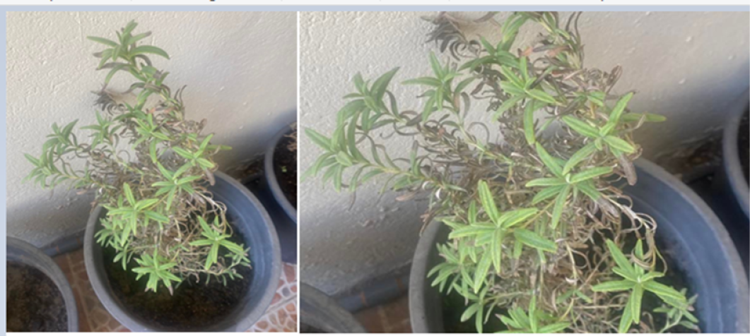
Rosemary seedlings exhibiting different symptoms of Fusarium wilt, including crown and root rot, and darkened vascular tissues of leaves (Figure 1), were sampled and collected randomly from different plant nurseries located in Al-Diwaniyah city of Al-Qadisiyah province of Iraq. Plant specimens were assembled and sent to the laboratory for pathogens’ isolation under cool conditions. The infected roots were chopped into tiny pieces, each measuring 0.5 to 1.0 cm in length, and were surface-sterilized for 2 min using 0.1% sodium hypochlorite and then washed with sterilized distilled water. Disinfected filter papers were used to dry the treated samples, which were then incubated on potato dextrose agar (PDA) medium in sterilized glass Petri-plates (60 cm diameter) at 25°C for a period of 7 days. To create a genetically pure culture, the hyphal extremity of the mycelia was re-cultured on PDA. The identification of the cultured pathogens was done by studying morphological characteristics of isolates under a compound microscope (up to 100×) .
Molecular detection and identification
Morphological and genetic traits were used to identify the F. solani, F. chlamydosporum, and A. terreus that were isolated from the infected rosemary samples. Using the collected mycelia, genomic DNA extraction and PCR amplification were carried out. ZR-Fungal/Bacterial/Yeast DNA MiniPrep Kit (ZYMO, USA) and FavorPrep Tissue Genomic DNA Extraction Mini Kits (Favorgen Biotech Corp, Ping-Tung, Taiwan) were used to extract the fungal DNA. An ITS gene fragment was amplified using ITS1/ITS4 forward “TCC GTA GGT GAA CCT TGC GG 3” and the reverse primer “TCC TCC GCT TAT TGA TAT GC 3” ; .
In brief, a PCR reaction volume of 25 μl contained 5 µl Taq PCR PreMix (Intron, Korea), 1.5 µl target DNA, 1 µl of primer (10 pmol), and distilled water. PCR amplification was performed on a thermal cycler (Gene Amp, PCR systems 9700; Advanced Biosystem) using the following thermal protocol. Three min of denaturation at 94°C, thirty-five cycles of dehydration at 94°C for forty-five sec, one min of annealing at 52°C, one min of extension at 72°C, and a final seven-min incubation step at 72°C. The final PCR products were visualized under a UV lamp on a 1.5% agarose gel with Red Safe Stain (20,000× Intron).
Sequencing of the PCR products of each fungal isolate was carried out by Macrogen, Inc. (Seoul, Republic of Korea) using an ABI 3730 XL Automatic Sequencer. Using previously deposited sequences from the National Center for Biological Information (NCBI), BioEdit Sequencing Alignment Editor (Version 7.1; DNASTAR, Madison, WI, USA) aligned pertinent isolated sequences. As indicated in Table 1, the isolated sequences of F. solani, F. chlamydosporum, and A. terreus used in this study were uploaded to GenBank with accession codes MN960013.1, KX783373.1, and MH918670.1, respectively.
Results and Discussion
Cultivation of rosemary, one of the popular medicinal and ornamental plants worldwide, is hampered by many soilborne diseases. Among these, Fusariosis is the most destructive disease often caused by Phytophthora, Fusarium, and Rhizoctonia species ; . Particularly, Fusarium species impose substantial damage to different plants, including rosemary ; .
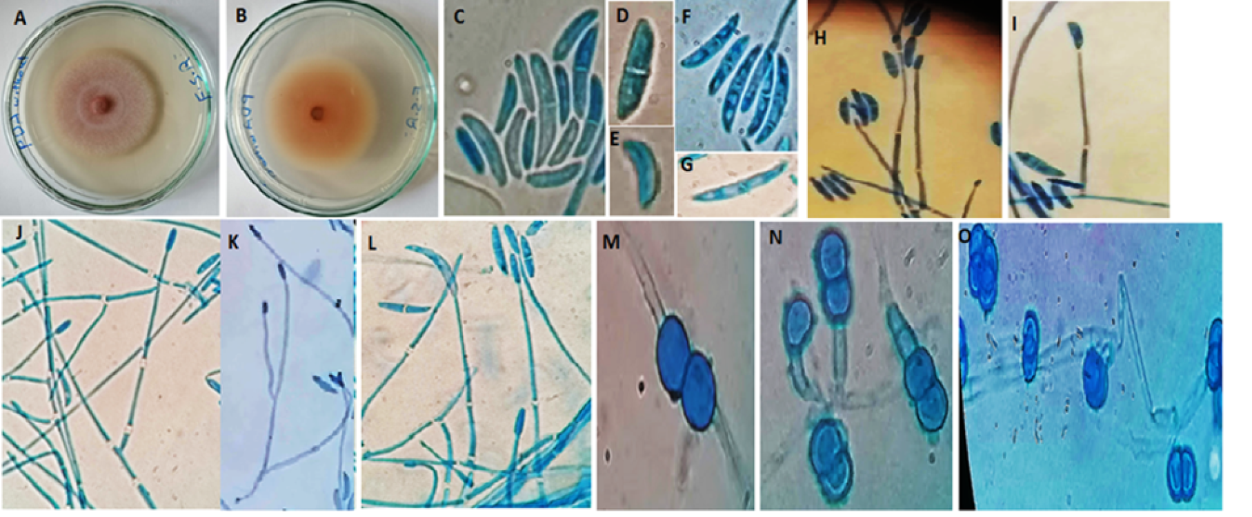
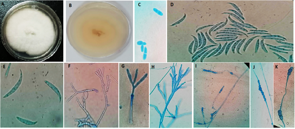
In this study, pathogenic fungal isolates of the wilted rosemary plants were cultured on PDA and were identified on the basis of their morpho-cultural traits as shown in Figure 2, 3, and 4. Results of morphological
diagnosis showed two species of Fusarium that caused fusariosis in rosemary plants. The first species was Fusarium solani, which distinguished itself with its appearance in PDA at 25°C, and showed pink upper-surface colonies and yellowish-brown pigmentation on the lower surface (Figure 2 A–B) with a radius of 50.5 mm after 7 days of growth. Microconidia observed were of oval or ellipsoid shape, consisting of a single cell. Additionally, there were sub-cylindric or slightly curved microconidia, consisting of two cells (Figure 2 C–E). The aerial conidiophores exhibited a lengthy and unbranched morphology that was somewhat narrow towards the apex. These structures were monophyletic and produced a plentiful supply of 0–1 conidia, which were clustered in a false head. (Figure 2 H–J). Macroconidia were slender to relatively straight, 3–5-septa (Figure 2 F–G). Lateral-branched aerial conidiophores producing 1–4 septate conidia (Figure 2 K–L). Intercalary chlamydospores present in hyphae were typically characterized by smooth walls and a globose shape, and were found in pairs (Figure 2 M). Terminal chlamydospores from conidiophores were smooth-walled and globes singly, in pairs, or in clusters (Figure 2 N–O). Similar features of mycelial growth and colony patterning of F. solani on PDA medium are reported by Zidan et al. (2022) and Valadez-Moctezuma et al. (2025).
The second species was F. chlamydosporum, characterised by a white cottony upper surface of colony and creamy-yellow pigmentation of the lower surface on PDA after 7 days (Figure 3 A–B) with a colony radius of 70 mm. Spindle-shaped microconidia with 0–1 septation (Figure 3 C) and typical sickle-shaped macroconidia with 3–5 septa (Figure 3 D–E) were observed. Microconidiophores were observed to manifest as both unbranched and branched monophialides, as well as polyphialides conidogenes, as depicted in Figure 3 (F–H). In addition, globose-shaped clamydospores were observed and were formed in either singular or short chain formations, as shown in Figure 3 (I–K). Current results confirmed the findings of Gupta and Misra (2012) and Lazreg et al. (2013), and Ibrahim et al. (2016).
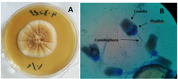
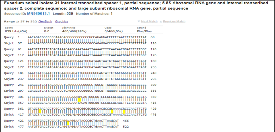
In addition to Fusarium species, A. terreus, a significant saprophytic filamentous fungus found in the soils, was also characterised by cinnamon colored colony (Figure 4). Animals, plants, and humans can all be harmed by the filamentous fungus A. terreus . More recently, A. terreus has also been found causing foliar blight of potato and may infect other crops . ; ; . Morphological characters of both Fusarium species were confirmed by ITS gene-based molecular markers . The size of the PCR products was about 600 bps. Molecular phylogenetic methods have been extensively employed to facilitate the accurate species identification of the Fusarium genus ;. Using the NCBI blast web, the sequence alignment of the F. solani isolate revealed a 99% identity with the F. solani Chinese isolate (accession No. MN960013.1),
with six variation sites, i.e., K (G,T) > T and M (A,C) > A in 382 bp, 384 bp, but in 423 bp, 459 bp, and 465 bp. The study revealed evidence of both transition mutations, which involve the substitution of a two-ring purine for a one-ring pyrimidine or vice versa. Specifically, the locations 423, 459, and 465 bps exhibited the occurrence of transition R (A, G)>A, R (A, G)>A, and R (A, G)>A, respectively. Additionally, transversions were also observed in locations 382, 383, and 497 bps (Table 1 and Figure 5).
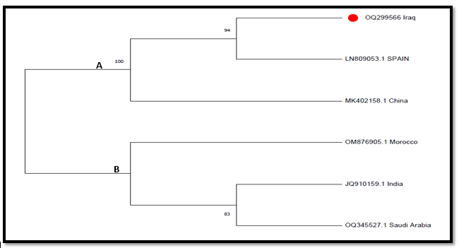
A phylogenetic tree of the Iraqi isolate of F. solani was drawn to illustrate the relationship between the species that were deposited in the GenBank database based on the ITS gene region by using the Mega 6 program, which illustrated the relationship of F. solani with five sequences of global isolates in GenBank, which were divided into two main nodes (A and B). A node included three isolates 100% similarity, but two isolates, Iraqi isolate (OQ299566.1) and Spanish isolate (LN809053.1), with 94% compatibility. B node contained three isolates, i.e., OM876905.1, JQ910159.1, and OQ345527.1 (Figure 6).
Similarly, a comparison between the sequence of F. chlamydosporum Iraqi isolate (OQ301746.1) and F. chlamydosporum Chinese isolate (KX783373.1) showed 100% identity, with no evidence of substitution in their respective positions (Table 1).
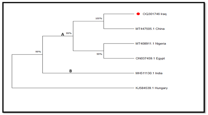
The phylogenetic tree of F. chlamydosporum Iraqi isolate (OQ301746.1) illustrates its relationship with five sequences of global isolates deposited to the GenBank database (Figure 7). Relationships
Table 1: Kind of ITS region polymorphism for Aspergillus terreus, Fusarium species, and Iraqi isolates. Transition & transversion mutations are compared to sequences in the GenBank database.
| Iraqi isolates | Identity | GenBank isolates | Sequence ID with compare | Nucleotide | Location | Type of situation |
| F.solani OQ299566.1 | 99% | F. solani isolate 31 | MN960013.1 | (G,T)K >T | 382bp | Transversion |
| M (A,C) > A | 383bp | Transversion | ||||
| R(A,G) >A | 423bp | Transition/ Transversion | ||||
| K(G,T) >T | 459bp | Transition/ Transversion | ||||
| R(A,G) >A | 465bp | Transition/ Transversion | ||||
| K(G,T) >T | 497bp | Transversion | ||||
| F.chlamydosporum OQ301746.1 | 100% | F.chlamydosporum isolate ZB11263540 | KX783373.1 | - | - | - |
| A.terreus OQ299570.1 | 100% | A. terreus strain CA-1 | MH918670.1 | - | - | - |
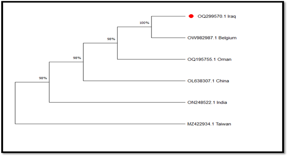
and Chinese isolate (MT447505.1), with 100% compatibility. Second internal nodes were included Nigerian isolate (MT408911.1) and Egyptian isolate (ON037459.1) with 99% compatibility. Finally, the second internal node (B) were Indian isolate (MH511130.1) compatible with four previous isolates with 99% compatibility. The second branch included a Hungarian isolate (KJ584539.1) showing 99% compatibility with five isolates (Figure 7). Moreover, the identity ratio of the sequences of Iraqi isolate of A. terreus (OQ299570.1) was 100% with A. terreus Indian isolate (MH918670.1) (Table 1). Dendrogram produced by the analysis of ITS sequencing data depicted that the Iraqi isolate of A. terreus (OQ299570.1) had a compatibility of 100% with Belgium isolate (OW982987.1) (Figure 8).
Conclusions and Recommendations
Based on overall study results, it is concluded that Fusariosis or Fusarium wilt affecting the rosemary cultivation and production in the nurseries of the sampled area of Iraq is primarily caused by two species of pathogenic fungal isolates, i.e., F. solani and F. chlamydosporum. Moreover, the findings corroborate the practical significance and implications of morpho-molecular diagnosis of plant pathogens. These results would aid in understanding the key factors that contribute to the spread of Fusarium wilt of rosemary plants, and in figuring out the most effective ways to control it in light of the present study’s results.
Acknowledgments
The authors are grateful to the Department of Plant Protection, Agricultural College, the University of Al-Qadisiyah, Al-Diwaniyah-Iraq for supporting and guiding the research process.
Novelty Statement
This study proposes Fusarium wilt is a major and destructive disease hampering rosemary growing in different areas in Iraq.
Author’s Contribution
All authors contributed to data collection and edited the draft and final versions of the manuscript.
Generative AI and AI-assisted technology statement
The authors declare that no generative AI and AI assisted technology was used in the creation of this manuscript.
Conflict of interest
The authors have declared no conflict of interest.
Figures
References
- Al-Baldawy, M.S.M., D.T. Al-Ameri, S.T. Hasan and A.S. Alhasan. 2020. The efficiency of powder and extract of Cassia senna plant and alum in inhibition the growth of Aspergillus ochraceus and Fusarium sp. and their mycotoxins. Int. J. Agric. Stat. Sci., 16: 1323–1328.
- Al-Taae, A.K. and H.H.W. Al-Taae. 2020. First record of root rot of rosemary (Rosmarinus officinalis) caused by Rhizoctonia solani in Iraq. Plant Arch., 20(1): 1094–1098.
- Ashrafi, S.J. and H. Saremi. 2012. Cultivation of rosemary as an ornamental–medicinal plant and managing its root rot disease in northeast of Iran. J. Med. Plants Res., 6(47): 5789–5796. https://doi.org/10.5897/JMPR12.878[CrossRef]
- Ashrafi, S.J., M.F. Rastegar and H. Saremi. 2010. Rosemary wilting disease and its management by soil solarization technique in Iran. Afr. J. Biotechnol., 9(42): 7048–7057.
- Bakrim, S., D. Taha, I. Mssillou, T. Benali, A. Khalid, R. Soltane and A. Bouyahya. 2025. Rosmanol: A natural bioactive compound unveiling multifaceted nutritional, pharmacological and health advantages. J. Funct. Food., 134: 107054. https://doi.org/10.1016/j.jff.2024.107054[CrossRef]
- Becer, E., E.M. Altundağ, M. Güran, H.S. Vatansever, S. Ustürk, D.Y. Hanoğlu and K.H.C. Başer. 2023. Composition and antibacterial, anti-inflammatory, antioxidant and anticancer activities of Rosmarinus officinalis L. essential oil. S. Afr. J. Bot., 160: 437–445. https://doi.org/10.1016/j.sajb.2023.06.019[CrossRef]
- Chandana, H.S., N.S.R. Bh, C.K. Kb, S.K. Prajwal and A.V. Machanoor. 2024. Morphological and molecular characterisation of Fusarium wilt pathogen isolated from sunhemp. J. Exp. Agric. Int., 46(9): 546–554. https://doi.org/10.9734/JEAI/2024/v46i92503[CrossRef]
- Chehri, K., B. Salleh and L. Zakaria. 2015. Morphological and phylogenetic analysis of Fusarium solani species complex in Malaysia. Microb. Ecol., 69: 457–471. https://doi.org/10.1007/s00248-014-0480-1[CrossRef]
- Ekwomadu, T.I. and M. Mwanza. 2023. Fusarium fungi pathogens: Identification, adverse effects, disease management and global food security. Agric., 13(9): 1810. https://doi.org/10.3390/agriculture13091810[CrossRef]
- Gupta, V.K. and A.K. Misra. 2012. Fusarium chlamydosporum causing wilt disease of guava in India. Arch. Phytopathol. Plant Prot., 45(20): 2425–2428. https://doi.org/10.1080/03235408.2012.705419[CrossRef]
- Hammer, M. and W. Junghanns. 2020. Rosmarinus officinalis L.: Rosemary. Med. Aromat. Stimul. Plants, Springer, Cham: 501–521. https://doi.org/10.1007/978-3-030-38796-2_25[CrossRef]
- Harish, J., P.P. Jambhulkar, R. Bajpai, M. Arya, P.K. Babele, S.K. Chaturvedi and D.K. Lakshman. 2023. Morphological characterization and molecular identification of Fusarium spp. in maize. Front. Microbiol., 14: 1121781. https://doi.org/10.3389/fmicb.2023.1121781[CrossRef]
- Ibrahim, N.F., M.H. Mohd, N.M.I.M. Nor and L. Zakaria. 2016. Pathogenicity of Fusarium semitectum and F. chlamydosporum in pineapple. Malays. J. Microbiol., 12(2): 164–170.
- Jambhulkar, P.P., M. Sharma, D. Lakshman and P. Sharma. 2015. Soil suppressiveness against Fusarium and related pathogens. Org. Amend. Soil Suppress. Plant Dis. Manag., Springer: 95–123. https://doi.org/10.1007/978-3-319-23075-7_6[CrossRef]
- Lazreg, F., L. Belabid, J. Sanchez, E. Gallego, J.A. Garrido-Cardenas and A. Elhaitoum. 2013. First report of Fusarium chlamydosporum on Aleppo pine. Plant Dis., 97(11): 1506. https://doi.org/10.1094/PDIS-02-13-0185-PDN[CrossRef]
- Leslie, J.F. and B.A. Summerell. 2006. The Fusarium Laboratory Manual. Blackwell Publishing Ltd.
- Lo, A.H., Y.C. Liang, S.Y. Lin-Shiau, C.T. Ho and J.K. Lin. 2002. Carnosol suppresses iNOS via NF-κB down-regulation. Carcinogen., 23(6): 983–991. https://doi.org/10.1093/carcin/23.6.983[CrossRef]
- Louis, B., P. Roy, S.D.W. Sayanika and N.C. Talukdar. 2013. Aspergillus terreus causing foliar blight of potato. Plant Pathol. Quar., 3(1): 29–33.
- McGovern, R.J. 2023. Diseases of rosemary. Handb. Veg. Herb Dis., Springer: 1–11. https://doi.org/10.1007/978-3-031-26549-7_15[CrossRef]
- Mekonnen, M. and B. Manahile. 2017. Yield loss in rosemary and sage caused by Fusarium oxysporum. Afr. J. Agric. Res., 12(19): 1669–1673. https://doi.org/10.5897/AJAR2017.12309[CrossRef]
- Mekonnen, M., A. Wariyo and G. Hilu. 2019. Antifungal activities of essential oils. Adv. Crop Sci. Tech., 7: 419. https://doi.org/10.4172/2329-8863.1000419[CrossRef]
- Nilsson, R.H., M. Ryberg, K. Abarenkov, E. Sjökvist and E. Kristiansson. 2009. ITS region for fungal community characterization. FEMS Microbiol. Lett., 296(1): 97–101. https://doi.org/10.1111/j.1574-6968.2009.01618.x[CrossRef]
- Nji, Q.N., O.O. Babalola and M. Mwanza. 2023. Soil Aspergillus species. J. Fung., 9(7): 766. https://doi.org/10.3390/jof9070766[CrossRef]
- Pennerman, K.K., G. Yin, A.E. Glenn and J.W. Bennett. 2020. Aspergillus pathogenicity factors. BMC Microbiol., 20: 342. https://doi.org/10.1186/s12866-020-02022-0[CrossRef]
- Pintore, G., M. Usai, P. Bradesi, C. Juliano, G. Boatto, F. Tomi and J. Casanova. 2002. Antimicrobial activity of rosemary oils. Flavour Fragr. J., 17(1): 15–19. https://doi.org/10.1002/ffj.1041[CrossRef]
- Rafie, H., H. Soheila and E. Grant. 2017. Therapeutic potential of rosemary. J. Herb. Med., 3(2): 8–14.
- Rashid, T.S. 2025. Trichoderma harzianum as biocontrol agent. J. Phytopathol., 173(4): e70130. https://doi.org/10.1111/jph.70130[CrossRef]
- Saremi, H., S.M. Okhovvat and S.J. Ashrafi. 2011. Fusarium diseases and soil solarization. Afr. J. Biotechnol., 10(80): 18391–18398.
- Schoch, C.L., K.A. Seifert, S. Huhndorf, V. Robert, J.L. Spouge, C.A. Levesque and M.M. White. 2012. ITS region as fungal barcode. Proc. Natl. Acad. Sci. USA, 109(16): 6241–6246. https://doi.org/10.1073/pnas.1117018109[CrossRef]
- Sharma, L. and G. Marques. 2018. Fusarium as an entomopathogen. Pathogens., 7(4): 93. https://doi.org/10.3390/pathogens7040093[CrossRef]
- Stoeva, D., D. Gencheva, R. Yordanova and G. Beev. 2023. Genetic diversity of Fusarium spp. Acta Microbiol. Bulg., 39(4): 376–385.
- Valadez-Moctezuma, E., S. Samah, A.E. Rangel-Castillo, J. García-Cariño, K. Juárez-Campos, A. Vázquez-Reyes and R. Salas-Flores. 2025. Fusarium species complexes. Acta Biol. Colomb., 30(3).
- Zarrin, M., F. Ganj and S. Faramarzi. 2016. ITS PCR-RFLP analysis of Fusarium. Biomed. Rep., 4(4): 471–474. https://doi.org/10.3892/br.2016.610[CrossRef]
- Zidan, L., W. Naffaa, A. Saleh and D. Jawdat. 2022. Identification of Fusarium spp. in wheat. Arch. Phytopathol. Plant Prot., 55(1): 76–88. https://doi.org/10.1080/03235408.2021.1984681[CrossRef]
Services
Article usage statistics
Loading…