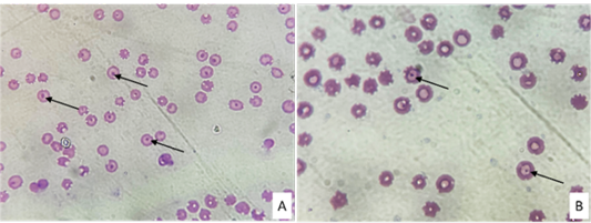
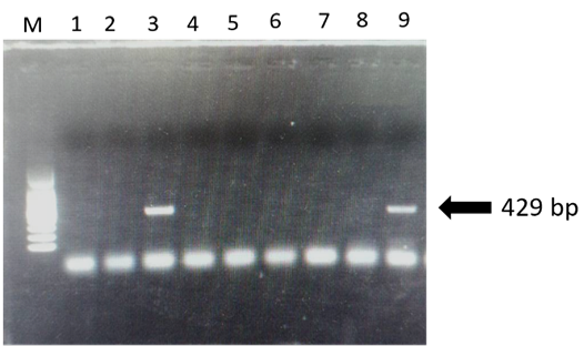
Conventional microscopy revealed that three samples were positive (Figure 1), whereas molecular detection of 429 bp of the 16S rRNA gene shared by all Anaplasma spp., 2 out of 59 sampled cattle (3.38%) were found that positive for Anaplasma spp. infection of which two were positive from microscopy (Figure 2).
By means of the hematology profile, the results indicated that the infected cattle have been shown to have severe hemolytic anemia.
Based on the serum biochemical profile, the infected cattle showed high levels of ALT, AST, ALP, total bilirubin, and BUN, whereas total protein and albumin were lower than.
Overall, the very first molecular detection of A. marginale infection associated with hemolytic anemia in naturally infected cattle was revealed.
Selected from the article's own text.
Abstract
Vector-borne pathogens have become a major problem since different species of cattle, e.g. indigenous, crossbred dairy, and crossbred beef, are raised together on most farms. Anaplasma spp. are obligate intracellular rickettsial vector-borne pathogens that impact on livestock farmers with major economic constraints and eventually threaten human health in cases of zoonotic species involvement. Therefore, our study aimed to identify Anaplasma spp. infection in a bovine host using both microscopy and molecular techniques. A total of 59 samples were collected from Mingaladon township, Yangon region, and the presence of infection was assessed using a Giemsa-stained thin blood smear and PCR of 16S rRNA gene amplification. Both microscopic and PCR-positive samples were further analyzed for hematobiochemical alteration. As a results, Anaplasma spp. was detected in 3.38% (2/59) of sampled animals. Hematology results revealed severe anemia with a low hemoglobin level together with a low PCV and total platelet count whereas the MCV and total WBC count were shown to be higher. Elevation of some enzymes such as ALT, AST, GGT, total bilirubin, and blood urea nitrogen (BUN) occurred by blood biochemical analysis, whereas total protein and albumin were low. To our best, this study is the first molecular evidence for the presence of Anaplasma marginale infection in cattle in Myanmar, with a sequence similarity range between 98.8 and 100%. By understanding one of the major tick-borne pathogens (TBPs) in Myanmar, possible control measures might be implemented not only to minimize the transmission but also to increase the farm productivity.
The livestock population plays a major role in country economic development in terms of the fact that 10-12% of GDP of the Myanmar economy comes from the agricultural and livestock sectors (https://www.statista.com/statistics/1061469/myanmar-growth-rate-gdp-livestock-fishery-sector/). Even though the livestock industry serves as living insurance for farmers, the challenges of antimicrobial resistance, parasite diseases, and climatic disasters have increased as huge negative impacts. Nowadays, cattle are affected by various pathogens worldwide. Haemoprotozoan and rickettsial diseases are considered major threats to livestock health and productive performance in both tropical and subtropical regions . Anaplasmosis is commonly known as “gall sickness,” which is caused by an obligate intraerythrocytic rickettsia microorganism (order Rickettsiales, family Anaplasmataceae). All ranges of ruminants can be infected by Anaplasma spp., whereas cattle seem to be more susceptible than others. Bovine anaplasmosis is an important tickborne disease that is caused by major species of A. marginale, A. bovis, A. centrale, A. phagocytophilum, A. capra, and A. platys , from which human cases have been reported by A. phagocytophilum, A. capra, and A. platys . Parasite infestation results in a wide range of clinical symptoms, from asymptomatic to severe fatal hemolytic anemia, hepatosplenomegaly, decreased milk production, abortion, and susceptibility to other pathogens. Nowadays, hematobiochemical analyses have been extensively used to determine the underlined clinical status and to generate a more reliable diagnosis . Once cattle have overcome the acute stage, they later develop long-lasting immunity and continue to harbor the infection for the rest of their lives, which serves as reservoir for susceptible animals . Of vector-borne transmission, 20 various ticks play a critical role as reservoirs for Anaplasma spp. infection, in which Boophilus microplus is known to be a major transmitting agent (. Possibly mechanical transmission by means of biting flies or blood-contaminated fomites has been reported . Until now, bovine anaplasmosis has been widespread globally and has been reported to be endemic in certain areas of Asia and Africa ; ; Ybanez and Inokuma, 2016).
Giemsa-stained thin blood smear is a gold standard microscopic test for diagnosis because it is affordable and easy, despite recent advances in the diagnosis of Anaplasma spp. infection. However, either false positive or false negative results may come from staining artifacts or a low sensitivity percentage in pre-symptomatic and carrier animals . Therefore, molecular methods have been applied for conclusive diagnosis with high sensitivity and specificity results and definite species identification . Apart from that, hematobiochemical analyses provide valuable information relating to hemoparasites infection, which can lead to the interpretation of relevant diagnoses, drug efficacy, and disease progress . Once red blood cell damage occurs the intravascular/ extravascular, and immune-mediated severe hemolytic anemia have been reported in Anaplasma spp. infected cattle . Among cattle hemoparasites, Babesia and Thelaria infections have been reported in certain areas of Myanmar , 2016), whereas nothing has been informed about bovine anaplasmosis. The aim of this study was, therefore, to determine the presence of Anaplasma spp. infection using combination of techniques of both microscopy and molecular methods altogether with hematobiochemical alteration in clinically healthy cattle in order to promote specific management strategies.
Materials and Methods
In the present study, a total of 59 cattle blood samples were collected with convenience sampling. Briefly, about 5 ml of whole blood from the jugular vein of each cattle was collected into vials containing ethylenediaminetetraacetic acid (EDTA) anticoagulated tubes (Sigma-Aldrich Co. LLC, Saint Louis, Missouri, USA). The questionnaire survey was made based on the information regarding animal age (<1 year, 1–3 years, 3–5 years), breed (indigenous, crossbred, beef), gender (male, female), tick infestation (present, absent), management practice (poor, good, excellent), and acaricidal treatment (yes, no). Giemsa-stained (Sigma-Aldrich) thin blood smears were prepared immediately, as described by . In detail, blood smears were prepared and fixed in methanol for 5 mins. After that, the labeled smear was stained with a 5% Giemsa solution for 30 mins and air-dried. Finally, the labeled thin blood smears were observed under a light microscope (Olympus, Japan) at 1000x magnification using oil immersion for the presence of Anaplasma inclusion bodies. In order to perform molecular detection, the EDTA blood samples were kept at -20°C prior to DNA extraction. To analyze the hematobiochemical alteration of the PCR-positive sample, sera were isolated by centrifugation at 3000 x g for 5 mins at 4°C. Hematological parameters were determined by an automated hemoanalyzer (IDEXX VetAutoread Hematology Analyzer, USA), and the blood biochemical profile was determined using a VetScan VS2 chemical analyzer (Zoetis, UK).
Genomic DNA was extracted using the DNeasy blood and tissue kit (Qiagen, Hilden, Germany) in accordance with the manufacturer’s instructions. The eluted DNA was stored at -20°C until use. The concentration and purity of the extracted DNA were measured by a nanodrop spectrophotometer (Thermofisher Scientific Inc., Waltham, USA). DNA samples with A260/A280 ratios between 1.7 and 2.2 were further analyzed. Polymerase chain reaction (PCR) was performed to amplify the 16S rRNA gene of expected size 429 bp shared by all Anaplasma spp. (F, 5ʹ-TACCTCTGTGTTGTAGCTAACGC-3ʹ; R, 5ʹ-CTTGCGACATT GCAACCTATTGT-3ʹ) as described by . PCR was performed under the following cycling conditions: Initial denaturation at 96°C for 5 min, followed by 35 cycles of 10 s at 96°C of denaturation, annealing at 55°C for 35 s and final extension at 72°C for 5 min. After 1.5% agarose gel electrophoresis and ethidium bromide staining, PCR products were visualized under UV transillumination. Sequences were analyzed using the BioEdit version 7.2 sequence alignment software (www.mbio.ncsu.edu/BioEdit/bioedit.htm), and sequences alignment was performed using Clustal W software version 2.0 . The resulting sequences were compared with the reference sequences in the GenBank database using the BLAST program of the National Center for Biotechnology Information (https://www.ncbi.nlm.nih.gov/). Phylogenetic trees were constructed using the neighbor-joining program in MEGA 11.0 software .
Results and Discussion
In the present study, a total of 59 cattle blood samples were collected from Mingaladon township, Yangon region. Conventional microscopy revealed that three samples were positive (Figure 1), whereas molecular detection of 429 bp of the 16S rRNA gene shared by all Anaplasma spp., 2 out of 59 sampled cattle (3.38%) were found that positive for Anaplasma spp. infection of which two were positive from microscopy (Figure 2). Sequencing exhibited that the two sequenced samples were identified as A. marginale, with the sequence homology range between 98.8 and 100% compared with other sequences. The two sequences obtained in this study were deposited in the GenBank database under accession numbers OR654955 and OR654956. In addition, the two PCR-positive animals were in the age group of 1-3 years and are crossbred female dairy cattle, in which no ticks were infested during sample collection. Considering management practices, these positive cattle were from the same farm, whereas they were not raised under good hygienic measures, which might have led to breeding ground for ticks and were closely related to other livestock farms.
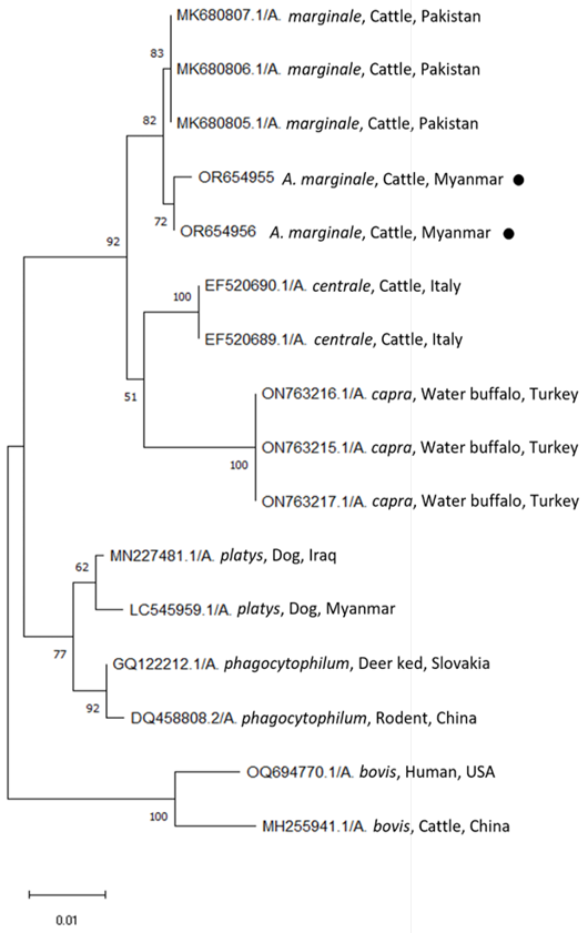
Phylogenetic analysis of the two partial 16S rRNA gene sequences (OR654955 and OR654956) was performed by aligning with selected Anaplasma spp. sequences in GenBank. As a result, the OR654955 and OR654956 sequences were closely related to A. marginale, followed by A. centrale, A. capra, A. platys, A. phagocytophilum, and A. bovis deposited in GenBank (Figure 3). The resulting Myanmar cattle isolates in the present study show 99.53% and 99.76% homology to sequences from A. marginale
strains originating from cattle in Pakistan (MK680805-MK680807), and 98.35% homology with sequences from A. centrale strains originating from cattle in Italy (EF520689 and EF520690). Interestingly, the two isolates found in this study were closely related to A. centrale, as they were in the same sister taxon as A. centrale isolates from cattle from Italy. However, in Myanmar, the information on A. centrale infection in livestock animals has not been updated yet. They were 97.64% sequence homology with A. capra isolated from water buffalo (ON763215-ON763217) from Turkey and 96.93% homology with A. platys sequence (LC545959) from dog from Myanmar. Previously, A. platys was reported from in dogs from Nay Pyi Taw, Myanmar, in 2020 with a low prevalence (0.25%). It was proposed that the Rhipicephalus sanguineus tick might be the main vector, as this is the dominant tick species found in Myanmar. Therefore, transboundary movement of animals could be a major transmission of sharing the same vector within the regions .
The two positive samples from both conventional microscopy and PCR were supposed to determine hematobiochemical alteration. By means of the hematology profile, the results indicated that the infected cattle have been shown to have severe hemolytic anemia. The hematological parameters of Hb, PCV, and total platelet count were remarkably lower, whereas the MCV and total WBC count were apparently higher in the infected cattle. In the present study, low hemoglobin levels and a remarkable increased of MCV in Anaplasma spp. infected cattle revealed evidence of hypochromic macrocytic anemia; hence, the type of anemia that occurred in Anaplasma spp. infection could depend the disease severity . Similar findings of hypochromic macrocytic anemia have been reported in A. marginale-infected cattle . In addition to this, normocytic forms of anemia have been reported in acute anaplasmosis, whereas chronic cases with macrocytic progresses in Anaplasma spp. infected cattle Anton and Solcan (2022). Therefore, the infected cattle in this study seemed to have chronic cases of infection. In line with previous reports, Anaplasma spp. could reduce erythrocyte life span and favor erythrocyte destruction, which in turn leads to severe hemolytic anemia . Contrast to Jurkovic et al. (2020), there was no clinical manifestation in Anaplasma spp.-infected cattle has been reported. In general, thrombocytopenia may occur relating to severe vasculitis, immune-mediated destruction, and pathogens of mainly blood sucking parasites (Rodriguez et al., 2018). However, other factors such as hormonal imbalance, nutritional deficiency, drugs intoxications, and environmental stress could not be ruled out in cases of erythrocyte destruction .
Based on the serum biochemical profile, the infected cattle showed high levels of ALT, AST, ALP, total bilirubin, and BUN, whereas total protein and albumin were lower than
Table 1: Hematobiochemical parameters of two A. marginale infected cattle.
Clinical parameters
Diagnosed values
Elevated/ Decreased
Reference range (George et al., 2010; Kaneko et al., 2008)
Hb (g/dL)
5.31 - 5.84
Decreased
8.5 - 12.2
PCV (%)
19.02 - 20.21
Decreased
22 - 33
MCV (fL)
60.07 - 60.25
Elevated
38 - 50
MCH (pg)
16.71 - 16.34
Normal
14 - 18
MCHC (g/dL)
36.1 - 39.2
Normal
36 - 39
Granulocyte count (x 103 cells/µL)
9.9 - 10.4
Elevated
1.8 - 7.2
Lymphocyte count (x 103 cells/µL)
5.9 - 6.2
Elevated
1.6 - 5.6
Monocyte count (x 103 cells/µL)
1.57 - 2.12
Elevated
0 - 0.8
Total platelets count (x 103 cells/µL)
137 - 142
Decreased
193 - 637
Albumin (g/dL)
2.47 - 2.88
Decreased
3.03 - 3.55
Globulin (g/dL)
2.01 - 2.8
Decreased
3 - 3.48
ALT (IU/L)
62 - 65
Elevated
11 - 40
AST (IU/L)
148 - 150
Elevated
78 - 132
ALP (IU/L)
525 - 610
Elevated
0 - 488
Total bilirubin (mg/dL)
1.8 - 2
Elevated
0.01 - 0.5
BUN (mg/dL)
48 - 50
Elevated
14 - 37
Total protein (g/dL)
3.4 - 3.6
Decreased
6.7 - 8.8
normal, relating to excessive erythrocytes destruction, which later succumbed to hepatocellular damage (Anton and Solcan, 2021) (Table 1). These findings are in line with previous findings by Ashuma et al. (2013), from which impairment of the hepatocellular system and erythrocyte destruction in Anaplasma spp. infected cattle might result in elevated bilirubin levels because of retention in the biliary system and excessive destruction of erythrocytes in the reticuloendothelial system. High levels of BUN in infected animals could be due to renal ischemia, nephrosis, and dehydration, as these clinical symptoms have been reported in A. marginale infected cattle . Resulting in decrease the capacity of protein synthesis due to hepatocellular damage, which can further lead to hypoalbuminemia, as previously described by Anton and Solcan (2022). Human infections have been reported by A. phagocytophilum, A. capra, and A. platys (Vanstreels et al., 2018) with mild to moderate clinical symptoms, from which high liver enzymes were important predictors of anaplasmosis. Even though bovine anaplasmosis has been shown to be clinical in most cases, the infected cattle from the study area shown to be infected sub-clinically. Despite the identified species in the study area was revealed as A. marginale, it is important to figure out the risk of zoonotic anaplasmosis in different geographical areas of Myanmar.
Even though, we could not calculate the significance due to the sample size limitation, management practices were shown to be important fact because cattle raised together with other livestock animals in poor hygienic practices were found to be more infected in the present study. The occurrence of A. marginale infection in the present study showed a relatively lower percentage compared to previous studies in other countries, even though the same analytic techniques were used: 72.6%, 43%, 23.2%, 11.2%, and 8.21% in Malaysia , Philippines , Thailand , China , and Bangladesh , respectively. Different geographical distribution and detection methods could produce different results hence, the main vector population could also be different .
Conclusions and Recommendations
Overall, the very first molecular detection of A. marginale infection associated with hemolytic anemia in naturally infected cattle was revealed. Moreover, the remarkable changes in the hematobiochemical profile indicated that clinical bovine anaplasmosis might be a differential diagnosis in the presence of severe hemolytic anemia. However, our study still has limitations for interpretation of results since only one pathogen has been targeted; hence, the hematobiochemical alterations might also come out with other hemoparasite infections. Besides, the sampling area could not be representative of the whole Myanmar, and consequently, the assessment for bovine anaplasmosis still needs to be updated. Therefore, further studies with large sample sizes and an overall seasonal assessment of the vector population might investigating the association of potential risks with the presence of infection.
Acknowledgements
We are grateful to Dr. Khin Su Hlaing, Dr. Zin Hnin Thaw and Dr. Paik Htwe for their help in the fieldwork. The authors would also thank the farmers for their collaboration in sample collection. The authors acknowledged Dr. Arin Ngamniyom from Major Environment, Faculty of Environmental Culture and ECO tourist, Srinakharinwirot University, Thailand for providing molecular facilities throughout the study. As this is collaborative work, we don’t have any funding acquisition.
Novelty Statement
This study is the first molecular detection of bovine anaplasmosis associated with hemolytic anemia as it is considerable economic loss in livestock industry.
Author’s Contribution
BKS wrote the main manuscript text and prepared figures and table. BKS, SLYM, TWN and NN were responsible for conceptualization, investigation, draft revision, analyses, original draft editing and author proof revision. BKS and TWN were responsible for methodology. HS was responsible for supervision. All authors have reviewed the manuscript.
Funding
This research did not receive any specific grant from funding agencies in the public, commercial, or not-for-profit sectors.
Abbreviations
16S rRNA: 16S of ribosomal RNA; A. marginale: Anaplasma marginale; A. capra: Anaplasma capra; A. phagocytophilum: Anaplasma phagocytophilum; A. centrale: Anaplasma centrale; A. bovis: Anaplasma bovis; A. platys: Anaplasma platys; PCR: polymerase chain reaction
Conflict of interest
The authors declare that the research was carried out without any commercial or financial affiliations that could be interpreted as a possible source of conflicting interests.
Figures
Figure 1:.
A and B showed Anaplasma inclusions (arrowhead) in the erythrocytes of Giemsa-stained thin blood smear.
Figure 2:.
Gel electrophoresis results for PCR of Anaplasma spp. (a): M = 100bp marker, lane 3 and 9 = positive samples, and lanes 1, 2, 4, 5, 6, 7 and 8 are negative samples.
Figure 3:.
Phylogenetic trees based on the 16S rRNA sequences of Anaplasma spp. were compared with the neighbor-joining method using the MEGA software (version 11) with maximum likelihood analysis based on the general time-reversible model. Phylogenetic tree showing taxonomic relationships of resulted two sequences; OR654955 and OR654956 (solid red circle) detected from cattle in Yangon region and 14 reference sequences of 16S rRNA of all Anaplasma spp. which can be infected to cattle. The relationships were inferred based on 16S rRNA data, including 1 from dog in Myanmar and 13 reference sequences of other Anaplasma spp. originated from different countries. For each sequence, the accession numbers are followed by full species names, the host and the country of origin.
References
Abdullah DA, Ali FF, Jasim AY, Ola-Fadunsin SD, Gimba FI, Ali MS (2020). Clinical signs, prevalence, and hematobiochemical profiles associated with Anaplasma infections in sheep of North Iraq. Vet. World, 13(8): 1524. https://doi.org/10.14202/vetworld.2020.1524-1527[CrossRef]
Al-Hosary A, Răileanu C, Tauchmann O, Fischer S, Nijhof AM, Silaghi C (2020). Epidemiology and genotyping of Anaplasma marginale and co-infection with piroplasms and other Anaplasmataceae in cattle and buffaloes from Egypt. Parasit. Vectors, 13(1): 1-11. https://doi.org/10.1186/s13071-020-04372-z[CrossRef]
Anton A, Solcan G (2022). A case study of photosensitivity associated with Anaplasma spp. infection in cattle. Animals, 12(24): 3568. https://doi.org/10.3390/ani12243568[CrossRef]
Ashuma AS, Singla LD, Kaur P, Bal MS, Batth BK, Juyal PD (2013). Prevalence and haemato-biochemical profile of Anaplasma marginale infection in dairy animals of Punjab (India). Asian Pac. J. Trop. Med., 6: 139–144. https://doi.org/10.1016/S1995-7645(13)60010-3[CrossRef]
Bawm S, Htun LL, Maw NN, Ngwe T, Tosa Y, Kon T, Kaneko C, Nakao R, Sakurai T, Kato H, Katakura K (2016). Molecular survey of Babesia infections in cattle from different areas of Myanmar. Ticks Tick-Borne Dis., 7(1): 204-207. https://doi.org/10.1016/j.ttbdis.2015.10.010[CrossRef]
Bawm S, Shimizu K, Hirota JI, Tosa Y, Htun LL, Maw NN, Thein M, Kato H, Sakurai T, Katakura K (2014). Molecular prevalence and genetic diversity of bovine Theileria orientalis in Myanmar. Parasitol. Int., 63(4): 640-645. https://doi.org/10.1016/j.parint.2014.04.009[CrossRef]
Chien NTH, Nguyen TL, Bui KL, Van Nguyen T, Le TH (2019). Anaplasma marginale and A. platys characterized from dairy and indigenous cattle and dogs in northern Vietnam. Korean J. Parasitol., 57: 43. https://doi.org/10.3347/kjp.2019.57.1.43[CrossRef]
Corona B, Dasiel OD, Yai YA, Ifonso P, Vega E, Díaz A, Martinez S (2014). Tendencies in diagnostic of bovine anaplasmosis. Rev. Salud Anim., 36(2): 73–79.
Das D, Sarma K, Roychoudhury P, Chethan GE, Ravindran R, Islam SJ, Prasad H, Rajesh JB, Behera B, Choudhury FA (2021). Gross and histopathological findings of naturally occurring Anaplasma marginale infection in cattle. Indian J. Anim. Res., https://doi.org/10.18805/IJAR.B-4283[CrossRef]
Das D, Sarma K, Eregowda CG, Roychoudhury P, Rajesh JB, Behera P, Prasad H, Lalrinkima H, Aktar F, Bora N, Deka C (2022). Naturally occurring Anaplasma marginale infection in cattle: Molecular prevalence and associated risk factors, haemato-biochemical alterations, oxidant/antioxidant status and serum trace mineral levels. Microb. Pathog., 167: 105575. https://doi.org/10.1016/j.micpath.2022.105575[CrossRef]
El-Hamiani KS, Daminet S, Duchateau L, Elhachimi L, Kachani M, Sahibi H (2021). Epidemiological and clinicopathological features of Anaplasma phagocytophilum infection in dogs: A systematic review. Front. Vet. Sci., 8: p.686644. https://doi.org/10.3389/fvets.2021.686644[CrossRef]
Fazio F, Casella S, Giannetto C, Giudice E, Piccione G (2016). Erythrocyte osmotic fragility in response to a short road transport in cattle, horses, and goats. J. Vet. Behav. Clin. Appl. Res., 12: 82–84. https://doi.org/10.1016/j.jveb.2015.11.003[CrossRef]
Galay RL, Llaneta CR, Monreal MKFB, Armero AL, Baluyut ABD, Regino CMF, Sandalo KAC, Divina BP, Talactac MR, Tapawan LP, Mojares MCL (2021). Molecular prevalence of Anaplasma marginale and Ehrlichia in domestic large ruminants and Rhipicephalus (Boophilus microplus) ticks from Southern Luzon, Philippines. Front. Vet. Sci., 8: 746705. https://doi.org/10.3389/fvets.2021.746705[CrossRef]
Hmoon MM, Htun LL, Thu MJ, Chel HM, Thaw YN, Win SY, Chan SN, Khaing Y, Thein SS, Bawm S (2021). Molecular prevalence and identification of Ehrlichia canis and Anaplasma platys from dogs in Nay Pyi Taw Area, Myanmar. Vet. Med. Int., pp. 1-7. https://doi.org/10.1155/2021/8827206[CrossRef]
Jalali SM, Ghorbanpour M, Jalali MR, Rasooli A, Safaie P, Norvej F, Delavari I (2018). Occurrence and potential causative factors of immune-mediated hemolytic anemia in cattle and river buffaloes. Vet. Res. Forum, 9(1): 7.
Jurković D, Mihaljević Ž, Duvnjak S, Silaghi C, Beck R (2020). First reports of indigenous lethal infection with Anaplasma marginale, Anaplasma bovis and Theileria orientalis in Croatian cattle. Ticks Tick-Borne Dis., 11(5): 101469. https://doi.org/10.1016/j.ttbdis.2020.101469[CrossRef]
Knowles TG, Edwards JE, Bazeley KJ, Brown SN, Butterworth A, Warriss PD (2000). Changes in the blood biochemical and haematological profile of neonatal calves with age. Vet. Rec., 147: 593–598. https://doi.org/10.1136/vr.147.21.593[CrossRef]
Kumar S, Tamura K, Jacobsen IB, Nei M (2001). MEGA 2: Molecular evolutionary genetics analysis software. Arizona State University, Tempe. https://doi.org/10.1093/bioinformatics/17.12.1244[CrossRef]
Lankester MW, Scandrett WB, Golsteyn-Thomas EJ, Chilton NC, Gajadhar AA (2007). Experimental transmission of bovine anaplasmosis (caused by Anaplasma marginale) by means of Dermacentor variabilis and D. andersoni (Ixodidae) collected in western Canada. Can. J. Vet. Res., 71(4): 271.
Larkin MA, Blackshields G, Brown NP, Chenna R, McGettigan PA, McWilliam H, Clustal W (2007). Clustal X version 2.0, Bioinformatics 23: 2947–2948. https://doi.org/10.1093/bioinformatics/btm404[CrossRef]
Sajid MS, Siddique RM, Khan SA, Zafar I, Khan MN (2014). Prevalence and risk factors of anaplasmosis in cattle and buffalo populations of district Khanewal, Punjab, Pakistan. Glob. Vet., 12(1): 146–153.
Maharana BR, Tewari AK, Saravanan BC, Sudhakar NR (2016). Important hemoprotozoan diseases of livestock: Challenges in current diagnostics and therapeutics: An update. Vet. World, 9(5): 487. https://doi.org/10.14202/vetworld.2016.487-495[CrossRef]
Mannan A, Alim MA, Manik MB, Rahman KMU, Siddiki MAHAZ (2021). Prevalence of anaplasmosis in cattle from Chattogram Division of Bangladesh. Bangladesh J. Vet. Anim. Sci., 9(1): 67-73. https://doi.org/10.60015/bjvas/V09I1A9[CrossRef]
Nasreldin N, Ewida RM, Hamdon H and Elnaker YF (2020). Molecular diagnosis and biochemical studies of tick-borne diseases (anaplasmosis and babesiosis) in Aberdeen Angus Cattle in New Valley, Egypt. Vet. World, 13(9): 1884. https://doi.org/10.14202/vetworld.2020.1884-1891[CrossRef]
Noaman V, Shayan P (2010). Comparison of microscopy and PCR-RFLP for detection of Anaplasma marginale in carrier cattle. Iran. J. Microbiol., 2(2): 89.
Ola-Fadunsin SD, Gimba FI, Abdullah DA, Sharma RSK, Abdullah FJF, Sani RA (2018). Epidemiology and risk factors associated with Anaplasma marginale infection of cattle in Peninsular Malaysia. Parasitol. Int., 67(6): 659-665. https://doi.org/10.1016/j.parint.2018.06.013[CrossRef]
Park J, Han DG, Ryu JH, Chae JB, Chae JS, Yu DH, Park BK, Kim HC, Choi KS (2018). Molecular detection of Anaplasma bovis in Holstein cattle in the Republic of Korea. Acta Vet. Scand., 60(1): 1-5. https://doi.org/10.1186/s13028-018-0370-z[CrossRef]
Doyle RL, França RT, Oliveira CB, Rezer JF, Klafke GM, Martins JR, Santos AP, do Nascimento NC, Mesick JB, Lopes ST, Leal DB (2016). Cattle experimentally infected by Anaplasma marginale: influence of splenectomy on disease pathogenesis, oxidative profile, and antioxidant status, Microb. Pathog., 95: 193–199. https://doi.org/10.1016/j.micpath.2016.04.011[CrossRef]
Roland L, Drillich M, Iwersen M (2014). Hematology as a diagnostic tool in bovine medicine. J. Vet. Diagn. Invest., 26(5): 592-598. https://doi.org/10.1177/1040638714546490[CrossRef]
Rodríguez Y, Rojas M, Gershwin ME, Anaya JM (2018). Tick-borne diseases and autoimmunity: A comprehensive review. J. Autoimmun., 88: 21-42. https://doi.org/10.1016/j.jaut.2017.11.007[CrossRef]
Saetiew N, Simking P, Saengow S, Kurajog B, Yimming B, Saeng-chuto K, Chimnoi W, Kengradomkil C, Yangtara S, Suksai S, Thaprathom N (2014). Seasonal effect on Anaplasma marginale infections of beef cattle in previously flooding areas. In Agricultural sciences: Leading Thailand to world class standards. Proc. 52nd Kasetsart Univ. Annu. Conf. Anim. Vet. Med., 2: 267-277.
Selim A, Manaa E, Abdelhady A, Said MB, Sazmand A (2021). Serological and molecular surveys of Anaplasma spp. in Egyptian cattle reveal high A. marginale infection prevalence. Iran. J. Vet. Res., 22(4): 288.
Vanstreels RET, Yabsley MJ, Parsons NJ, Swanepoel L, Pistorius PA (2018). A novel candidate species of Anaplasma that infects avian erythrocytes. Parasit. Vectors, 11(1): 1-7. https://doi.org/10.1186/s13071-018-3089-9[CrossRef]
Yang J, Li Y, Liu Z, Liu J, Niu Q, Ren Q, Chen Z, Guan G, Luo J, Yin H (2015). Molecular detection and characterization of Anaplasma spp. in sheep and cattle from Xinjiang, northwest China. Parasit. Vectors, 8: 1-7. https://doi.org/10.1186/s13071-015-0727-3[CrossRef]
Ybañez AP, Inokuma H (2016). Anaplasma species of veterinary importance in Japan. Vet. World, 9(11): 1190. https://doi.org/10.14202/vetworld.2016.1190-1196[CrossRef]