
Fasciolosis is a parasitic disease of livestock and is widespread throughout the world. As a neglected tropical disease, fasciolosis can spread from animal to human raising zoonotic public health implications. In order to ascertain the prevalence of Fasciola gigantica, causative agent of fasciolosis, we investigated routinely slaughtered animals (cows and buffaloes) in Iraq. In the central slaughterhouse of Nasiriyah, 58 F. gigantica adults were collected from the livers of cows and buffaloes. We observed that buffaloes were infected with fasciolosis at a rate of 15.29%, higher than the incidence of fasciolosis in cows (10.66%). We recorded a greater prevalence of fasciolosis in females than in males, as the prevalence of bovine fasciolosis in females and males reached 15% and 9.09%, respectively. The infection rate in slaughtered buffalo for males and females was 13.33% and 20%, respectively. The results of the current research also showed that the infection rate in the age group <1.5 reached 10.34%, while the infection rate in the age group 1-2.5 was about 13.79%, and the infection rate in the age group 2-2.5 was about 15. 51%. In the age group between 2.5-3.5, the percentage was 22.72%, while for the oldest age group it was >3.5, with a rate of 30.30%, which is the highest. Taken together, finding of this study provide clear distribution of fasciolosis across animals, ages and sex. These findings facilitate better control measures, assess efficacy of existing deworming application and to delineate the causation of dropping animal productivity.
Special Issue: Emerging and Re-Emerging Animal Health Challenges in Low and Middle-Income Countries · Volume 12, Issue 1, 28 November 2024 · Open Access
Fasciola gigantica Parasitic Infection in Slaughtered Cows and Buffaloes
· ·
Affiliations & Notes ⌄
- 1Department of Biology, College of Education, University of Al-Qadisiyah, Iraq
- 2Directorate of Education Dhi Qar, Ministry of Education, Iraq
- 3Veterinary Directorate Dhi Qar Veterinary Hospital. *Correspondence | Hind Abdulzahra Abdulkadhim Alshaibani, Department of Biology, College of Education, University of Al-Qadisiyah, Iraq
- 4Email: abduljalil.aziz@qu.edu.iq
Article Info ⌄
- Journal:
- Journal of Animal Health and Production
- Published:
- 28 November 2024

From the article
- The total infection rate of Fasciolosis in the slaughtered cows was 10.66%, and the infection rate in buffaloes was 15.29% (Table 1, Figure 1).
- Based on the analysis, we noticed that buffaloes were infected with fasciolosis at a rate of 15.29%, higher than the incidence of fasciolosis in cows, which amounted to 10.66%.
- Our study also recorded a greater prevalence of fasciolosis in females than in males, as the prevalence of bovine fasciolosis in females and males reached 15% and 9.09%, respectively.
- The infection rate in slaughtered buffaloes for males and females, respectively, reached 13.33% and 20%.
Selected from the article's own text.
Abstract
Introduction
Fasciolosis is a parasitic disease caused by two species of liver flukes known as Fasciola (F) hepatica and F. gigantica, which are widespread worldwide (Mas‐Coma et al., 2009). Fasciolosis is a neglected tropical zoonotic disease that occasional affects humans. According to the World Health Organization, human infections were considered only of secondary importance until 1990 , however, it has now become evident that fasciolosis is one of the economically impactful disease of livestock and impose public health risks.
The public health importance of this disease was highlighted when considering cases of severe pathogenicity, even including fatal cases . Global estimates indicate that there are about 17 million infected people around the world. This number is an underestimate due to the expansion of the spread areas. F. gigantica and F. hepatica, two species of the genus Fasciola, are most commonly causing Fasciolosis in domestic and wild ruminants but are also causative agents of Fasciolosis in humans. Fasciolosis reduces the productivity of animal industries, imposing an economic burden of at least $3.2 billion annually, worldwide .
Foodborne diseases represent a major public health problem and are the leading cause of morbidity and mortality world wide . Among foodborne pathogens, fasciolosis caused by F. hepatica and F. gigantica is of major concern ; . The life cycle of F. gigantica begins when adult worms invade the body of the livestock species. Adult worms live in the liver and lay their eggs in the intestines. Eggs are excreted in the stool. The eggs hatch in the water into Miracidia and then swim to find the snails. Different species of snails are vectors that play a major role in the life cycle of F. gigantica . Miracidia remain in the body of the snail for two weeks, then transform into Sporocysts that reproduce asexually to produce Rediae and become cercariae. The Cercaria larva then develops a tail and turns into Metacercaria, after which it leaves the snail’s body to attach to aquatic plants. When cattle feed on plants contaminated with Metacercaria, they penetrate the intestinal wall and reach adulthood in the liver of the cattle . Animals become infected when they consume feed or water containing the infective larval stage of parasites, called metacercariae, of Fasciola spp. ; . Humans can acquire infections by consuming raw or undercooked liver products, as well as contaminated crops and water (Mas‐Coma et al., 2009; .
Fasciolosis has acute and chronic clinical stages, such as inflammation of the liver and bile ducts, anemia, anorexia, general malaise, jaundice, and eventually death . Fasciolosis causes significant financial loss due to the high cost of anthelmintics needed for treatment, stigmatization of infected livers, loss of production due to death, decreased production of meat, milk, and wool, slowed weight gain, and disturbance of fertility ; .
Most animals in developing countries, fasciolosis is common infections which is mainly attributed to weak or non-existent control measures, this complicates the task of studying the disease from an epidemiological and public health perspective (Isah and Control, 201). This study was setup to interrogate the prevalence of fasciolosis in Iraq to better guide control measures, efficacy of existing deworming application and to delineate the causation of dropping animal productivity ; ; ; ; Hijazi et al., 2023; ; .
Materials and Methods
Sample collection
The study was conducted in the period from January 2023 to October 2023, during which the data was collected from cattle slaughtered in the central slaughterhouse of Nasiriyah. The study was carried out on buffaloes and cows in the slaughterhouses through periodic visits. A total of 470 heads of livestock species were examined in this slaughterhouse, designated for slaughter, from different areas of Dhi Qar Governorate in southern Iraq. A total of 58 adult F. gigantica were collected from the livers of cows and buffaloes through the process of examining samples slaughtered in the slaughterhouse.
Diagnosis
We based the diagnosis of the samples on the external appearance of the worms including morphological characteristics such as shape and size, and based on characteristics reported earlier . These include classification of F. gigantica (relatively large in size and more leaf-like), as the size of the F. gigantica worm was larger than F. hepatica, which can reach 7.5 cm in length, is grayish brown in color, and becomes fully mature in the bile ducts.
The age of cattle was estimated on the basis of teeth. The body condition of each cow or buffalo was also recorded before slaughtering the animal according as suggested before . Sex-related information was recorded based on previous guidelines ; . The livers of each studied animals were carefully examined by palpation and incision was made into each liver and bile duct for the presence of lesions suggestive of exogenous F. gigantica infection and were sliced for confirmation. The infected livers were then collected along with adult parasites (.
We also relied on the molecular diagnosis referred to in our previous research, which was registered in the NCBI-BLAST GenBank under the serial numbers OQ152484, OQ152485, OQ152486, OQ152487, OQ152488.
Results and Discussion
Approximately 300 cows and 170 buffaloes were slaughtered in the central slaughterhouse of Nasiriyah on a regular basis and from different regions. The total infection rate of Fasciolosis in the slaughtered cows was 10.66%, and the infection rate in buffaloes was 15.29% (Table 1, Figure 1).
Table 1: The total number of fasciolosis rates in slaughtered animals.
| Slaughtered animals | Total number | Infections | Percentage % |
| Cows | 300 | 32 | 10.66 |
| Buffalo | 170 | 26 | 15.29 |
Based on the analysis, we noticed that buffaloes were infected with fasciolosis at a rate of 15.29%, higher than the incidence of fasciolosis in cows, which amounted to 10.66%. The reason for this may be that the buffalo live in the marshes south of the city of Nasiriyah and near a water source that causes Lymnea spp. snails to gather. Which are the intermediate hosts for this parasite that increases the risk of Fasciolosis, as buffalo pick up more metacercarial cysts released by snails while grazing on the banks of rivers and near water sources. The results of our research are consistent with the findings reported previously ; ; Arif et al., 2023; ; ; Husseun et al., 2022).
Animals that spend time grazing near the banks of rivers and lakes are more susceptible to infection with F. gigantica as reported earlier . It has previously been identified that a high frequency of Fasciolosis was recorded during the rainy season in the state of Adamawa, northeastern Nigeria. At the beginning of the rainy season, when snails Lymnea spp., the intermediate host for Fasciola, emerge from diapause and release huge numbers of metacercarial cysts ; . Animals become infected by ingesting these parasites while grazing. In addition, it has been noted that Metacercaria were abundant on plants around the banks of rivers, lakes and streams during rainy and early dry seasons (24). Metacercaria can attach to plants that live in aquatic areas such as rice and waste plants in rivers .
Our study also recorded a greater prevalence of fasciolosis in females than in males, as the prevalence of bovine fasciolosis in females and males reached 15% and 9.09%, respectively. The infection rate in slaughtered buffaloes for males and females, respectively, reached 13.33% and 20%. Females had a higher incidence of infection than males, as shown in Table 2.
Table 2: Percentage of Fasciolosis infection by sex of slaughtered cattle.
| Slaughtered animals | Sex | Total number | Infections | Percentage % |
| Cows | Female | 80 | 12 | 15 |
| Male | 220 | 20 | 9.09 | |
| Buffalo | Female | 50 | 10 | 20 |
| Male | 120 | 16 | 13.33 |
The results of our study are consistent with previous finding . Where females were more affected by fasciolosis than males. It is also consistent with the findings of researchers ; ; who found that the infection rate is higher in females than in males.
It has been identified that majority of infected animals were females ; . The reason for this is that purchasing females is cheaper than purchasing males. The reason may also be due to the fact that animal breeders keep them for a longer period on the farm for breeding purposes and milk production. However, finding of our studies contradict with some of the results reported earlier where no clear difference of infectivity in male and female is reported ; ; .
The greater incidence of fascioliasis in females is thought to be the result of biological, physiological and behavioral differences between the sexes . The reason for the increase in the infection rate in females and the decrease in males may also be due to the fact that farmers usually graze female cows in open fields. Therefore, they are more susceptible to infection than males, which are usually inside barns. The reason may also be that butchers slaughter old cows, which increases their risk of infection.
As for the age of slaughtered livestock, the oldest age group was the most infected compared to the lowest age group. The results of the current research showed that the average infection rate in the age group less than < 1.5 reached 10.34%, while the infection rate in the age group 1.5-2. About 13.79%, and the infection rate in the age group 2-2.5 was about 15.51%. In the age group between 2.5-3.5, the percentage was 22.72%, while the oldest age group was > 3.5, with a rate of 30.30%, which is the highest infection rate (Table 3).
The results of our current study are consistent with the findings of previous studies ; who noted that the prevalence of fasciolosis increases with age. This is because older animals had a longer time to become infected with the disease, and thus many worms accumulate in their livers compared to younger animals. Our results are also consistent earlier reports where it has been shown that older animals experienced more grazing activities, as the probability of developing Fasciolosis increases when the plant is contaminated with metacercariae .
Table 3: Age groups affected by Fasciolosis.
| Age | Total number | Percentage % |
| < 1.5 | 6 | 10.34 |
| 1.5–2 | 8 | 13.79 |
| 2–2.5 | 9 | 15.51 |
| 2.5–3.5 | 15 | 22.72 |
| > 3.5 | 20 | 30.30 |
However, our study differs from some of the findings , where it was demonstrated that the presence of fasciolosis in small animals compared to large ones. It is plausible that the developed immune system and acquired immunity in adults would lead to development of resistance.
Taken together, finding of this study provide clear distribution of fasciolosis across animals, ages and sex. These findings facilitate better control measures, assess efficacy of existing deworming application and to delineate the causation of dropping animal productivity.
Acknowledgements
We would like to thank and appreciate the Nasiriyah Central Slaughterhouse for helping us collect samples.
Novelty Statement
There are no comprehensive studies on the prevalence of Fasciola in economically important animals in Iraq. Therefore, we conducted this study because of its importance to the country’s economy.
Author’s Contribution
We did this work in solidarity, as Dr. Hind played an active role in investigating research and studies on this parasite, and Dr. Abdul Jalil and Dr. Mohammad collected samples, diagnosed them, and studied them morphologically.
Conflict of interest
The researchers did not encounter any significant problems due to the active participation in carrying out the research project.
Figures

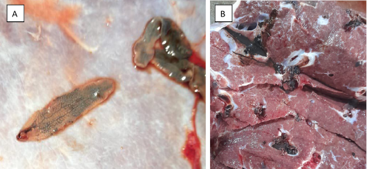
Fasciola gigantica (A) Fasciola gigantica morphology. (B) Buffalo liver infected with Fasciola gigantica.
References
- Abubakar Z, Ombugadu R, Tongjura J, Amuga G, Yako AJNAOP, Sciences A (2019). Coprological detection of bovine fasciolosis in federal capital territory, Abuja, Nigeria. J. Basic Appl. Sci., 2: 67-74. https://doi.org/10.46912/napas1i.2019110[CrossRef]
- Adedokun OA, Ayinmode AB, Fagbemi BO (2008). Seasonal prevalence of Fasciola gigantica infection among the sexes in Nigerian cattle.
- Ahmad I, Yakubu Y, Chafe UM, Bolajoko BM, Muhammad UJVPRS, Reports (2020). Prevalence of fasciolosis (Liver flukes) infection in cattle in Zamfara, Nigeria: A slaughterhouse surveillance data utilizing postmortem examination. Vet. Parasitol. Reg. Stud. Rep., 22: 100483. https://doi.org/10.1016/j.vprsr.2020.100483[CrossRef]
- Al-Jassani MJ, Sayah MA, Qasim MT, Kadhim AJ, Muhammad EH (2022). Isolation and evaluation of antibacterial agents produced by soil Bacillus sp. and study some of their immunological parameters. Rev. Electron. Vet., 23(4): 105-111.
- Al-Safi MT, Qasim MT (2023). Study of some genetic and molecular markers for some rheumatoid arthritis patients in Iraq.
- Ardo M, Aliyara Y, Lawal H, Barkindo AJIJoLR (2013). Economic assessment of bovine fasciolosis in some selected abattoirs of Adamawa state, Nigeria. Int. J. Livest. Res., 3(2): 1-8.
- Arif A, Alameri AA, Tariq UB, Ansari SA, Sakr HI, Qasim MT, Karampoor S (2023). The functions and molecular mechanisms of Tribbles homolog 3 (TRIB3) implicated in the pathophysiology of cancer. Int. Immunopharmacol., 114: 109581. https://doi.org/10.1016/j.intimp.2022.109581[CrossRef]
- Bargues MD, Valero MA, Trueba GA, Fornasini M, Villavicencio AF, Guamán R, De Elias-Escribano A, Pérez-Crespo I, Artigas P, Mas-Coma SJA (2021). DNA multi-marker genotyping and CIAS morphometric phenotyping of Fasciola gigantica sized flukes from Ecuador, with an analysis of the Radix absence in the New World and the evolutionary lymnaeid snail vector filter. Anim., 11(9): 2495. https://doi.org/10.3390/ani11092495[CrossRef]
- Bashar BS, Kareem HA, Hasan YM, Ahmad N, Alshehri AM, Al-Majdi K, Qasim MT (2022). Application of novel Fe3O4/Zn-metal organic framework magnetic nanostructures as an antimicrobial agent and magnetic nanocatalyst in the synthesis of heterocyclic compounds. Front. Chem., 10: 1014731. https://doi.org/10.3389/fchem.2022.1014731[CrossRef]
- Cárdenas-Perea M, Gutiérrez-Dávila A, Tamariz-Cruz OJRdGdM (2015). Human fasciolosis diagnosed in the acute phase: A first clinical report in Mexico. Rev.gastroenterol. México., 81(2): 111-113. https://doi.org/10.1016/j.rgmxen.2015.08.006[CrossRef]
- Chen, M, Mott KJTdb (1990). Progress in assessment of morbidity due to Fasciola hepatica infection: A review of recent literature. WHO., 87(4).
- Damwesh S, Ardo MJVJoVS (2012). Epidemiological studies of bovine fasciolosis in Yola modern abattoir, Adamawa state, Nigeria. J. Vet. Sci., 9(1): 54-60.
- Gupta J, Suliman M, Ali R, Margiana R, Hjazi A, Alsaab HO, Ahmed M (2023). Double-edged sword role of miRNA-633 and miRNA-181 in human cancers. Pathol. Res. Pract., pp. 154701. https://doi.org/10.1016/j.prp.2023.154701[CrossRef]
- Hjazi A, Ahsan M, Alghamdi MI, Kareem AK, Al-Saidi DN, Qasim MT, Mirzaei R (2023). Unraveling the impact of 27-hydroxycholesterol in autoimmune diseases: Exploring promising therapeutic approaches. Pathol. Res. Pract., pp. 154737. https://doi.org/10.1016/j.prp.2023.154737[CrossRef]
- Hjazi A, Nasir F, Noor R, Alsalamy A, Zabibah RS, Romero-Parra RM, Akram SV (2023). The pathological role of CXC chemokine receptor type 4 (CXCR4) in colorectal cancer (CRC) progression; special focus on molecular mechanisms and possible therapeutics. Pathol. Res. Pract., pp. 154616. https://doi.org/10.1016/j.prp.2023.154616[CrossRef]
- Hussein HA, Khudair SA, Alwan M, Aljawahiry T, Qasim TM, Pavlova VI (2022). Impact of pollution caused by salmon breeding centers on river water quality. Caspian J. Environ. Sci., 20(5): 1039-1045.
- Isah UMJPe, Control (2019). Studies on the prevalence of fascioliasis among ruminant animals in northern Bauchi state, North-Eastern Nigeria. Parasite Epidemiol Control., 5: e00090. https://doi.org/10.1016/j.parepi.2019.e00090[CrossRef]
- Jean-Richard V, Crump L, Abicho AA, Naré NB, Greter H, Hattendorf J, Schelling E, Zinsstag JJBvr (2014). Prevalence of Fasciola gigantica infection in slaughtered animals in south-eastern Lake Chad area in relation to husbandry practices and seasonal water levels. BMC Vet. Res., 10(1): 1-8. https://doi.org/10.1186/1746-6148-10-81[CrossRef]
- Jilo SA, Abadura SZ, Adem ME, Fereda AD, Kibi BD, Alemu HH, Ayele ST, Hussein SA, Gelchu AA, Usmael BJP (2021). Epidemiology of Fasciolosis in cattle in selected districts of bale zone, south eastern Ethiopia. Inter. J. Vet. Sci. and Anim. Husbandry., 400(192): 48. https://doi.org/10.22271/veterinary.2021.v6.i6a.471[CrossRef]
- Keiser J, Utzinger JrJCmr (2009). Food-borne trematodiases. Clin. Microbiol. Rev., 22(3): 466-483. https://doi.org/10.1128/CMR.00012-09[CrossRef]
- Khademvatan S, Majidiani H, Khalkhali H, Taghipour A, Asadi N, Yousefi EJCi, Microbiology and I. diseases (2019). Prevalence of fasciolosis in livestock and humans: A systematic review and meta-analysis in Iran. Microbiol. Infect. Dis., 65: 116-123. https://doi.org/10.1016/j.cimid.2019.05.001[CrossRef]
- Lafta HA, AbdulHussein AH, Al-Shalah SA, Alnassar YS, Mohammed NM, Akram SM, Najafi M (2023). Tumor-associated macrophages (TAMs) in cancer resistance; modulation by natural products. Current topics in medicinal chemistry. https://doi.org/10.2174/1568026623666230201145909[CrossRef]
- Lei Z, Alwan M, Alamir HTA, Alkaaby HHC, Farhan SS, Awadh SA, Nekuei A (2022). Detection of abemaciclib, an anti-breast cancer agent, using a new electrochemical DNA biosensor. Front. Chem., 10: 980162. https://doi.org/10.3389/fchem.2022.980162[CrossRef]
- Line K, Isupov MN, LaCourse EJ, Cutress DJ, Morphew RM, Brophy PM, Littlechild JAJSR (2019). X-ray structure of Fasciola hepatica Sigma class glutathione transferase 1 reveals a disulfide bond to support stability in gastro-intestinal environment. Sci. Rep. 9(1): 902. https://doi.org/10.1038/s41598-018-37531-5[CrossRef]
- Abbas MM, Abooud WK, Mohammed QA, Hasan A-ZS, Hussain A, Hameed MN, Batayneh AK (2022). Effects of various irrigation levels and biochar-based fertilizers on peanut production. J. Nutr., 13(4): 289-300. https://doi.org/10.47750/pnr.2022.13.04.076[CrossRef]
- Magaji, A., K. Ibrahim, M. Salihu, M. Saulawa, A. Mohammed and A. J. A. i. E. Musawa (2014). Prevalence of fascioliasis in cattle slaughtered in Sokoto metropolitan abattoir, Sokoto, Nigeria. Advan. Epidemiol. 2014: 1-5. https://doi.org/10.1155/2014/247258[CrossRef]
- Margiana R, Alsaikhan F, Al-Awsi GRL, Patra I, Sivaraman R, Fadhil AA, Hosseini-Fard S (2022). Functions and therapeutic interventions of non-coding RNAs associated with TLR signaling pathway in atherosclerosis. Cell. Signal., 100: 110471. https://doi.org/10.1016/j.cellsig.2022.110471[CrossRef]
- Mas‐Coma S, Valero MA, Bargues MDJAip (2009). Fasciola, lymnaeids and human fascioliasis, with a global overview on disease transmission, epidemiology, evolutionary genetics, molecular epidemiology and control. Advan. Parasitol. 69: 41-146. https://doi.org/10.1016/S0065-308X(09)69002-3[CrossRef]
- Mas-Coma S, Agramunt VH, Valero MAJAiP (2014). Neurological and ocular fascioliasis in humans. Adv.Parasitol., 84: 27-149. https://doi.org/10.1016/B978-0-12-800099-1.00002-8[CrossRef]
- Mason C, Offer J (2004). Fasciolosis associated with metabolic disease in a dairy herd and its effects on health and productivity.
- Nicholson M, Butterworth MJVp (1986). A guide to condition scoring of zebu cattle. International livestock center for Africa, Addis Ababa, Ethiopia: Northern Turkana, Kenya. Inter. Livestock Centre for Africa., 104: 85-91.
- Njoku-Tony R, Okoli GJJAS (2011). Prevalence of fascioliasis among slaughter sheep in selected abattoirs in Imo state, Nigeria. J Am Sci., 7(2): 361-366.
- Obialigwe TF, Pindar HM, Uko IB, Ekechukwu KCJM, Diseases I (2023). Prevalence of bovine fasciolosis in Jalingo abattoir, Taraba State, Nigeria. https://doi.org/10.21608/mid.2023.229531.1591[CrossRef]
- Rinca KF, Prastowo J, Widodo DP, Nugraheni YRJVW (2019). Trematodiasis occurrence in cattle along the Progo River, Yogyakarta, Indonesia. Vet. World., 12(4): 593. https://doi.org/10.14202/vetworld.2019.593-597[CrossRef]
- Sane S, Mahoori A, Abdulabbas HS, Alshahrani SH, Qasim MT, Abosaooda M, Darvishzadehdaledari S (2023). Investigating the effect of pregabalin on postoperative pain in non-emergency craniotomy. Clin. Neurol. Neurosurg., 226: 107599. https://doi.org/10.1016/j.clineuro.2023.107599[CrossRef]
- Shinggu P, Olufemi O, Nwuku J, Baba-Onoja E, Iyawa PJAiPM (2019). Liver flukes egg infection and associated risk factors in white Fulani cattle slaughtered in Wukari, southern Taraba State, Nigeria., https://doi.org/10.1155/2019/2671620[CrossRef]
- Shonhiwa AM, Ntshoe G, Essel V, Thomas J, McCarthy KJIJoID (2019). A review of foodborne diseases outbreaks reported to the outbreak response unit, national institute for communicable diseases, South Africa, 2013–2017. Inter. J. Infect. Dis., 79: 73. https://doi.org/10.1016/j.ijid.2018.11.186[CrossRef]
- Spithill TW, Smooker PM, Bruce DJF (1999). 15 Fasciola gigantica: Epidemiology, control, immunology and molecular biology. Fasciolosis., 50(2): 465-525.
- Tenorio JCB, Molina ECJVIS (2021). Monsters in our food: Foodborne trematodiasis in the Philippines and beyond: https://doi. org/10.12982/VIS. 2021.038.” Vet. Integr. Sci., 19(3): 467-485. https://doi.org/10.12982/VIS.2021.038[CrossRef]
- Tietz Marques SM, Scroferneker MLJPL (2003). Fasciola hepatica infection in cattle and buffaloes in the State of Rio Grande do Sul, Brazil. Parasitol. latinoamericana., 58(3-4): 169-172. https://doi.org/10.4067/S0717-77122003000300015[CrossRef]
- Tsegaye B, Abebaw H, Girma SJJoVM, Health A (2012). Study on coprological prevalence of bovine fasciolosis in and around Woreta, Northwestern Ethiopia. J. Vet. Med. Anim. Health., 4(7): 89-92.
- Ulayi B, Umaru-Sule B, Adamu S (2007). Prevalence of Dicrocoelium hospes and Fasciola gigantica infections in cattle at slaughter in Zaria, Nigeria.
- Urquhart GM, Armour J, Duncan J, Dunn A, Jennings F (1987). Veterinary parasitology.
- Valinata S, Susilo J, Pramono B, Karmil TF, Hamzah A, Gani FA, Jalaluddin M, Harris AJJMV (2020). Incidency and fasciolosis risk factors in cows in Pringsewu District, November 2019-January 2020. J. Med. Vet., 14(1). https://doi.org/10.21157/j.med.vet..v14i1.17403[CrossRef]
- Vassilev G (1999). Prevalence of internal parasite infections of cattle in the communal farming areas of Mashonaland East Province, Zimbabwe.
- Vázquez AA, Alda P, Lounnas M, Sabourin E, Alba A, Pointier JP, Hurtrez-Boussès SJCR (2019). Lymnaeid snails hosts of Fasciola hepatica and Fasciola gigantica (Trematoda: Digenea): A worldwide review. CABI Reviews. (2018): 1-15. https://doi.org/10.1079/PAVSNNR201813062[CrossRef]
- Yatswako S, Alhaji, Control (2017). Survey of bovine fasciolosis burdens in trade cattle slaughtered at abattoirs in north-Central Nigeria: The associated predisposing factors and economic implication. Parasit. Epidemiol. Cont., 2(2): 30. https://doi.org/10.1016/j.parepi.2017.02.001[CrossRef]
Services
Article usage statistics
Loading…