
Lentinula edodes C91-3 is an edible mushroom with demonstrated medicinal activity against various types of cancer in vitro and in vivo. The gene for Latcripin-5 (LP-5) was obtained from Lentinula edodes strain C91-3 and inserted into a pET32a (+) vector, which was then, expressed in the Rosetta gami (DE3) strain. The purified LP-5 protein was analyzed using SDS-PAGE and western blot. The optimal solubilization parameters were found to be 0.6 mM IPGT for 6 h incubation at 37℃. The solubilized protein was refolded using a refolding buffer and then dialyzed and concentrated using a dialysis buffer and PEG 20000, respectively. Phyre2 bioinformatics tool was used for protein modelling. The concentrated LP-5 protein was tested for its effect on the viability and cytotoxicity of various cancerous and non-cancerous cell lines using a cell counting kit-8 (CCK-8) assay. The LP-5 protein had the lowest IC50 value against the liver cancer line HepG2, at 58.15 μg/ml. Dose- and time-dependent morphological changes, such as cell shrinkage, blebbing formation, and cell fragmentation, were observed in treated cells. Apoptosis markers were evaluated using qPCR, flow cytometry, and western blot, and it was found that LP-5 protein increased the expression of Bax, Caspass-3, -8, -9, Cytochrome-C, and PARP, while decreasing the expression of Bcl2. Cell cycle arrest was also analyzed through qPCR, flow cytometry. Western blot results showed upregulated p21 and p27while CDK2, CDK4, CDK6, Cycline D1, and Cycline E1 were downregulated. These results suggest that LP-5 protein has potential as an anticancer agent against liver cancer cells.
Research Article · Volume 56, 22 March 2025 · Open Access
Expression, Characterization and Purification of Latcripin-5 from Lentinula edodes Strain C91-3 and its In Vitro Anticancer Activities
· · · · · · · ·
Affiliations & Notes ⌄
- 1Department of Microbiology, College of Basic Medical Science, Dalian Medical University, Dalian 116044, PR China 2Department of Biochemistry and Molecular Biology, College of Basic Medical Science, Dalian Medical University, Dalian 116044, PR China 3Department of Zoology, Abbottabad University of Science and Technology, Havelian KPK Pakistan * Corresponding author: dyzhongmt@163.com, nazia.murtaza@gmail.com
Article Info ⌄
- Journal:
- Pakistan Journal of Zoology
- Published:
- 22 March 2025

From the article
- The LP-5 protein showed higher inhibitory effects against liver cancer cells (58.15µg/ml) as compare to other cells in this study (Fig. 5A).
- LP-5 treated cells showed a significant up-regulation in expression of Bax, caspas-3, 8, -9, Cytochrome-C, and PARP while down-regulation in expression of Bcl-2 (Fig. 6B).
- This suggests that the LP-5 damage destabilize liver cancer cells by external stress (Fig. 6C).
- Various in-vitro analyses against liver cancer cell line HepG2 suggests that LP-5 inhibit proliferation, promoted apoptosis, and stopped the cell cycle at the G1 phase.
Selected from the article's own text.
Abstract
Introduction
Cancer is one of the leading public health issues with the high rate of morbidity and mortality worldwide. According to Global cancer statistics (2020) liver cancer is the 6th commonly diagnosed cancer worldwide and it is account for third leading cause of death . In 2019, the incidence and mortality rate of liver cancer increased (WHO, 2020). Liver cancer which ranks 5th in terms of global burden and reported as second on the basis of mortality in male population . In spite of advancement in liver cancer treatment and prognosis have been reported in recent past years, although the effectiveness is still unsatisfactory needs improvement . Mushroom natural products and compound has been reported for their pharmacological efficacy against various types of disorders includes cancer ; . Medicinal and edible mushroom has been reported to have various potentials i.e., cytotoxic, antiproliferative, and antioxidant ; ; ; ; ; . The mushrooms extracts consist of polysaccharides, proteins, polysaccharide-protein complex, steroids, dietary fibers, and phenols ; ; . Lentinula ededes inhabitant of warm and moist climate has been studied for its medicinal effect. Various studies have reported that extracts sterols, lipid, terpenoids, polysaccharide, and mycelia from L. edodes have not only anti-cancer activity but also anti-viral, anti-bacterial, and anti-fungal effects ; .
The L. edodes recombinant proteins have been cloned and expressed using a variety of expression techniques ; ; ; . Our research group reported that L. edodes strain C91-3 protein components have anticancer potential both in vivo and in vitro models. For the de novo characterization of the L. edodes strain C91-3 transcriptome, a total of 57 million nucleotides were read and assembled into 18,120 unigene through next-generation sequencing (NGS) (deep solexa/Illumena). Thousands of genes were identified which are still under investigation for various therapeutic applications ; .
The Latcripin-5 (LP-5) has not been reported for its anti-cancer activity against liver cancer cell lines HepG2. LP-5 protein was obtained through a prokaryotic expression system followed by suitable purification techniques . The aim of this study was to evaluate the anti-cancer role of LP-5 in liver cancer cell line HepG2 and facilitate more effective choice of therapy for liver cancer treatment. This was noted that LP-5 plays a significant role by enhancing cell cycle arrest and apoptosis in liver cancer cell line HepG2.
Materials and Methods
The shiitake mushroom strain of Lentinula edodes C91-3 was obtained from the Chinese general microbiological culture collection center (CGMCC). The bacterial strain of E. coli JM109, PCR cloning kit, Plasmid Purification kit, 3′- full RACE Core Set 2.0, 5′-Ful RACE kit, DNA ligation kit and Molecular enzymes and primers EcoR 1 and Xho1 used in this research project were ordered from Takara, Dalian, China. The expression vector pET32a (+) was purchased from Invitrogen. 6x-Hist-tag monoclonal antibody and horseradish peroxidase-Rabbit Anti-mouse IgG (H+L) was from Proteintech Group, Inc. The bicinchoninic acid (BCA) protein assay kit, was purchased from NaijiangKeyGen Biotech Co. Carbancelline, chloramphenicol, tetracycline, kanamycin sulphate, phenyl methane sulfonyl fluoride and isopropyl β-D-1-thiogalactopyranoside (IPTG) were ordered from Tiagen Biotech Co., LTd. The sypro orange protein gel stain was bought from Sigma Aldrich. The common integrated potato culture medium, Luria Agar and Luria broth were prepared in the Microbiology department Dalian Medical University. Anti-microbial agent (100µg/ml streptomycin, 100 units/ml penicillin), Trypsin with EDTA and without EDTA was purchased from Thermo Fisher scientific; Incwhile the RIPA buffer was from Proteintech. Polyvinylidene fluoride (PVDF) membranes were obtained from Millipore, Billerica. Bcl-2 (12789-1-Ap), Bax (23931-1-Ap), Caspase-3 (19677-1-Ap), Caspase-9 (10380-1-Ap), PARP (13371-1-Ap), p21 (10355-1-Ap), and GAPDH along secondary anti-Rabbit antibodies were bought from Protein-tech (Wuhan, China). Cyclin-D1 (60186-1-Ig) purchased from Thermo Fisher Scientific, Waltan, MA, USA). CDK6 (D4S8S), p21 (12D1), and Cycline-E1 (HE12), mouse antibody along with anti-mouse secondary antibody purchased from cell signaling USA. Annexin V-FITC/PI kit was acquired from Invitrogen.
Bioinformatics analysis of LP5
The nucleotide sequences of LP-5 of Lentinula edodes strain C91-3were analyzed by NCBI database (https://www.ncbi.nlm.nih.gov) (Accession no. KF772202.1) and amino acid sequence with protein ID (AHB815431). Functional domain and structural integrity analysis were studied on European Bioinformatics Institute (EBI) tools (https://https://www.ebi.ac.uk/). ExPASy server were used for determination of theoretical molecular Weight and pI value (MW=44.3Kd and pI value=6.0 (https://web.expasy.org/compute_pi/) , 1994). The phyre2 server was used to predict the tertiary structure of the LP-5 functional domain (https://www.sbg.bio.ic.ac.uk/). Using the ExPASy database, the amino acid composition of the LP-5 functional domain was examined (http://www.expasy.org/).
Complimentary DNA synthesis, cloning of LP-5 gene
Total RNA was extracted from L. edodes C91-3 mycelium by Trizol method. Using Oligo 6.0 software, primers were built for the 3′-RACE and 5′-RACE in response to sequencing data. The LP-5 gene was amplified using upstream primer with EcoR1 restriction endonuclease (RE) site (5’-GCGAATTCACCAACAATGCATCCGGTG-3’) and downstream primer with XhoI RE site (5’-GCTCGAGATTACAAGAGCGCTCAGTA-3’). Thirty cycles of PCR reaction were performed under the following conditions: 98°C for 10 sec, 56°C for 10 sec, 72°C for 1 min, 72°C for 5 min, and 4°C for 1 min. The amplified sequence was verified using the ABI PRIMTM 3730XL DNA Sequencer. The Mini BEST 1% Agarose Gel DNA Extraction Ver. 3.0 kit was used to purify amplified complementary DNA. Following the manufacturer’s instructions, the purified clone DNA was inserted into the Eco RI/XhoI sites of the pET32a (+) vector through the Infusion HD Cloning kit. The pET32a (+) vector was successfully introduced into E. coli JM109 competent cells. DNA sequencing was used to confirm positive white colonies. The recombinant plasmid pET32a (+)-LP-5 was extracted and transformed into Rosetta-gami (DE3).
Expression and purification of LP-5
A single colony of transformants was cultured overnight in Luria Broth with antibiotics (chloramphenicol 34 µg/ml, tetracycline 12.5 µg/ml, kanamycin sulphate 15 µg/ml, and carbancelline 100 µg/ml) on an orbital shaker at 37˚C for the preliminary expression of LP-5 until the optical density reached 600 nm. Cultured cells were grouped in to two: control and test groups. IPTG (0.6 mM) was added to the test group only, both control group and tested group were incubated for 6 h at 37oC on the shaker to induced fusion protein expression. Then bacterial culture was centrifuged at 3,824 g at 4oC for 5 min. A single PBS (pH 7.4) wash was performed on the bacterial cell pellet before it was resuspended in lysis buffer. The lysis buffer containing mixture was sonicated as: 10 cycles in ice for 1 min at 4oC for 1 h. Centrifugation was performed on the lysed and sonicated bacterial suspension at 15,297 g for 20 min at 4°C. Both supernatant and sediments were analyzed using Hist-Tagged protein through western blot technique.
Three distinct parameters (IPTG concentration, temperature, and incubation time) were evaluated in order to optimize the LP-5 expression conditions. The culture was treated with various IPTG concentrations (0.05, 0.1, 0.2, 0.3, 0.4, 0.5, 0.6, 0.7, 0.8, 0.9, and 1 mM) for different incubation period (1, 2, 3, 4, 5 and 6 h) and at 20, 30, 37, 40, and 50°C temperatures, respectively. The optimum expression of LP-5 protein was observed at IPTG concentration (0.6 mM) on 37 °C for 6 h. Each 10 µl supernatant and pellet was analyzed by 12% SDS-PAGE electrophoresis while protein quantification and size were measure through Image Lab Software.
At post lysis the pellet (contained inclusion body) were three times washed with washing buffer at 4 °C on 15,294 g for 20 min followed by reconstitution in double distilled water. Solubilization buffers (20 mM Na3PO4, 20 mM KH2PO4, 20 mM Tris HCl and deionized H2O, and pH 8.0) containing 3M urea were used to dissolve the homogeneous suspension of LP-5 protein containing inclusion bodies (10 mg/ml). Various pH (5, 6, 7, 8, and 9), temperature (24, 20, 4, -20, -40, and -80°C) and solvents (PBS, NaP, Tris, ddH2O and KP) were optimized. Using Ni magnetic beads, (Tomos Biotools Shanghai Co., Ltd., Shanghai, China) the solubilized protein supernatant was stirred for h at 4 °C. Washing buffer (500 mM NaCl, 50 mM Imidazole, 20 mM NaH2PO4, 3M Urea pH 8.0) was applied three times at 4°C to remove unbound proteins. Then samples were washed, and protein was eluted through elution buffer. The BCA technique was used to assess the pure protein content. Protein (10 µl) was screened by 12% SDS-PAGE and western blot.
Refolding of LP-5 functional domain protein
The purified LP-5 functional domain protein was refolded through utilization of four dialysis buffer: Dialysate 1 (2 mM reduced glutathione (GSH) 4 M urea, and 0.2 mM oxidized glutathione (GSSH) and phosphate-buffered saline (PBS), Dialysate 2: (2 mM GSH. 3 M urea, and 0.2 mM GSSG in PBS), Dialysate 3 (0.2mM GSSH and 2 mM GSH in PBS), and Dialysate 4 (PBS) at 4˚C. Each concentration of dialysate was used three times and the dialysate was changed every eight hours.
Dialysis and concentration of LP-5 protein
Refolded LP-5 protein was poured into dialysis membrane (MWCO 14 kDa; Biosharp, Hefei, China) while the dialysis was changed twice at 4°C for 12 h. The dialyzed protein was concentrated using PEG 20,000 (Sigma-Aldrich, Merck Millipore, Darmstadt, Germany), the BCA protein assay kit was used to measure LP-5 quantity and precise band size was determined 12% SDS-PAGE electrophoresis.
Cell culture
Seven human cancerous cells were used i.e., HepG2 (liver cancer), A549 and H1299 (lung cancer), SGC-7901 and BGC-823 (gastric cancer), MDA-MB-231 and MCF-7 (brest cancer) and a single normal cell line HaCaT (human keratinocytes). Among these cell lines i.e., HepG2, MBA-MD-231, MCF-7, and HaCaT were culture in Dulbecco’s modified Eagle’s medium (DMEM). While A549, H1299, SGC-7901, and BGC-823 were culture in RPMI-1640 medium containing 10% fetal bovine serum (FBS). The culture cells were maintained in 5% carbon dioxide at 37°C and 95% humidity in the incubator.
Cell viability assay
According to the manufacturer’s instructions, the CCK-8 kit was used to assess cell viability, proliferation, and cytotoxicity. Briefly, 1×10⁵ cells were seeded overnight in 96-well plates and cultured at 37°C. After that, cells were exposed to various doses of LP-5 protein (0, 25, 50, 75, 100, 125, and 150 g/ml) and incubated for 24, 48, and 72 h at 37oC. Cells were finally incubated for 4 hs at 37°C with 10 µl of CCK-8 reagent applied to each well of 96-well plates. After incubation, the absorbance was measured at 450 nm via microplate reader. Cell viability was calculated using the formula below.

Colony formation assay
The LP-5 was applied to the 1×10³ liver cancer cells on a 6-well plate in several concentrations (0, 75, 100, and 125µg/ml). After 15 days of incubation at 37°C and 5% CO2 under 95% humidified atmosphere. The cells were subsequently rinsed three times with PBS, fixed for a half-h in a 4% paraformaldehyde (PFA) solution, and stained for 5 min with Giemsa strain. The colonies of cultured cells were observed and counted under the microscope.
Western blotting
The liver cancer cell lines HepG2 control and treated with LP-5 at concentrations: 75, 100, and 125µg/ml were incubated for 48 h then gently rinsed with ice-cold PBS. RIPA buffer was used to lyses the cells and extract the protein. The extracted proteins were quantified with a BCA kit. Thirty microgram (30µg/ml) protein was added to each well of SDS-PAGE gel (12%) then transferred to the polyvinylidene fluoride (PVDF) membrane. The next step is to block PVDF membrane by incubating in 1xTBST with 5% skimmed milk for 2 h at room temperature. Then PVDF membranes were washed and incubated with primary antibodies at 4 °C. that secondary antibody were added to the membranes and incubated for two hs at room temperature after three rounds of 1x TBST washing. After adding the ECL the blot were then subjected to chemiluminescent gel imaging device for visualisation (ChemiDcoTM XRS Imager Bio-Rad).
Apoptosis analysis by flowcytometry
The liver cancer cell lines were treated with LP-5 for 48 hs, after treatment the cells were washed with PBS. The collected cells were stained with PI and Annexin V-FITC according to the instructions of manufacturer. The apoptosis rate was calculated using a FACS-Calibur Cytometer (BD Accuri C6 Biosciences, Heidelberg, Germany).
Cell cycle arrest by flowcytometry
The liver cancer cells (Control and LPS-5 treated) were collected after 48 h and washed with PBS, the cells were then incubated overnight at 4°C in 70% ethanol for fixation. After fixation the Cells (1x10⁶ cells/ml) were collected and incubated for 30 min at 37°C. Propidium Iodide50 µg/ml and RNase 5µ g/mL RNase cell were added to cells. Samples were analyzed by using FACS-Calibur Cytometer.
Quantitive polymerase chain reaction (q-PCR)
The cell cycle and apoptosis regulatory mRNA expression were determined by q-PCR. Total RNA was extracted from LP-5 treated and control group cells after 48 h of treatment with IC50 concentration through TRIzol method according to standard protocols. Using a Transgene reverse transcriptase reagent kit with gDNA removal agent, 1 µg of RNA was utilized to produce cDNA. The cDNA was quantified through RT master mix (Transgene) using a real-time PCR system (Step One TM Applied, Singapore). For SYBER reverse transcription, the procedure was as: 15 min at 42°C, 5 sec at 85°C, 30 sec at 94°C, 40x (94°C for 5 second, 60°C for 15 sec, 72°C for 10 sec). The reaction was performed in triplicate, while GAPDH was used as internal control. Using the 2- Δcct approach, the relative gene expression of mRNA was determined.
Statistical analysis
The statistical analysis was performed using Graph Pad Prism 5.0 and Microsoft Excel 2007 software. Test of significance i.e. One-way analysis of variance (ANOVA), Tukey’s multiple comparison was applied. Linear regression was calculated for the half-maximal inhibitory concentration (IC50) of LP-5.
Results
Structural analysis of LP-5
For modelling, the 3-dimensional structure of the LP-5 phyre2 server was used, which uses the Markov method to generate a model based on published structure and use it as a template. The structure of LP-5 was developed by providing a sequence of amino acids to the phyre2 server under intense mode. Phyre2 server used hydrolase protein as a template based on sequence identity and maximum coverage and generated a protein structure with maximum confidence (Fig. 1A). The structure generated via phyre2 was further referred to by minimizing the energy via YASARA energy minimization software, Ramachandran plot was used. The secondary structure revealed the presence of 7 beta-hairpin loops, which houses 10 beta-sheets and 1 gamma turn. 11 beta-sheets and 1 gamma turn exist outside of the beta-hairpin loop (Fig. 1B). Validation of 3D structure generated via phyre2 server through Ramachandran plot. More than 90% residues were favoured region and 8.2% were in allowed region. Plot on left side showing all residues combine while smaller right side plot shows major amino acids on phi and psi scale (Fig. 1C).
LP-5 vector construction
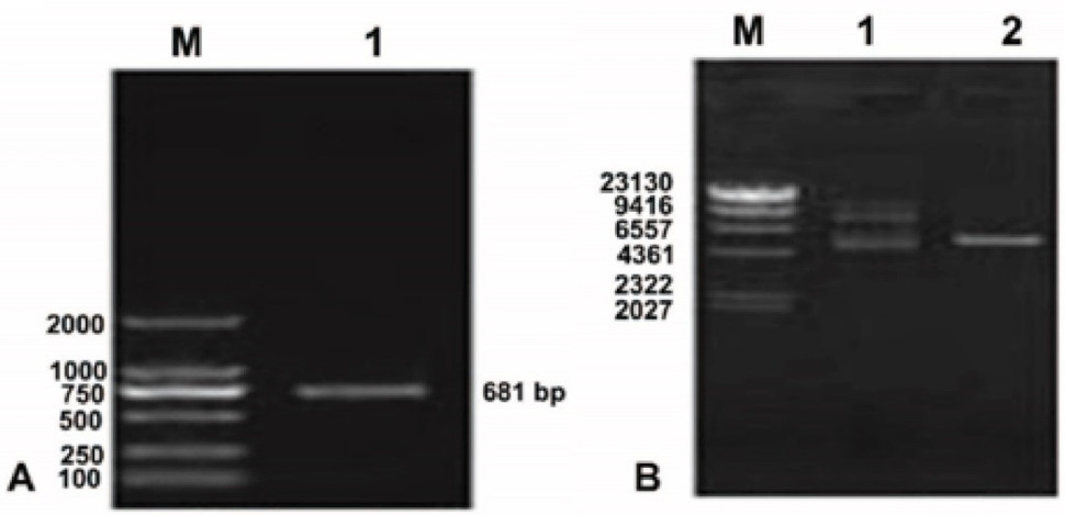
The agarose gel electrophoreses results shown that the target LP-5 gene band was noted at 681bp position (Fig. 2A). Successful digestion was carried out and analyzed via agarose gel electrophoresis. The band was observed at 6.4kbp after double digestion which showed a complete and successful construction of LP-5 vector (Fig. 2B).
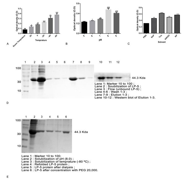
Various parameters were considered for the optimal expression of LP-5, which included temperature, IPTG concentration, and induction times. The growth of E. coli was carried out in a range of temperature from 20, 30, 37, 40 and 50˚C and while optimum growth was noted at 37˚C (Fig. 3A). At 37˚C, the optimal IPTG concentration and time of induction were determined. 0.6mM IPTG concentration was found optimal across the range 0.05, 0.1, 0.2, 0.3, 0.4, 0.5, 0.6, 0.7, 0.8, 0.9 and 1 mM concentration (Fig. 3B). Furthermore, the induction and expression of LP-5 was at maximum at 6 h of time duration across 1, 2, 3, 4, 5, and 6 h of time period (Fig. 3C). At these optimal conditions cell were collected, lysed, and centrifuged. The pellet containing inclusion bodies (IBs) were analyzed for further analysis to confirm the induce and uninduced LP-5 protein (Fig. 3D).
Solubilzation of LP-5 inclusion bodies (IBs)
LP-5 protein was obtained from IBs. Various temperatures were used in studies ranging from 20, 24, 04, -20, -40 and -80˚C. The LP-5 protein was observed to have the maximum solubility on -80˚C (Fig. 4A). The different pH was used in the study range i.e., 5, 6, 7, 8, and 9. The best and efficient solubility was observed of inclusion bodies at 8.0 pH (Fig. 4B). Various solvents were used for the solubility of LP-5 protein. Results indicated that the buffer consisting of 3M Urea, 20mM Tris-HCl under pH 8.0 was the best solubilized medium to elute the LP-5 proteins from IBs (Fig. 4C).
Purification and refolding of LP-5 protein
Nickel magnetic beads were used to purify the LP-5 protein the binding buffer (500 mM NaCl, 20 mM NaH2PO4, 5 mM Imidazole, 3M Urea pH 8.0) was used for binding of protein at 4oC under constant shaking condition. Elution buffer (20 mM NaH2PO4, 500 mM NaCl, 500 mM Imidazole, 3M Urea pH 8.0) was used to elute His-tagged LP-5 protein. After eluting the His-Tag LP-5 protein the band was observed at 44.3 KDa. The LP-5 protein was also confirmed by western blot and expected band of 44.3KDa size was observed (Fig. 4D). The protein was properly refold and then expressed in the active form. In our study the mild solubilzation condition were used to preserve the active structure of the LP-5 protein for in vitro refolding. PEG 20,000 was used to obtained concentrated refolded protein (Fig. 4E). This concentrated protein was extensively used in our study for different biological assays.
Anticancer activity of LP-5
The CCK-8 assay was performed to evaluate the cytotoxic effect of LP-5 in panel of cancerous and a normal cells lines (HepG2, A549, H1299, SGC-7901, BGC-823, MCF-7, MDA-MB-231 and HaCat) with varying concentration of LP-5 ranging from 0 to 150 ug/ml for 48 h IC50 values were calculated for all cell lines. IC50 of LP-5 was 58.1ug/ml for Liver cancer HepG2 cell line which was lower among the other cancerous and normal cell lines. Similarly, LP-5 inhibited cell proliferation (50%) in normal cell line i.e., IC50 value 201.1 ug/ml which is higher than HepG2 (Fig. 5A). The CCk-8 assay was performed for cell viability for HepG2 liver cancer cell lines at three various time period (0, 24, 48 and 72 h) and also in dose dependent manner of LP-5 ranging from 0, 25, 50, 75, 100, 125 and 150ug/ml and the minimum viable cell was noted in 125 and 150 ug/ml (Fig. 5B). Cell proliferation assay was used to assess the rate
of cell growth inhibition at dosage 0, 75, 100 and 125 ug/ml, and time interval 0, 24, 48 and The HepG2 cell treated with LP-5 showed a response in dose and time dependent manner while higher number of cells were inhibited at higher concentration after 72 h (Fig. 5C). The HepG2 morphological changes were examined under the phase contrast microscope. The morphological changes were noted in the shape; size and volume of the cells compared with control group (Fig. 5D).
Apoptosis activity of LP-5 protein in HepG2
The number of colonies is inversely proportional to the concentration of LP-5 (0, 75, 100 and 125µg/ml) as compared with control group (Fig. 6A). Apoptosis related mRNA and protein expression after 48 h of treatment with LP-5 was investigated. The mRNA expression of Bax, Bcl2, caspase-3, -8, -9, Cytochrome-C and PARP detected through qRT-PCR, the expression of all these proteins were confirmed by western blot. These results showed down-regulation of Bcl2 while significant upregulation in mRNA expression levels of Bax, Caspase-3, -8, -9, Cytochrome-C and PARP (Fig. 6B). Confirmation of apoptosis was carried out via flow cytometry by utilization of twoapoptotic markers AV and PI at 48 h on dose dependent manner (0, 75, 100 and 125µg/ml).
In HepG2 treated cells with LP-5 as compared to the control group, it was shown that the rate of apoptosis increased significantly (Fig. 6C). The increased significant level of expression was observed on western blotting an apoptotic related proteins i.e., Bax as well as Caspase-3, -9, Cytochrome-C and PARP while Bcl2 expression levels was down regulated with the increasing concentration of LP-5 protein (Fig. 6D).
Cell cycle arrestment activity of LP-5 in HepG2
Treatment for 48 h of HepG2 cells with LP-5 (IC50 concentration) resulted in the up-regulation of CDKs inhibitory genes p21, p27 and down regulation of Cyclin-D1, Cyclin-E1, CDK2, CDK4, and CDK6 (Fig. 7A). Flow cytometric analysis of cell cycle represented that significant population of cells were arrested in G1 phase depend on dosage and time. The increase trend in the G1 phase of cell cycle in HepG2 was 52.4%, 58.8% and 60.6% at LP-5 concentrations (75, 100 and 125µg/ml) respectively, in comparison with control group, however in S phase mild increase 38.6% was noted at dose of 75µg/ml as compare to control group and then decrease trend was observed i.e., 30.6% and 28.6% at concentration of 100µg/ml and 125µg/ml (Fig. 7B). Cell cycle regulation related genes (p21, CDK6, Cyclin-D1, and Cyclin-E1 in HepG2) were analyzed through western blot. Results revealed that p21 was up-regulated while CDK6, Cyclin-D1 and Cyclin-E1 was down-regulated (Fig. 7C).
Discussion
Liver cancer is the 3rd leading cancer which causes death across the Globe . therefore, it is needed to disclose novel therapeutics bioactive natural products with having potential effect against liver cancer therapy. L. edodes is one of the edible and medicinal mushroom. Various studies have been conducted for its utilization as treatment of various diseases. L. edodes consist of numerous bioactive molecules with a pharmacological potential against hypertension, hepatic disorder, cancer and enhanced immunity ; . LP-1 and LP-7A was reported to have anti-cancer effect on gastric cancer cell line BGC-231 and SGC-7901 ; . In this study LP-5 gene was successfully cloned in prokaryotic expression system, using pET32a (+) in E. coli Rosetta gami (DE3) under the optimized condition. Inclusion bodies (IBs) contain the expressed protein. Various factors were considered into account for the solubilzation of the IBs, such as solvents, temperature and pH. Many studies have been reported to show effective anticancer activity of LP proteins extracted from L. edodes. LP-4 protein was reported to successfully inhibit the proliferation of human liver cancer cell . Similarly, LP-1, LP-3, LP-13, and LP-15, was also showed promising effects against human Lung cancer cells ; ; ; . LP-7A was also reported possessing antiproliferative characteristics against breast cancer cells . LP-11 and Lp16-PSP was reported as effective against human acute Promyelocytic Leukaemia HL-60 ; . IC50 was evaluated for anti-cancer effect of LP-5 protein on various human cancerous and normal cells line which includes (HepG2, A549, H1299, BGC-231, SGC-7901, MDA-MB-231 and MCF-7), and normal cells (HaCat). These cells were selected randomly on the base of the severity that fall in the list of most fatal cancer types of 2020 . The LP-5 protein showed higher inhibitory effects against liver cancer cells (58.15µg/ml) as compare to other cells in this study (Fig. 5A). Similar result of LP-1 was reported for IC50 against liver cancer cell line i.e., 71 µg/ml. The HepG2 cell treated with LP-5 showed a response in dose and time dependent manner while higher numbers of cells were inhibited at higher concentration after 72 h (Fig. 5B). For cell growth inhibition rate, proliferation assay was used. LP-5 treated HepG2 was significantly inhibited on base dosage and time. Similar results shown that the polysaccharide from Pleurotus ostreatus and LP-4 from L. edodes inhibit cell viability of liver cancer HepG2 cells ; (Fig. 5C). After the treatment of liver cancer cell with LP-5, cells morphological changes was observed in shape, size and volume with as compare with control group (Fig. 5D). We further analyzed the colony inhabitation of liver cancer line HepG2 via colony formation assay and noted that colonies were decrease with increase of concentration of LP-5 (Fig. 6A). Similar studies were reported that chlorogenic acid and dioscin significantly inhibit the colonies in HepG2 liver cancer cell in concentration dependent manner and reported less colonies in higher concentration ; . The LP-5 apoptosis effect on various apoptosis related genes i.e. Bcl-2, Bax, caspase-3, -8,-9, cytochrome-C and PARP were evaluated post treatment by qRT-PCR method. LP-5 treated cells showed a significant up-regulation in expression of Bax, caspas-3, 8, -9, Cytochrome-C, and PARP while down-regulation in expression of Bcl-2 (Fig. 6B). Previous studies also reported that mRNA expression level of Bax, capase-3, 8, 9, cytochrome-C, and PARP increases while Bcl-2 mRNA expression level was decreased which led the human leukemic cancer, breast cancer and Jurkat T-cell, cervical adenocarcinoma and hepatocellular carcinoma to apoptosis ; Kiddane et al., 2022; ; ; . Bcl-2 plays a vital role in apoptosis through maintaining mitochondria in active state. The downregulation of anti-apoptotic protein i.e., Bcl-2 and upregulation of pro-apoptotic proteins i.e., Bax, caspase-3, capase-9 and cytochrome-C, initiate apoptotic process via mitochondrial disintegration . Pro-apoptotic (Bax) and anti-apoptotic (Bcl-2) protein ratio modulate the release of cytochrome-C results in a cell death phase via mitochondrial pathway . In our study increasing the dose LP-5 protein decreases the expression level of Bcl-2 and upregulate the expression level of Bax. These observations indicated that the LP-5 induced apoptosis in HepG2 cells was triggered by downregulation of Bcl-2 and the upregulation of Bax, caspases-3 and -9, PARP and releases cytochrome-c from mitochondria (Fig. 6d). Previous studies reported that cytochrome-C play key role in the activation of caspases which results in apoptosis ; . Our findings showed that the LP-5 decreases mitochondrial membrane potential in Liver cancer cells. Liver cancer cell apoptosis after treatment with LP-5 was examined through flow cytometry. A significant percentage of apoptotic cells in late apoptotic stage were noted. However, there were more necrotic cells found in the HepG2 cell treatment groups. This suggests that the LP-5 damage destabilize liver cancer cells by external stress (Fig. 6C). Other studies confirmed our results which demonstrated that mycelia extracted polysaccharide-zinc complex and LP-15 from medicinal mushroom and marine natural products induced apoptosis in liver, and lung cancer cells, in dose-dependent manner , 2016; ; .
In order to identify the LP-5 effect on HepG2 cell cycle arrest at G1 phase, mRNA expression was evaluated for CDK2, CDK4, CDK6, Cyclin-E, Cycline-D1, p21 and p27 at post treatment via qPCR method. Treatment with LP-5 down regulates the mRNA expression of CDK2, CDK4, CDK6, Cycline-E1 and Cycline-D1 while up-regulate of p21 and p27 (Fig. 7A). Similarly, other studies reported that LP16-PSP, Asparagus officialis, and Cordycepin treated cancerous cells i.e., HL-60, human endometrial carcinoma ishikawa cells and leukaemia cells showed changes in mRNA expression of cell cycle related genes ; ; . Scientific reports represented that cell cycle dysregulation cause’s inhibition of cell growth or apoptosis and being considered as a marker for chemotherapeutic and chemo preventive agents . In order to study whether LP-5 induce cell cycle disturbance in HepG2 cells, flow-cytometric analysis using propidium iodide (PI) and RNase as staining agent was performed, following LP-5 treatment for 48 h and the cell cycle was arrested at G1 phase (Fig. 7B). Our study was consistent with previously reported studies, as Sdy-1, Terminalia bentzoe extract, Astrakurkurol, medicinal mushrooms, LP-7A and Olenaolic acid arrested cell cycle in G/G0 phase in liver, cervical, colon, gastric cancerous cell lines and hepatocellular carcinoma ; ; ; ; ; . The increased expression of cyclin-cdk complex inhibitor p21 and reduced Cyclin (Cyclin-D1 and Cyclin-E1) and CDK6 expression in HepG2 cell lines (Fig. 7C). Tamarix articulata, was reported that arrest cell cycle at G0/G1 phase, up-regulated p21, p27, and down-regulated p53, Cyclin D1, Cycline-E1 and CDK family CDK2, CDK4, and CDK6 in hepatocellular carcinoma .
Conclusions
The present study suggested that the LP-5 protein has a potential anticancer activity. This study provides production and purification method of LP-5 protein. Various in-vitro analyses against liver cancer cell line HepG2 suggests that LP-5 inhibit proliferation, promoted apoptosis, and stopped the cell cycle at the G1 phase. We recommend that further detailed in-vivo animal model studies will develop as preclinical experiments.
Acknowledgments
We acknowledge Miss Hu Jie for her support in the experiments. We also acknowledge the International Education College of Dalian Medical University, PR China, and the Chinese Scholarship Council for providing assistance.
Institutional review board statement
This study was approved by the Ethical Research Committee of Dalian Medical University.
Ethical statement
This study was ethically approved from Basic Medical Collage, Department of Microbiology Dalian Medical University, Dalian, People Republic of China.
Data availability statement
All the data is presented in this study. The additional data is available from the first or corresponding author upon a reasonable request.
Statement of conflicts of interest
The authors have declared no conflict of interest.
Figures
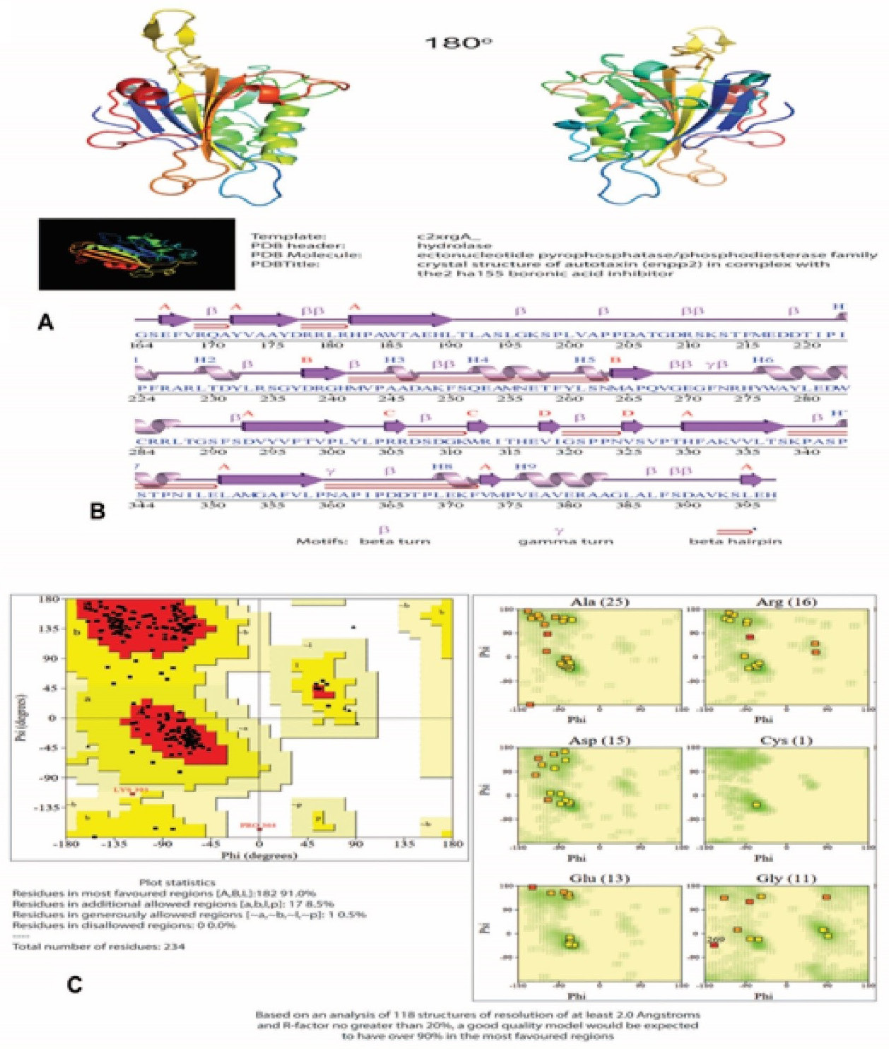
Structure modeling of LP-5: 3-dimemtional structure (side view at 180 degree) genrated via phyre2 using hydrolase as template (A). Secondary structure of LP-5 showing various motifs (B). Ramachandran plot validation (C).
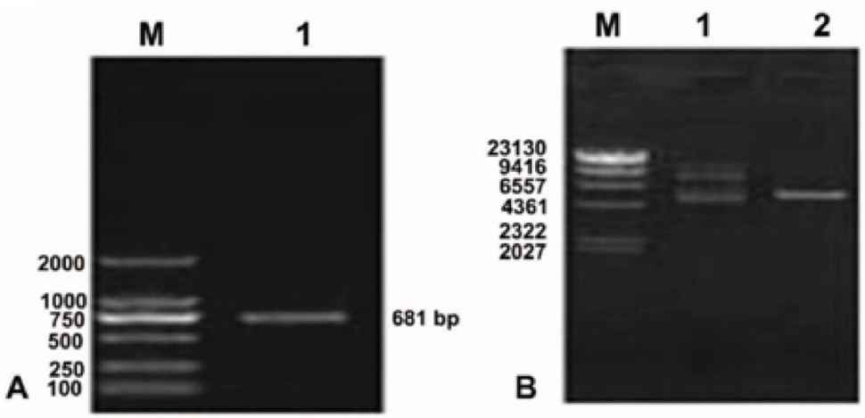
Lane 1, PCR products of LP 5-RCC1 cDNA. (B) Plasmid pET-32a(+)-LP-5 functional domain cDNA. M, DNA Marker DL8000; Lane 1, plasmid pET 32a(+) LP-5 extracted from JM109 cells; Lane 2, double restriction enzyme digestion of plasmid pET 32a(+) LP 5 RCC1 extracted from JM109 cells.
Efficient production of LP-5 protein. Effect of temperature (A), IPTG concentration (B), and induction time on LP-5 production (C). SDS-PAGE analysis of LP-5 under optimized condition (D). Statistical analysis was performed using multiple t test via GraphPad Prism 6.0 (* p < 0.05, ** p < 0. 01, and p *** < 0.001).
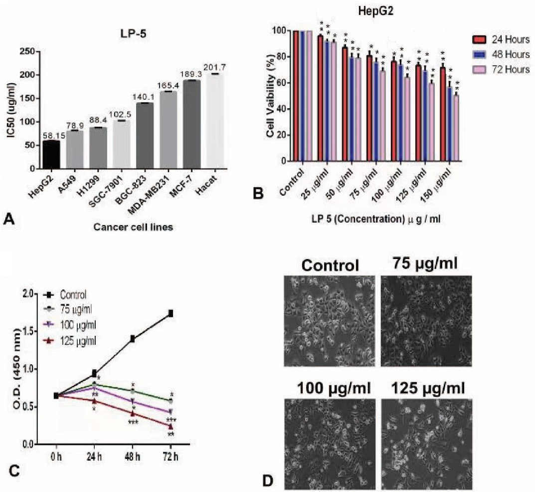
Effect of LP-5 on the viability and proliferation of HepG2 cells. (A) IC50 values of LP-5 against a panel of cancerous and normal cell lines were evaluated and results are expressed in three independent experiments at 48 h. (B) HepG2 cells treated with different concentration of LP-5 for 24, 48 and 72 h to determine cell viability. Results are expressed as a percentage of corresponding control (C) Cell proliferation assay performed by CCK-8, with a low dose of LP-5 at different time intervals for HepG2. Results are presented in three independent experiments (D) Morphological changes in HepG2 cell lines were observed under a phase contrast microscope after 48 h treatment with a mentioned concentration of LP-5 (0, 75, 100 and 125 g/ml) at 40x magnification.Statistical analysis was performed using multiple t test via GraphPad Prism 6.0 (* p < 0.05, ** p < 0. 01, and p *** < 0.001).
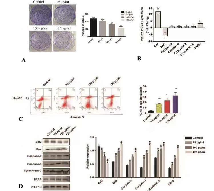
Colony formation by liver cancer cells line HepG2 treated with LP-5 (0, 75, 100 and 125 g/ml) (A). Analysis of q RT-PCR apoptosis related genes (Bax, Bcl2, caspase-3, capase-8 and caspas 9, Cytochrome-C, and PARP ) (B). Evaluation of apoptosis via flow cytometry in LP-5 treated liver cancer cells lines HepG2 dose and time dependent-manner (0, 75, 100 and 125 g/ml and 48 hours). Quadrant 2 (Q2) denotes late apoptosis; Quadrant 3 (Q3) indicate early apoptosis, while Q4 represent vialble cells (C). Evaluation of apoptosis related markers (Bax, Bcl2, caspase-3, caspase-9, Cytochrome-C and PARP) in HepG2 cells treated with LP-5 (0, 75, 100 and 125 g/ml and 48 h incubation time) (D). Statistical analysis was performed using multiple t test via GraphPad Prism 6.0 (* p < 0.05, ** p < 0. 01, and p *** < 0.001).
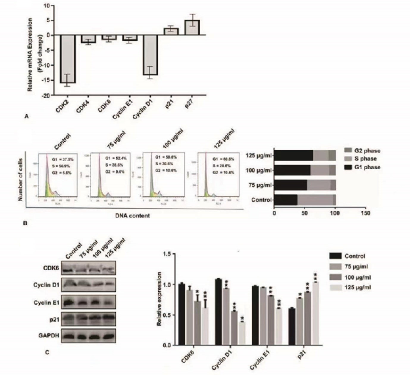
Analysis expression of q RT-PCR cell cycle releted genes (Cyclin E1, Cyclin D1, CDK6 and p21) treated HepG2 cells with LP-5 (A). Cell cycle anylasis of liver cancer cell lines HepG2 treated with LP-5 (0, 75, 100 and 125 g/ml and 48 h incubation time) (B). Detection of cell cycle related markers Cyclin-E1, Cyclin D1, CDK6 and P21 expressions in HepG2 liver cancer cells with LP-5 dose and time dependent mannaer (0, 75, 100 and 125 g/ml and 48 h incubation time) (C). Statistical analysis was performed using multiple t test via GraphPad Prism 6.0 (* p < 0.05, ** p < 0. 01, and p *** < 0.001).
References
- Alnuqaydan, A.M. and Rah, B., 2021. Tamarix articulata inhibits cell proliferation, promotes cell death mechanisms and triggers g0/g1 cell cycle arrest in hepatocellular carcinoma cells. Fd. Technol. Biotechnol., 59: 162-173. https://doi.org/10.17113/ftb.59.02.21.6904[CrossRef]
- Ann, X.H., Lun, Y.Z., Zhang, W., Liu, B., Li, X.Y., Zhong, M.T. and Huang, M., 2014. Expression and characterization of protein latcripin-3, an antioxidant and antitumor molecule from Lentinula edodes c91-3. Asian Pac. J. Cancer Prevent., 15: 5055-5061. https://doi.org/10.7314/APJCP.2014.15.12.5055[CrossRef]
- Arora, S. and Tandon, S., 2015. Mushroom extracts induce human colon cancer cell (colo-205) death by triggering the mitochondrial apoptosis pathway and go/g1-phase cell cycle arrest. https://doi.org/10.1155/2014/129697[CrossRef]
- Bassil, N.M., Abdel-Massih, R., El-Chami, N., Smith, C.A. and Baydoun, E., 2012. Pleurotus ostreatus and ruscus aculeatus extracts cause non-apoptotic jurkat cell death. J. Pl. Stud., 1: 14. https://doi.org/10.5539/jps.v1n1p14[CrossRef]
- Batool, S., Joseph, T.P., Hussain, M., Vuai, M.S., Khinsar, K., Din, S.R.U. and Zhang, W., 2018. Lp1 from Lentinula edodes c91-3 induces autophagy, apoptosis and reduces metastasis in human gastric cancer cell line sgc-7901. Int. J. mol. Sci., 19: 2986. https://doi.org/10.3390/ijms19102986[CrossRef]
- Bisen, P., Baghel, R.K., Sanodiya, B.S., Thakur, G.S. and Prasad, G., 2010. Lentinus edodes: A macrofungus with pharmacological activities. Curr. med. Chem., 17: 2419-2430. https://doi.org/10.2174/092986710791698495[CrossRef]
- Bjellqvist, B., Basse, B., Olsen, E. and Celis, J.E., 1994. Reference points for comparisons of two-dimensional maps of proteins from different human cell types defined in a ph scale where isoelectric points correlate with polypeptide compositions. Electrophoresis, 15: 529-539. https://doi.org/10.1002/elps.1150150171[CrossRef]
- Bjellqvist, B., Hughes, G.J., Pasquali, C., Paquet, N., Ravier, F., Sanchez, J.C. and Hochstrasser, D., 1993. The focusing positions of polypeptides in immobilized ph gradients can be predicted from their amino acid sequences. Electrophoresis, 14: 1023-1031. https://doi.org/10.1002/elps.11501401163[CrossRef]
- Chen, B., Zhou, S., Zhan, Y., Ke, J., Wang, K., Liang, Q. and Wei, X., 2019. Dioscin inhibits the invasion and migration of hepatocellular carcinoma hepg2 cells by reversing tgf-β1-induced epithelial-mesenchymal transition. Molecules, 24: 2222. https://doi.org/10.3390/molecules24122222[CrossRef]
- Dias, D.A., Urban, S. and Roessner, U., 2012. A historical overview of natural products in drug discovery. Metabolites, 2: 303-336. https://doi.org/10.3390/metabo2020303[CrossRef]
- Din, S.R.U., Nisar, M.A., Ramzan, M.N., Saleem, M.Z., Ghayas, H., Ahmad, B. and Huang, M., 2021. Latcripin-7a from Lentinula edodes c91-3 induces apoptosis, autophagy, and cell cycle arrest at g1 phase in human gastric cancer cells via inhibiting pi3k/akt/mtor signaling. Eur. J. Pharmacol., 907: 174305. https://doi.org/10.1016/j.ejphar.2021.174305[CrossRef]
- Din, S.R.U., Zhong, M., Nisar, M.A., Saleem, M.Z., Hussain, A., Khinsar, K.H. and Li, X., 2020. Latcripin-7a, derivative of Lentinula edodes c 91–3, reduces migration and induces apoptosis, autophagy, and cell cycle arrest at g 1 phase in breast cancer cells. Appl. Microbiol. Biotechnol., 104: 10165-10179. https://doi.org/10.1007/s00253-020-10918-z[CrossRef]
- Dong, M., He, X. and Liu, R.H., 2007. Phytochemicals of black bean seed coats: Isolation, structure elucidation, and their antiproliferative and antioxidative activities. J. Agric. Fd. Chem., 55: 6044-6051. https://doi.org/10.1021/jf070706d[CrossRef]
- Galluzzi, L., Vitale, I., Aaronson, S.A., Abrams, J.M., Adam, D., Agostinis, P. and Andrews, D.W., 2018. Molecular mechanisms of cell death: Recommendations of the nomenclature committee on cell death 2018. Cell Death Differ., 25: 486-541. https://doi.org/10.1038/s41418-018-0102-y[CrossRef]
- Gao, Y., Padhiar, A.A., Wang, J., Zhang, W., Zhong, M., Liu, B. and Huang, M., 2018. Recombinant latcripin 11 of Lentinula edodes c91-3 suppresses the proliferation of various cancer cells. Gene, 642: 212-219. https://doi.org/10.1016/j.gene.2017.10.080[CrossRef]
- Guo, X., Wang, X., Liu, M., Chen, L., Chanda, W., Pahiar, A.A. and Huang, M., 2018. Expression and antitumor function of latcripin-4 rcc1 and ank domain protein on hepg2 from the shiitake medicinal mushroom, lentinus edodes c 91–3 (agaricomycetes), transcriptome. Int. J. Med. Mushrooms, 20: 1163-1172. https://doi.org/10.1615/IntJMedMushrooms.2018028430[CrossRef]
- Harvey, A.L., 2008. Natural products in drug discovery. Drug Discovery Today, 13: 894-901. https://doi.org/10.1016/j.drudis.2008.07.004[CrossRef]
- Ivanova, T., Krupodorova, T., Barshteyn, V., Artamonova, A. and Shlyakhovenko, V., 2014. Anticancer substances of mushroom origin. Exp. Oncol., 36: 58-66.
- Jiang, J. and Sliva, D., 2010. Novel medicinal mushroom blend suppresses growth and invasiveness of human breast cancer cells. Int. J. Oncol., 37: 1529-1536. https://doi.org/10.3892/ijo_00000806[CrossRef]
- Joseph, T.P., Chanda, W., Mohammad, A.F., Kanwal, S., Batool, S., Zhang, M. and Huang, M., 2017. Lp16-psp, a member of yjgf/yer057c/uk114 protein family induces apoptosis and p21waf1/cip1 mediated g1 cell cycle arrest in human acute promyelocytic leukemia (apl) hl-60 cells. Int. J. mol. Sci., 18: 2407. https://doi.org/10.3390/ijms18112407[CrossRef]
- Joseph, T.P., Chanda, W., Padhiar, A.A., Batool, S., LiQun, S., Zhong, M. and Huang, M., 2018. A preclinical evaluation of the antitumor activities of edible and medicinal mushrooms: A molecular insight. Integr. Cancer Ther., 17: 200-209. https://doi.org/10.1177/1534735417736861[CrossRef]
- Khinsar, K.H., Abdul, S., Hussain, A., Din, R., Lei, L., Cao, J. and Yi, X., 2021. Anti-tumor effect of polysaccharide from Pleurotus ostreatus on h22 mouse hepatoma ascites in-vivo and hepatocellular carcinoma in-vitro model. AMB Express, 11: 1-15. https://doi.org/10.1186/s13568-021-01314-5[CrossRef]
- Kiddane, A.T., Kang, M.J., Ho, T.C., Getachew, A.T., Patil, M.P., Chun, B.S. and Kim, G.D., 2022. Anticancer and apoptotic activity in cervical adenocarcinoma hela using crude extract of ganoderma applanatum. Curr. Issues mol. Biol., 44: 1012-1026. https://doi.org/10.3390/cimb44030067[CrossRef]
- Kim, S.H., Song, Y.S., Kim, S.K., Kim, B.C., Lim, C.J. and Park, E.H., 2004. Anti-inflammatory and related pharmacological activities of the n-buoh subfraction of mushroom phellinus linteus. J. Ethnopharmacol., 93: 141-146. https://doi.org/10.1016/j.jep.2004.03.048[CrossRef]
- Li, C., Huang, Q., Xiao, J., Fu, X., You, L. and Liu, R.H., 2016. Preparation of prunella vulgaris polysaccharide-zinc complex and its antiproliferative activity in hepg2 cells. Int. J. Biol. Macromol., 91: 671-679. https://doi.org/10.1016/j.ijbiomac.2016.06.012[CrossRef]
- Li, X., Zhong, M., Liu, B., Wang, X., Liu, L., Zhang, W. and Huang, M., 2014. Antiproliferative protein from the culture supernatant of Lentinula edodes c91-3 mycelia. J. Agric. Fd. Chem., 62: 5316-5320. https://doi.org/10.1021/jf500316f[CrossRef]
- Liang, Y.C., Zhong, Q., Ma, R.H., Ni, Z.J., Thakur, K., Khan, M. R. and Wei, Z.J., 2022. Apigenin inhibits migration and induces apoptosis of human endometrial carcinoma ishikawa cells via pi3k-akt-gsk-3β pathway and endoplasmic reticulum stress. J. Funct. Fds, 94: 105116. https://doi.org/10.1016/j.jff.2022.105116[CrossRef]
- Liu, B., Zhong, M., Lun, Y., Wang, X., Sun, W., Li, X. and Liu, L., 2012. A novel apoptosis correlated molecule: Expression and characterization of protein latcripin-1 from Lentinula edodes c91–3. Int. J. mol. Sci., 13: 6246-6265. https://doi.org/10.3390/ijms13056246[CrossRef]
- Liu, F.Y., Luo, K.W., Yu, Z.M., Co, N.N., Wu, S.H., Wu, P. and Kwok, T.T., 2009. Suillin from the mushroom suillus placidus as potent apoptosis inducer in human hepatoma hepg2 cells. Chem. Biol. Interact., 181: 168-174. https://doi.org/10.1016/j.cbi.2009.07.008[CrossRef]
- Liu, Y., Feng, Y., Li, Y., Hu, Y., Zhang, Q., Huang, Y. and Zhou, G., 2020. Chlorogenic acid decreases malignant characteristics of hepatocellular carcinoma cells by inhibiting dnmt1 expression. Front. Pharmacol., 11: 867. https://doi.org/10.3389/fphar.2020.00867[CrossRef]
- Luqmani, Y.A., 2005. Mechanisms of drug resistance in cancer chemotherapy. Med. Princ. Pract., 14 (Suppl 1): 35-48. https://doi.org/10.1159/000086183[CrossRef]
- Ma, L. and Li, W., 2014. Emodin inhibits lovo colorectal cancer cell proliferation via the regulation of the bcl-2/bax ratio and cytochrome c. Exp. Therapeut. Med., 8: 1225-1228. https://doi.org/10.3892/etm.2014.1900[CrossRef]
- Mbazima, V.G., Mokgotho, M.P., February, F., Rees, D.J.G. and Mampuru, L., 2008. Alteration of bax-to-bcl-2 ratio modulates the anticancer activity of methanolic extract of commelina benghalensis (commelinaceae) in jurkat t cells. Afr. J. Biotechnol., 7: 3569-3576.
- Nandi, S., Adhikary, A. and Acharya, K., 2022. Anti-cancer effect of astrakurkurol from a folklore tribal mushroom on human hepatocellular carcinoma cells via mediating cell cycle inhibition, apoptosis, and migration. J. Fd. Biochem., 46: e14021. https://doi.org/10.1111/jfbc.14021[CrossRef]
- Ren, X., Xie, X., Chen, B., Liu, L., Jiang, C. and Qian, Q., 2021. Marine natural products: A potential source of anti-hepatocellular carcinoma drugs. J. med. Chem., 64: 7879-7899. https://doi.org/10.1021/acs.jmedchem.0c02026[CrossRef]
- Resurreccion, N.G.U., Shen, C.C. and Ragasa, C.Y., 2016. Chemical constituents of lentinus edodes. Der. Pharma. Lett., 8: 117-120.
- Rummun, N., Rondeau, P., Bourdon, E., Pires, E., McCullagh, J., Claridge, T. D. and Neergheen, V.S., 2020. Terminalia bentzoe, a mascarene endemic plant, inhibits human hepatocellular carcinoma cells growth in vitro via g0/g1 phase cell cycle arrest. Pharmaceuticals, 13: 303. https://doi.org/10.3390/ph13100303[CrossRef]
- Sakamoto, Y., Watanabe, H., Nagai, M., Nakade, K., Takahashi, M. and Sato, T., 2006. Lentinula edode s tlg1 encodes a thaumatin-like protein that is involved in lentinan degradation and fruiting body senescence. Pl. Physiol., 141: 793-801. https://doi.org/10.1104/pp.106.076679[CrossRef]
- Singh, S.R., Kaur, P.H. and Kanwar, R.J., 2016. Mushroom lectins as promising anticancer substances. Curr. Protein Peptide Sci., 17: 797-807. https://doi.org/10.2174/1389203717666160226144741[CrossRef]
- Shi, L., Cao, H., Fu, S., Jia, Z., Lu, X., Cui, Z. and Yu, D., 2022. Cordycepin enhances hyperthermia-induced apoptosis and cell cycle arrest by modulating the mapk pathway in human lymphoma u937 cells. Mol. Biol. Rep., pp. 1-11. https://doi.org/10.1007/s11033-022-07705-6[CrossRef]
- Siegel, R.L., Miller, K.D. and Jemal, A., 2020. Cancer statistics, 2020. CA Cancer J. Clin., 70: 7-30. https://doi.org/10.3322/caac.21590[CrossRef]
- Sun, M., Zhou, D., Wu, J., Zhou, J. and Xu, J., 2022. Sdy-1 executes antitumor activity in hepg2 and hela cancer cells by inhibiting the wnt/β-catenin signaling pathway. Mar. Drugs, 20: 125. https://doi.org/10.3390/md20020125[CrossRef]
- Sun, Z., Han, Q., Duan, L., Yuan, Q. and Wang, H., 2018. Oridonin increases anticancer effects of lentinan in hepg2 human hepatoblastoma cells. Oncol. Lett., 15: 1999-2005. https://doi.org/10.3892/ol.2017.7485[CrossRef]
- Sung, H., Ferlay, J., Siegel, R.L., Laversanne, M., Soerjomataram, I., Jemal, A. and Bray, F., 2021. Global cancer statistics 2020: Globocan estimates of incidence and mortality worldwide for 36 cancers in 185 countries. CA Cancer J. Clin., 71: 209-249. https://doi.org/10.3322/caac.21660[CrossRef]
- Thohinung, S., Kanokmedhakul, S., Kanokmedhakul, K., Kukongviriyapan, V., Tusskorn, O. and Soytong, K., 2010. Cytotoxic 10-(indol-3-yl)-[13]cytochalasans from the fungus chaetomium elatum che01. Arch. Pharma. Res., 33: 1135-1141. https://doi.org/10.1007/s12272-010-0801-5[CrossRef]
- Tian, L., Wang, X., Li, X., Liu, B., Zhang, W., Cao, J. and Zhong, M., 2016. In vitro antitumor activity of latcripin-15 regulator of chromosome condensation 1 domain protein. Oncol. Lett., 12: 3153-3160. https://doi.org/10.3892/ol.2016.5106[CrossRef]
- Walsh, C.M., 2014. Grand challenges in cell death and survival: Apoptosis vs. Necroptosis. Front. Media SA. 2: 3. https://doi.org/10.3389/fcell.2014.00003[CrossRef]
- Wang, J., Zhong, M., Liu, B., Sha, L., Lun, Y., Zhang, W. and Ning, A., 2015. Expression and functional analysis of novel molecule latcripin-13 domain from Lentinula edodes c91-3 produced in prokaryotic expression system. Gene, 555: 469-475. https://doi.org/10.1016/j.gene.2014.11.014[CrossRef]
- Wang, X., 2001. The expanding role of mitochondria in apoptosis. Genes Dev., 15: 2922-2933.
- Wang, Y., Bao, L., Liu, D., Yang, X., Li, S., Gao, H. and Liu, H., 2012. Two new sesquiterpenes and six norsesquiterpenes from the solid culture of the edible mushroom Flammulina velutipes. Tetrahedron, 68: 3012-3018. https://doi.org/10.1016/j.tet.2012.02.021[CrossRef]
- Wasser, S.P., 2005. Shiitake (Lentinus edodes). Encyclopedia of dietary supplements. pp. 653-664.
- WHO, 2020. Global health estimates 2020: Deaths by cause, age, sex, by country and by region, 2000-2019. World Health Organization.
- Xu, X., Yang, J., Luo, Z. and Zhang, X., 2015. Lentinula edodes-derived polysaccharide enhances systemic and mucosal immunity by spatial modulation of intestinal gene expression in mice. Fd. Funct., 6: 2068-2080. https://doi.org/10.1039/C4FO01192A[CrossRef]
- Zakaria, Y., Rahmat, A., Pihie, A.H.L., Abdullah, N.R. and Houghton, P.J., 2009. Eurycomanone induce apoptosis in hepg2 cells via up-regulation of p53. Cancer Cell Int., 9: 1-21. https://doi.org/10.1186/1475-2867-9-16[CrossRef]
- Zhao, J., Chen, Y. and Kwan, H., 2000. Molecular cloning, characterization, and differential expression of a glucoamylase gene from the basidiomycetous fungus Lentinula edodes. Appl. environ. Microbiol., 66: 2531-2535. https://doi.org/10.1128/AEM.66.6.2531-2535.2000[CrossRef]
- Zhao, J. and Kwan, H.S., 1999. Characterization, molecular cloning, and differential expression analysis of laccase genes from the edible mushroom Lentinula edodes. Appl. environ. Microbiol., 65: 4908-4913. https://doi.org/10.1128/AEM.65.11.4908-4913.1999[CrossRef]
- Zhong, M., Liu, B., Wang, X., Liu, L., Lun, Y., Li, X. and Huang, M., 2013. De novo characterization of Lentinula edodes c91-3 transcriptome by deep solexa sequencing. Biochem. biophys. Res. Commun., 431: 111-115. https://doi.org/10.1016/j.bbrc.2012.12.065[CrossRef]
- Zhu, Y.Y., Huang, H.Y. and Wu, Y.L., 2015. Anticancer and apoptotic activities of oleanolic acid are mediated through cell cycle arrest and disruption of mitochondrial membrane potential in hepg2 human hepatocellular carcinoma cells retraction in/10.3892/mmr. 2021.12079. Mol. Med. Rep., 12: 5012-5018. https://doi.org/10.3892/mmr.2015.4033[CrossRef]
- Zu, C., Zhang, M., Xue, H., Cai, X., Zhao, L., He, A. and Zheng, X., 2015. Emodin induces apoptosis of human breast cancer cells by modulating the expression of apoptosis-related genes. Oncol. Lett., 10: 2919-2924. https://doi.org/10.3892/ol.2015.3646[CrossRef]
Services
Article usage statistics
Loading…