Maria Andonova1*, Valentina Urumova2, Dimitrichka Dimitrova3, Evgeni Slavov1, Petko Dzhelebov1, Tsvetan Chaprazov4, Ivan Borissov4
1Department of General and Clinical Pathology; 2Department of Veterinary Microbiology, Infectious and Parasitic Diseases; 3Department of Pharmacology, Animal Physiology and Physiological Chemistry; 4Department of Veterinary Surgery, Faculty of Veterinary Medicine, Trakia University, Stara Zagora 6000, Bulgaria.
*Correspondence | Maria Andonova, Trakia University, Stara Zagora, Bulgaria; Email: andonova_m@yahoo.com
Abstract
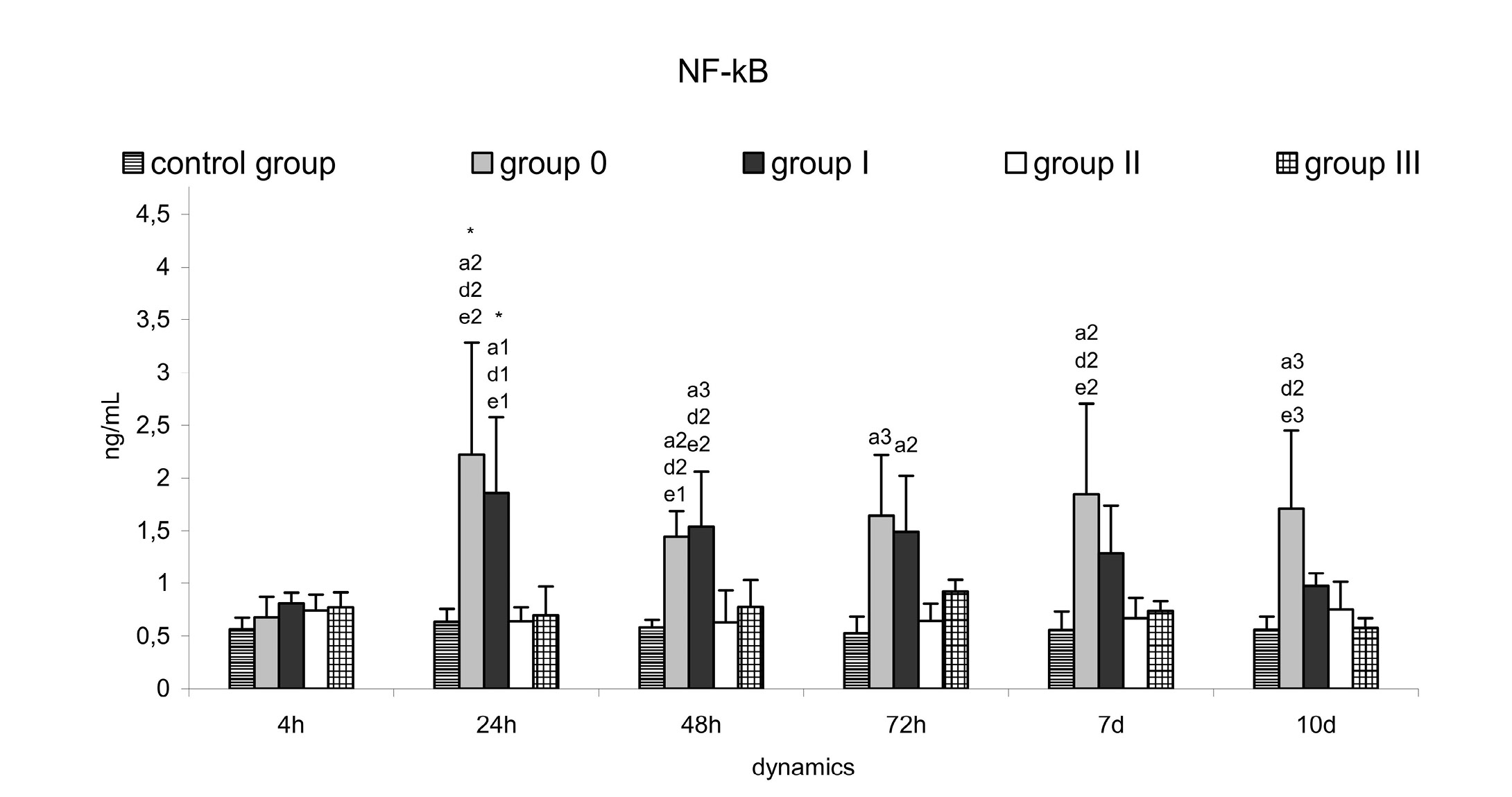
The optimisation of strategies for control of Pseudomonas aeruginosa skin infections in dogs includes minimization of induced hyper-inflammation. We aimed at monitoring changes in serum nuclear factor kappa B (NF-кB) and nitric oxide (NO) concentrations, and blood neutrophil/lymphocyte ratio (N/L ratio), to evaluate their potential as inflammatory markers throughout the course of therapy in canine skin infection experimentally induced by P. aeruginosa. A complementary therapy including antibiotic against the pathogen and NF-кB targeted host modulatory therapy by parthenolide from the phytopreparation Feverfew was applied. NF-кB was determined by ELISA kit. The NO assay was based on the Griess reaction. The N/L ratio was calculated on the basis of CBC counts. The infection was induced by P. aeruginosa (1×10⁸ CFU/mL) injected s.c. to 20 male dogs. Four experimental groups were formed (0 – untreated dogs and three treated groups: I – with antibiotic; II – with Feverfew and III – with antibiotic and Feverfew) and one control group. The results showed that NF-кB in group 0 was significantly higher than those in controls on hour 24 (p=0.0103), hour 48 (p=0.0001), hour 72 (p=0.0505), day 7 (p=0.0114), and day 10 (p=0.0094). The treatment with Feverfew (group II and III) achieved an efficient control on NF-кB. In groups I and II, biphasic increase in NO was observed, associated with the early – hour 4 (p=0.00007 for group I; p=0.0001 for group II vs. baseline) and late – day 14 (p=0.0092 for group II vs. controls) stage of infection development. By hour 24, N/L ratio in these groups increased (p=0.005 and p=0.0104 for groups I and II respectively vs. baseline). The established changes were generalised signs of inflammatory response accompanied by fever on hour 4 in infected dogs (p=0.0005 for group 0; p=0.0008 - I; p=0.0002–II; p=0.0047-III vs. controls).
Keywords | Pseudomonas aeruginosa, Canine skin infection, Inflammatory biomarkers, Therapy, Feverfew