J. Venkataravanappa Lokesh, Nitin. P. Kurade*, M.U. Shivakumar1, Anil Kumar Sharma, Swapan K. Maiti1
Division of Pathology, 1Division of Surgery, Indian Veterinary Research Institute, Izatnagar, 243 122 (U.P.), India
*Corresponding author: npkurade@yahoo.co.in
ABSTRACT
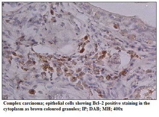
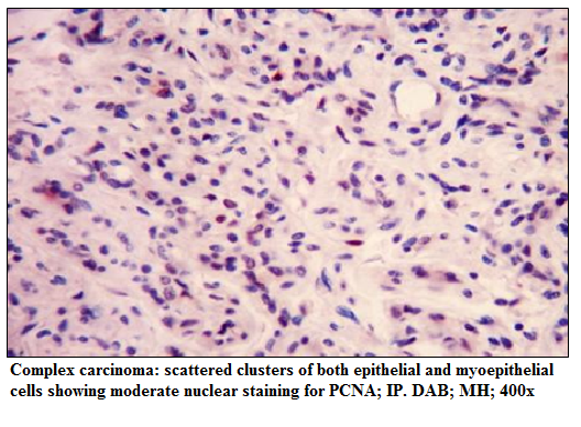
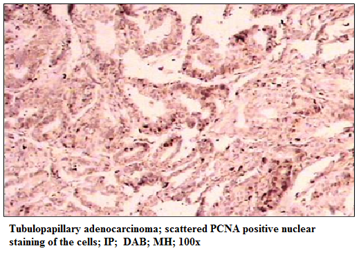
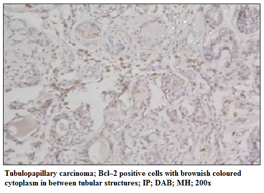
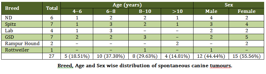
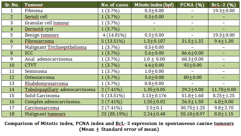
In the present study, the pathology of spontaneous canine tumours, their classifications, the expression of the BcL–2 as well as expression of the proliferation indices (mitotic and PCNA index) were investigated. A total of 27 canine tumour cases were studied and samples were collected in 10% buffered formalin. Organs wise distribution of tumour cases showed highest involvement of mammary gland (48.15%) followed by skin (29.62%), testicles (11.11%) and bone, muscle and eye (3.7% each). Histologically, 4 (14.81%) tumours were diagnosed as benign and 23 (85.19%) as malignant. Mitotic index was significantly low in benign tumours (0.3±0.00/hpf) compared to the malignant tumours (2.24±0.48/hpf). The highest Proliferating Cell Nuclear Antigen (PCNA) index was found to be in canine transmissible venereal tumour (92 %) followed by squamous cell carcinoma (86.6 %), anal adenocarcinoma (68.3±0.00), solid carcinoma (51.8±1.68), fibrosarcoma (31.5±1.32), carcinosarcoma (30.75±1.25) and tubulopapillary carcinoma (29.2±0.08). The BcL–2 immunostaining was membranous and cytoplasmic. It was mainly observed in non–proliferating and peripheral neoplastic cells. Out of 27 total tumours, only 9 showed the positive reaction to BcL–2 immunostaining. Benign tumour, fibroma showed high BcL–2 index (19.3±0.00) compared to malignant tumours (8.8±1.15). BcL–2 expression was noticed in both benign and malignant tumours and was prominent in comparatively less aggressive tumours showing mild PCNA immunostaining. Further studies, with more number of tumours, may elucidate the role of BcL–2 in progression of canine tumours.
Key Words: Canine tumours, Mitotic index, PCNA, BcL–2